BV1 Binocular and Spatial Vision
 Call Kai
 Learn
Learn Practice Test
 Spaced Repetition
 Match
Match Flashcards
 Knowt Play
1/86

There's no tags or description
Looks like no tags are added yet.
Name | Mastery | Learn | Test | Matching | Spaced | Call with Kai | Chat |
|---|
No analytics yet
Send a link to your students to track their progress
87 Terms
Why is binocular and spatial vision considered a core area of optometric expertise?
Optometrists specialize in functional vision assessment, including how the two eyes work together to perceive space. This expertise helps distinguish optometry from ophthalmology, which generally has less extensive training in binocular/spatial vision.
What are the two major types of two-eyed vertebrate vision?
Utrocular vision: the eyes function separately.
Ambiocular vision: the eyes work together, representing true binocular vision.
What is binocular vision?
Binocular vision is the coordinated behavior of the two eyes in the perception of space. It requires the eyes to work together to support spatial judgment and depth perception.
What are the major characteristics of utrocular vision?
Utrocular vision involves independent eye movements, complete crossing of the optic nerves, and is typically found in lower vertebrates.
What are the major characteristics of ambiocular vision?
Ambiocular vision involves coordinated eye movements, follows Hering’s law of equal innervation, has partial decussation of the optic nerves, and is typical of mammals.
How do optic nerve crossings differ between utrocular and ambiocular vision?
Utrocular vision: complete crossing of optic nerves.
Ambiocular vision: partial crossing/partial decussation of optic nerves.
What law is associated with ambiocular vision, and what does it mean?
Hering’s law of equal innervation: paired muscles in the two eyes receive equal neural input, allowing the eyes to move together in a coordinated way.
How does eye placement relate to visual function in prey vs predator animals?
Prey animals: usually have lateral eyes, giving a wide panoramic field for safety/detection of predators.
Predator animals: usually have frontal eyes, improving overlap between the two eyes, which supports better depth perception and visual acuity for hunting/offensive behavior.
Are frontal eyes required for ambiocular/binocular vision?
No. Frontal eyes facilitate ambiocular vision by increasing binocular overlap, but they are not strictly required for the two eyes to work together.
What is ocular laterality, and how is it measured?
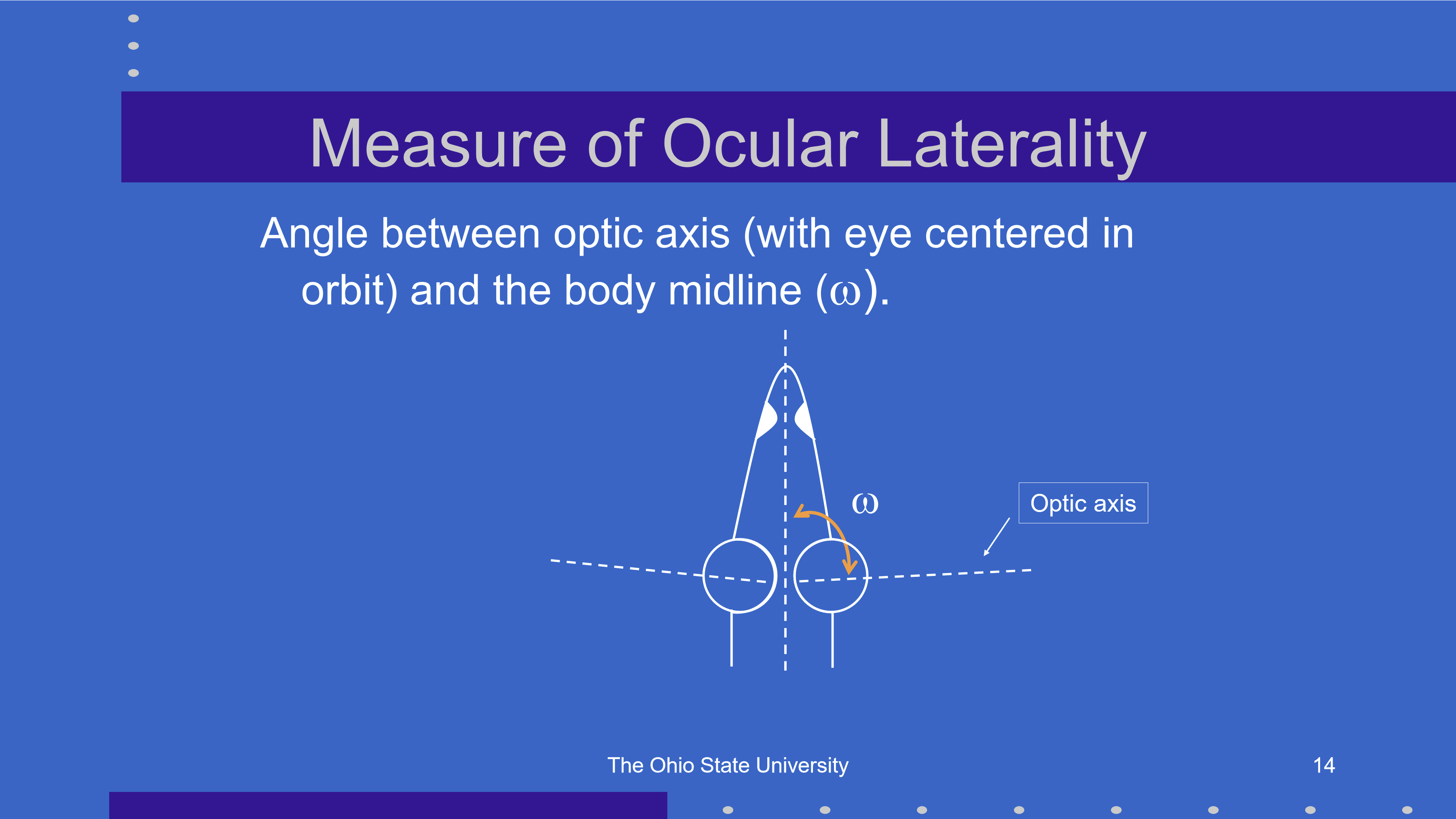
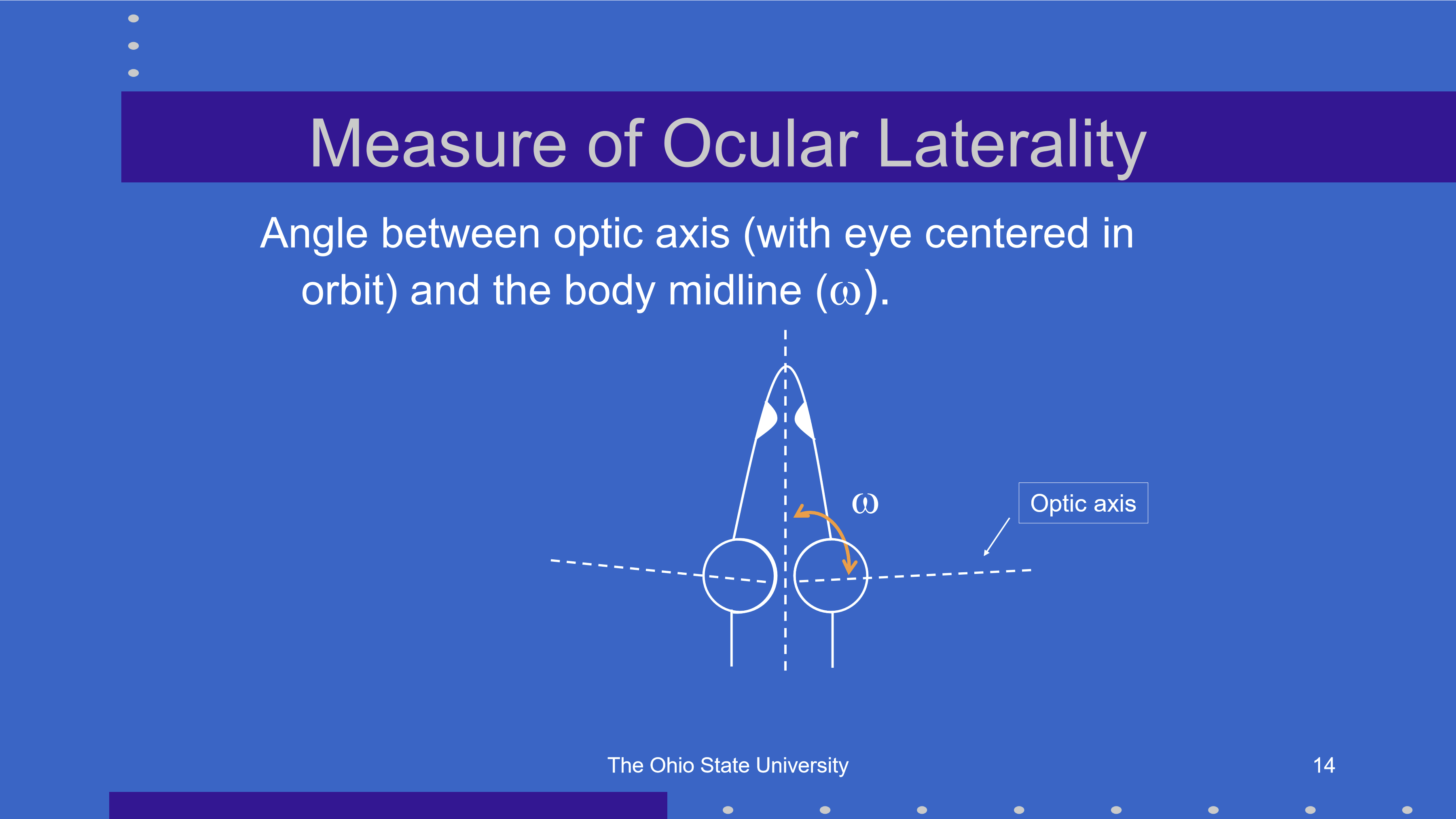
Ocular laterality is measured by the angle ω (omega) between the optic axis of the eye, with the eye centered in the orbit, and the body midline.
What does a larger ocular laterality angle generally indicate?
A larger ω angle means the eyes are positioned more laterally, typically increasing panoramic visual field but reducing binocular overlap/depth perception.
What does the angle γ represent in binocular/spatial vision?
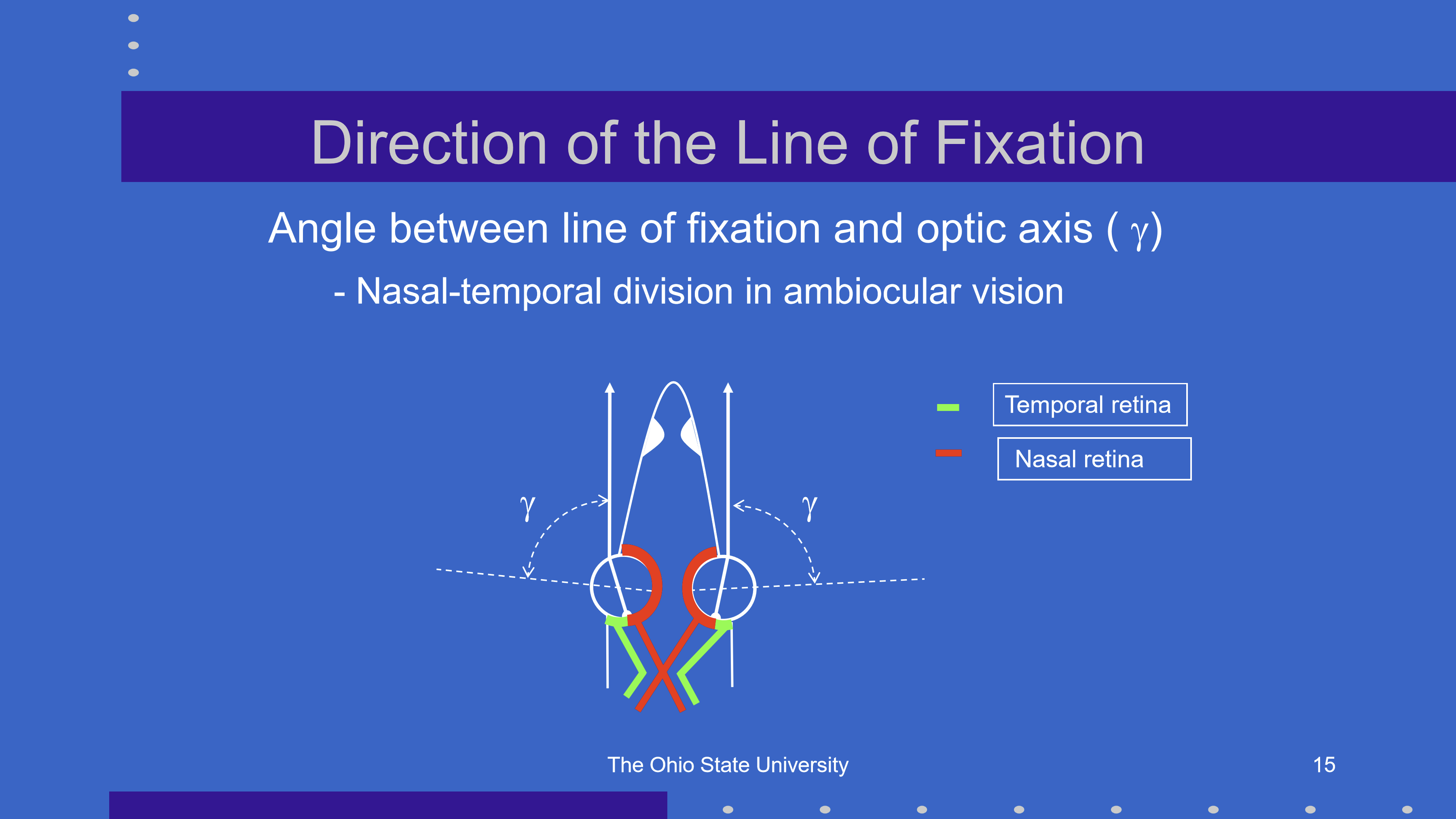
γ (gamma) is the angle between the line of fixation and the optic axis of the eye.
What retinal division is important in ambiocular vision?
Ambiocular vision involves a nasal-temporal retinal division, where nasal and temporal retinal regions contribute differently to binocular visual processing.
What is stereopsis, and why is it important in ambiocular vision?
Stereopsis is the specific sensation of depth produced by ambiocular/binocular vision. It allows more precise spatial judgments than monocular cues alone.
What makes stereopsis possible?
Stereopsis requires:
Coordinated eye movements:both eyes align on the target.
Retinal correspondence: matching retinal points in each eye represent the same visual direction.
Why can ambiocular vision improve visual acuity?
Frontal eyes tend to have a smaller γ angle, meaning the line of fixation is closer to the optic axis, producing better optical quality and sharper vision.
Was stereopsis likely the only reason ambiocular vision evolved?
Not necessarily. Ambiocular vision may have been an “afterthought,” while improved visual acuity from better optics may also have been a major advantage of ambiocular/frontal-eye systems.
How are visual directions organized in utrocular vision?
In utrocular vision, the visual directions from the two eyes are independent. Each eye assigns its own visual direction rather than combining both eyes into a single shared perceptual direction.
What is the perceptual consequence of utrocular vision?
Because the two eyes function independently, there is no common binocular visual direction. Objects seen by each eye may be perceived separately rather than fused into one unified spatial percept.
How are visual directions organized in ambiocular vision?
In ambiocular vision, the two foveas are assigned a common oculocentric direction, meaning the visual system treats both foveal images as pointing in the same perceived direction.
What law applies to visual direction in ambiocular vision?
Hering’s law of identical visual directions applies: corresponding points in the two eyes, especially the foveas, are perceived as having the same visual direction.
Why can ambiocular vision create visual confusion?
If different objects stimulate corresponding retinal points, the visual system may assign them the same perceived direction, producing confusion because two different objects appear in one visual location.
Why is bifoveal fixation necessary in ambiocular vision?
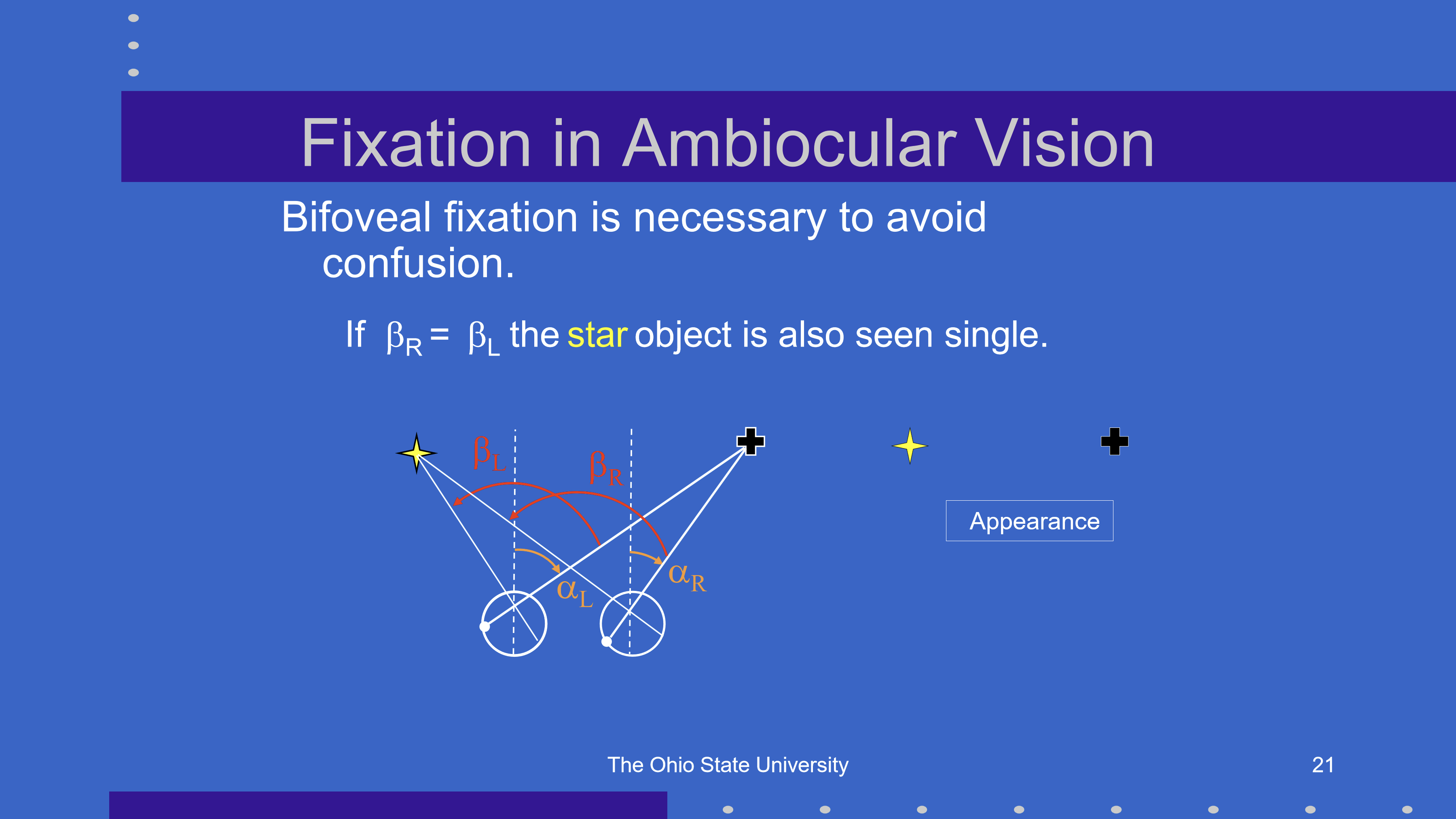
Bifoveal fixation is needed so the image of a single object falls on both foveas, allowing it to be perceived as single and avoiding visual confusion.
What condition allows the star object to be seen as single in ambiocular vision?
The star is seen single when the fixation angles are equal:
βR = βL
This means each eye is directed appropriately toward the same object, supporting single binocular perception.
What are the main ways monocular vision differs from binocular vision in humans?
Monocular and binocular vision differ in:
Depth perception
Brightness perception
Light adaptation
Light detection threshold
What is the major depth-perception advantage of binocular vision?
Binocular vision provides stereopsis, a precise sensation of depth based on comparing the two eyes’ images. Monocular vision can still use depth cues, but lacks true binocular stereopsis.
How does binocular depth perception compare with monocular depth perception at 1 meter?
Binocular depth perception has a much lower threshold than monocular depth perception, meaning it can detect much smaller depth differences.
At 1 m, binocular target separation threshold is much smaller than monocular.
Key idea: binocular vision is more sensitive for depth judgments because of stereopsis.
What does a lower depth threshold mean clinically/perceptually?
A lower threshold means better depth discrimination. The visual system can detect smaller separations between objects in depth.
Binocular < monocular threshold = better depth perception
This reflects the advantage of using retinal disparity between the two eyes.
How does perceived brightness with two eyes compare to one eye?
Perceived brightness with both eyes is usually about the same as with one eye, not twice as bright.
How are luminances from the two eyes combined in binocular brightness perception?
The visual system generally averages the luminance signals from the two eyes rather than summing them.
Not: right eye + left eye
Instead: roughly the average luminance of the two eyes
What is the distinction between binocular depth and binocular brightness processing?
Binocular vision greatly improves depth perception through stereopsis, but binocular brightness is usually not doubled because luminance signals are averaged rather than summed.
How does dark adaptation work between the two eyes?
The two eyes dark adapt independently. Patching one eye allows that eye to dark adapt without affecting the adaptation state of the other eye.
What is the clinical/perceptual significance of independent dark adaptation?
If one eye is kept in darkness, it becomes more sensitive to dim light while the uncovered eye remains adapted to the current lighting.
Are two eyes better than one for detecting dim light?
Yes. Using two eyes improves the probability of detecting a stimulus because of probability summation.
What is the probability summation equation for binocular light detection?
q = 1 − (1 − p)ⁿ
Where:
q = probability of detecting the stimulus
p = probability of detection by one eye
n = number of eyes/sensors
If each eye detects a threshold stimulus 50% of the time, what is the probability of detecting it with two eyes?
With two eyes:
q = 1 − (1 − 0.5)² = 0.75
So the probability of seeing the stimulus with two eyes is 75%.
How do binocular brightness perception and binocular light detection differ?
Brightness perception: luminance from two eyes is usually averaged, so the world does not look twice as bright.
Light detection threshold: two eyes improve detection probability through probability summation, making dim stimuli easier to detect.
What are the three main types of visual direction used to describe where something is seen?
Ocular/eye direction (α): direction relative to the individual eye
Oculocentric/relative direction (β): direction relative to the observer’s visual system
Egocentric/absolute direction (χ): direction relative to the body/self in space
What is the sign convention for visual direction measurements?
Right and up directions are positive (+)
Left and down directions are negative (−)
How do plus spectacle lenses affect perceived target direction and eye rotation?
Plus lenses create prismatic effects that shift the image, so the eye must rotate more than normal to fixate the target.
The patient must “pastpoint” farther because the object’s false location differs from its true location.
When wearing plus lenses, does the prism move the eye or the image?
The prism moves the image, not the eye. The eye then changes its rotation to look toward the shifted/false image location.
How do minus spectacle lenses affect perceived target direction and eye rotation?
Minus lenses create prismatic effects that shift the image, so the eye must rotate less than normal to fixate the target.
Plus lenses → eye rotates more
Minus lenses → eye rotates less
Compare the prismatic effects of plus vs minus spectacle lenses on eye rotation.
Plus lenses: require the eye to rotate more than normal
Minus lenses: require the eye to rotate less than normal
Both effects occur because the lens shifts the apparent/false location of the object.
How do patients adapt to the visual displacement caused by glasses?
Patients adapt by recalibrating limb proprioception to match the new visual input.
Glasses can shift perceived object location.
Over time, the patient’s motor system adjusts so reaching/pointing matches where objects appear.
Why can switching between glasses and contact lenses require adaptation?
Glasses produce prismatic/image displacement effects, while contact lenses usually move with the eye and create less of this displacement.
Over time, frequent switching may require the patient to maintain two separate visuomotor “matrices”: one for glasses and one for contact lenses.
How can traumatic brain injury affect adaptation to visual changes?
After TBI, visual adaptation is often poor, meaning patients may have difficulty recalibrating to altered visual input from glasses, contact lenses, or multifocals.
What visual changes are especially disorienting for patients with poor adaptation after TBI?
Commonly disorienting changes include:
Switching from glasses to contact lenses
Wearing bifocals
These can be difficult because they alter spatial perception and require visuomotor adaptation.
What are the three types of binocular direction?
Ocular direction: direction specified separately by each eye
Oculocentric direction: perceived direction relative to the visual system/cyclopean eye
Egocentric direction: perceived direction relative to the self/body in space
What question is binocular ocular direction trying to answer?
It asks how the separate ocular directions from each eye, αL and αR, are combined into one binocular direction.
What are the two main theories for how ocular directions combine in binocular vision?
Dominant eye theory: binocular direction follows the ocular direction of the dominant eye, either αL or αR.
Cyclopean theory: binocular direction is the average of the two eyes’ directions.
What does the cyclopean theory predict for binocular ocular direction?
Binocular direction is the average of the left and right eye directions:
αB = (αL + αR) / 2
This corresponds to the bisector of the angle of convergence.
How does dominant eye theory differ from cyclopean theory?
Dominant eye theory: one eye controls perceived direction.
Cyclopean theory: perceived direction is averaged between both eyes.
How are oculocentric directions from each eye combined in binocular vision?
Dominant eye theory: one eye’s oculocentric direction is suppressed, and perceived direction comes from the other eye.
Cyclopean theory: the two β values are averaged:
βB = (βL + βR) / 2
What does the cyclopean theory predict for binocular oculocentric direction?
The perceived oculocentric direction is the average of the left and right eye’s relative directions:
βB = (βL + βR) / 2
What information determines egocentric direction for each eye?
Egocentric direction combines:
Ocular direction (α) = where the eye is pointed
Oculocentric direction (β) = where the image falls relative to the eye
So for each eye:
Left eye: αL + βL
Right eye: αR + βR
How does dominant eye theory explain binocular egocentric direction?
Dominant eye theory says perceived egocentric direction comes from one eye only:
Either αL + βL
Or αR + βR
How does cyclopean theory explain binocular egocentric direction?
Cyclopean theory says binocular egocentric direction is the average of both eyes’ egocentric information:
ego = [(αL + βL) + (αR + βR)] / 2
Equivalent form:
ego = (αL + αR)/2 + (βL + βR)/2
What evidence supports cyclopean theory of binocular egocentric direction?
Placing a minus lens or prism before only one eye influences perceived egocentric direction.
This supports cyclopean theory because changing input to one eye still shifts binocular perceived direction, meaning both eyes contribute to the final direction judgment.
Why does a prism or minus lens before one eye argue against pure dominant eye theory?
If only the dominant eye determined direction, altering the nondominant eye should have little or no effect. But because changing one eye can shift egocentric direction, perceived direction is likely based on a binocular average, consistent with cyclopean theory.
What is the egocenter in binocular spatial vision?
The egocenter is the body reference point for egocentric direction: the point from which the visual system judges where objects are located relative to the self.
Where is the egocenter located according to dominant eye vs cyclopean theory?
Dominant eye theory: egocenter is located at the dominant eye.
Cyclopean theory: egocenter is located midway between the two eyes.
How can the egocenter be experimentally estimated?
By having a person mark points between fixed objects and their perceived locations. The pattern of marked points can be used to infer the body reference point for perceived direction.
What is the typical measured location of the egocenter?
The egocenter is usually found between the eyes, but about 10 cm behind the plane of the eyes.
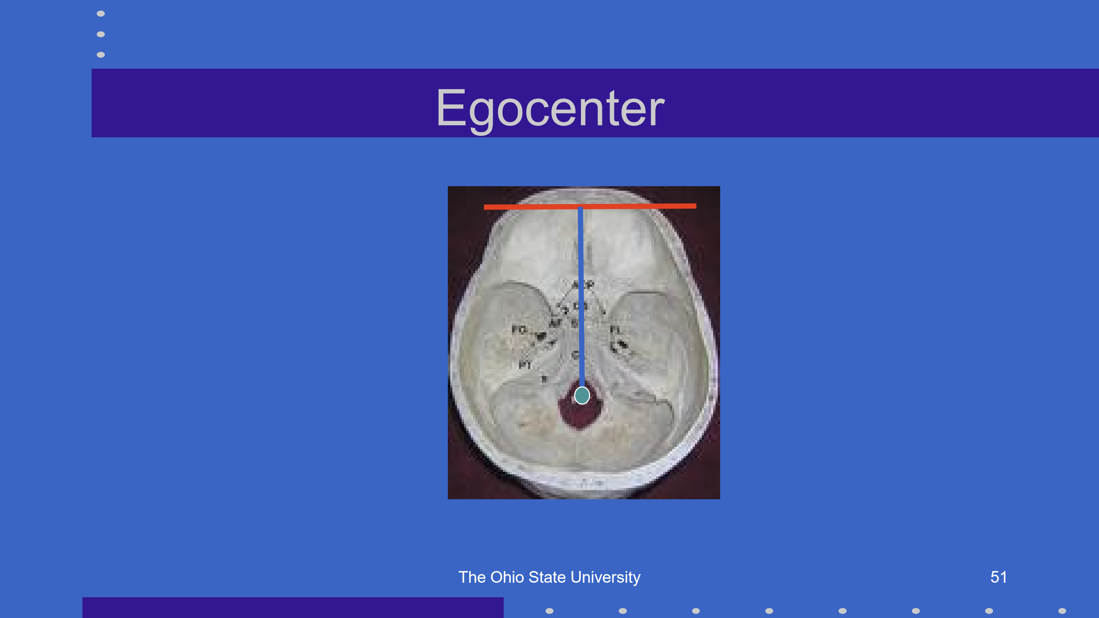
Why does the egocenter support cyclopean-style spatial perception?
Because perceived direction is referenced from a point near the midline between the eyes, not from a single dominant eye. This supports the idea that binocular spatial direction is based on an integrated/cyclopean reference system.
What are corresponding retinal points?
Corresponding points are retinal points, one in each eye, that have the same oculocentric direction.
Each eye has its own relative direction: βL and βR
Theoretically, corresponding points occur when:
βL = βR
Why is retinal correspondence important for binocular vision?
Retinal correspondence allows the brain to treat images from the two eyes as having the same visual direction, supporting single binocular perception.
Same β in each eye → same perceived direction
Mismatched/noncorresponding points → possible diplopia or spatial confusion
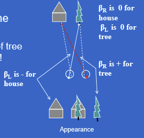
What does Hering’s window demonstrate?
Hering’s window demonstrates retinal correspondence and Hering’s law of identical visual directions: corresponding retinal points are perceived as having the same visual direction.
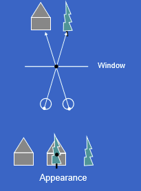
In Hering’s window, why do the tree and house appear in the same location?
When fixation is on the spot on the window, the two foveas have the same oculocentric direction, so different objects aligned with each fovea can appear in the same perceived location, causing visual confusion.
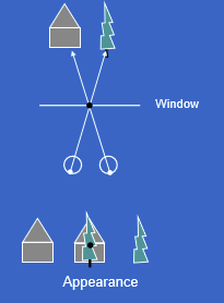
Why is diplopia present in Hering’s window?
Diplopia occurs because the tree and house do not fall on corresponding retinal points in the two eyes.
Corresponding points → single perception
Noncorresponding points → double vision/diplopia
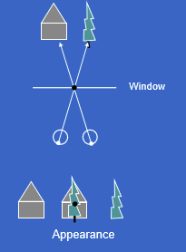
What is the key distinction between confusion and diplopia in Hering’s window?
Confusion: two different objects are perceived in the same location because they stimulate corresponding directions.
Diplopia: one object is seen twice because its images fall on noncorresponding retinal points.
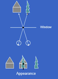
What happens during normal bifoveal fixation of an object?
With bifoveal fixation, the target object falls on both foveas and is seen singly. Other objects fall on matching corresponding retinal points, so they appear in their proper spatial locations.
Key condition: βR = βL
Why do the tree and house appear correctly in normal binocular vision?
Because their images fall on corresponding retinal point pairs in the two eyes. Corresponding points have the same oculocentric direction, allowing accurate single binocular perception.
What happens to fixation in right esotropia?
In right esotropia, the right eye turns inward, so the deviating right eye does not fixate the same object as the left eye.
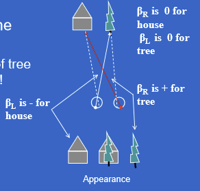
Why are perceived object locations wrong in right esotropia?
Because images fall on noncorresponding retinal points, so the brain assigns incorrect oculocentric directions. This produces abnormal perceived locations of objects.
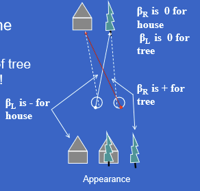
What visual symptoms can occur in right esotropia?
Right esotropia can cause both:
Confusion: different objects appear in the same perceived location
Diplopia: one object is seen twice because its images fall on noncorresponding retinal points
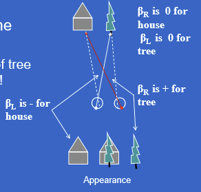
In right esotropia, why can confusion and diplopia occur together?
The deviating eye creates a mismatch between the two eyes’ retinal directions.
If two different objects stimulate corresponding directions → confusion
If one object stimulates noncorresponding points → diplopia
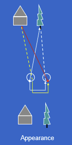
What is anomalous retinal correspondence (ARC) in right esotropia?
ARC is an adaptation where the visual system changes which retinal points are treated as corresponding.
In right esotropia, the left fovea corresponds to a nasal retinal point in the right eye instead of the right fovea.
This helps compensate for the deviating right eye.
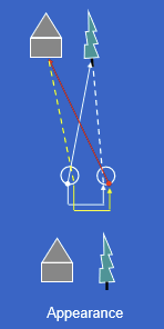
What is harmonious ARC?
Harmonious ARC occurs when the anomalous correspondence exactly offsets the strabismic deviation, allowing the two eyes to superimpose the fixated object correctly.
Result: objects appear in proper locations.
ARC can prevent confusion and diplopia.
Why might ARC not always be something to “fix”?
ARC is an adaptive sensory strategy that can reduce symptoms. If it prevents confusion and diplopia, disrupting it may make the patient more symptomatic.
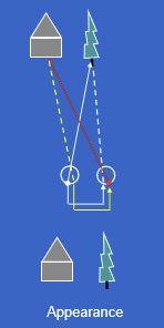
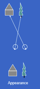
What would vision look like if there were no retinal correspondence?
Without correspondence, each eye would localize objects independently.
The tree and house could be correctly localized by each eye separately.
There would be no forced binocular matching of retinal points.
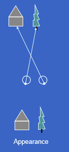
Why can lack of correspondence be confused with harmonious ARC?
Both can make objects appear correctly localized and may avoid diplopia/confusion.
Harmonious ARC: abnormal binocular correspondence compensates for deviation.
No correspondence/utrocular vision: eyes localize independently without binocular matching.
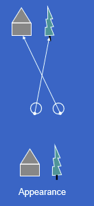
What is the key distinction between ARC and utrocular/no-correspondence vision?
ARC: the two eyes still have a binocular correspondence system, but it is shifted/adapted.
Utrocular vision: there is essentially no binocular correspondence, so each eye functions more independently.
What are corresponding retinal points?
Corresponding retinal points are retinal positions, one in each eye, that have identical oculocentric directions.
The two foveas are corresponding points.
For horizontal angles: βL = βR
What is the key condition for two retinal points to correspond?
They must have the same oculocentric/relative direction in each eye:
βL = βR
This means the brain treats those two retinal locations as representing the same visual direction.
What is retinal disparity?
Retinal disparity is the difference in oculocentric direction between the two eyes caused by stimulation of non-corresponding retinal points.
Formula:
retinal disparity = βL − βR
When is retinal disparity zero?
Retinal disparity is zero when the two eyes’ oculocentric directions are equal:
βL = βR → retinal disparity = 0
This means the images fall on corresponding retinal points.
What does retinal disparity measure perceptually?
It measures the angular separation of images between the two eyes.
High-yield idea:
Corresponding points → no disparity
Non-corresponding points → retinal disparity, which can contribute to depth perception or diplopia depending on size/context.