(2) BOOK 4: Bacteria

0.0(0)
Studied by 0 people
call kaiCall Kai
learnLearn
examPractice Test
spaced repetitionSpaced Repetition
heart puzzleMatch
flashcardsFlashcards
GameKnowt Play
Card Sorting

1/16

encourage image

There's no tags or description

Looks like no tags are added yet.

Last updated 2:54 PM on 10/9/26
Name
Mastery
Learn
Test
Matching
Spaced
Call with Kai
Chat

No analytics yet

Send a link to your students to track their progress

17 Terms

1
New cards

Bacteria Morphology

3 Common Groups of Bacteria Shape:

  • Coccus (spherical) (plural: cocci)

  • Bacillus (rod-shaped) (plural: bacilli)

  • Spirillum / Spirochete (spiral) (plural: spirilla)

* Cocci and bacilli can form clusters of various sizes
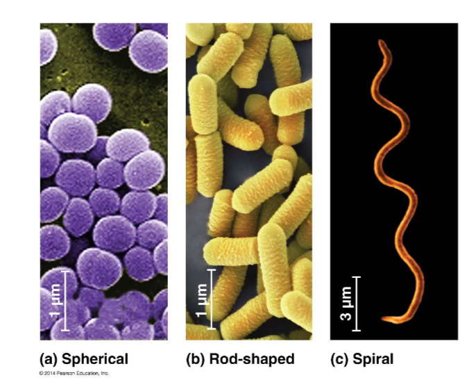

<p><span>3 Common Groups of Bacteria Shape: </span></p><ul><li><p><span>Coccus (spherical) (plural: cocci)</span></p></li><li><p><span>Bacillus (rod-shaped) (plural: bacilli)</span></p></li><li><p><span>Spirillum / Spirochete (spiral) (plural: spirilla)</span></p></li></ul><p><span>* Cocci and bacilli can form clusters of various sizes</span></p><p></p>
2
New cards

Bacteria Cell Ultrastructure

Overview:

  • Bacteria are single-celled microorganisms

  • They lack a nuclear membrane

  • They are capable of metabolic activities

  • They divide by binary fission

    • Many bacteria multiply at rapid rates

  • They exist widely in both parasitic and free-living forms


Sizes:

  • Vary from less than 1 micro m to approximately 10 micro m


Prokaryotes:

  • All bacterial cells are prokaryotic, and hence lack a true nucleus and membrane-bound organelles

  • This distinguishes them from eukaryotic cells, which possess a membrane-bound nucleus and organelles


<p><mark data-color="yellow" style="background-color: yellow; color: inherit;">Overview:</mark></p><ul><li><p>Bacteria are single-celled microorganisms</p></li><li><p>They lack a nuclear membrane</p></li><li><p>They are capable of metabolic activities</p></li><li><p>They divide by binary fission</p><ul><li><p>Many bacteria multiply at rapid rates</p></li></ul></li><li><p>They exist widely in both parasitic and free-living forms</p></li></ul><p></p><p class="has-focus is-empty"><mark data-color="yellow" style="background-color: yellow; color: inherit;">Sizes:</mark></p><ul><li><p class="has-focus is-empty">Vary from less than 1 micro m to approximately 10 micro m</p></li></ul><p class="has-focus is-empty"></p><p class="has-focus is-empty"><mark data-color="yellow" style="background-color: yellow; color: inherit;">Prokaryotes:</mark></p><ul><li><p class="has-focus is-empty">All bacterial cells are <strong>prokaryotic</strong>, and hence <strong>lack a true nucleus and membrane-bound organelles</strong></p></li><li><p class="has-focus is-empty">This distinguishes them from eukaryotic cells, which possess a membrane-bound nucleus and organelles</p></li></ul><p></p>
3
New cards

Bacteria Cell Ultrastructure — Cell Surface Structures — CELL WALL

Cell Wall:

  • All bacterial cells possess a cell wall composed of peptidoglycan

    • Peptidoglycan is a cross-linked polymeric mesh (VS cellulose cell wall in plants)

    • The glycan component is a linear polymer of alternating monosaccharide subunits of N-acetylglucosamine (NAG) and N-acetylmuramic acid (NAM)

      • This polymer is the carbohydrate “backbone” of the mesh

      • β(1,4)-glycosidic bond between the monomers make up the polysaccharide

      • The bonds are cleaved by the anti-bacterial enzyme lysozyme

    • The “peptido” portion of the polymer is a short string of amino acids that serves as to cross-link adjacent polysaccharide strands at the NAM subunits of the backbone, forming a network with high tensile strength

      • The presence of D-amino acids helps to render the bacterial cell wall resistant to host peptidases, such as those in the intestine


  • Gram-positive bacteria:

    • They have thick, multi-layered, peptidoglycan cell walls that are exterior to the membrane

    • The peptidoglycan in most Gram-positive species is covalently linked to teichoic acid, which is essentially a polymer of substituted glycerol units linked by phosphodiester bonds

    • The teichoic acids are major cell surface antigens


  • Gram-negative bacteria:

    • They have two membranes – an outer membrane and an inner (cytoplasmic) membrane

    • The peptidoglycan layer is located between the two membranes in the periplasmic space

    • The periplasmic space also contains enzymes and various other substances

    • In contrast to Gram-positive cells, the peptidoglycan layer of Gram-negative cells is thin, and the cells are consequently more susceptible to physical damage

    • The outer membrane is distinguished by the presence of various embedded lipopolysaccharides

      • The polysaccharide portion (O-polysaccharide) is antigenic, used to identify different strains and species

      • The lipid portion (called lipid A) is toxic to humans and animals

        • Since lipid A is an integral part of the membrane, it is called an endotoxin

        • As opposed to exotoxins, which are secreted substances


  • Gram staining procedure:

    • Initially uses the dye crystal violet, which is taken up by all bacteria

    • The cells are then treated with iodine as a mordant (a substance that sets the colour and makes it permanent), which helps the Gram-positive cells retain the crystal violet

    • When alcohol is added in the next step, the Gram-negative cells lose their colour but the Gram-positive cells retain the violet dye

    • As the now-colourless Gram-negative cells would be invisible under a light microscope at this point, they are counterstained with the red dye safranin


<p><strong>Cell Wall:</strong></p><ul><li><p>All bacterial cells possess a cell wall composed of <strong>peptidoglycan</strong></p><ul><li><p>Peptidoglycan is a <u>cross-linked</u> <u>polymeric mesh</u> (VS cellulose cell wall in plants)</p></li><li><p>The <strong>glycan</strong> component is a <strong>linear polymer of <u>alternating monosaccharide</u> subunits</strong> of <u>N-acetylglucosamine (NAG)</u> and <u>N-acetylmuramic acid (NAM)</u></p><ul><li><p class="has-focus">This polymer is the carbohydrate “backbone” of the mesh</p></li><li><p class="has-focus"><u>β(1,4)-glycosidic bond</u> between the monomers make up the polysaccharide</p></li><li><p class="has-focus">The bonds are cleaved by the anti-bacterial enzyme <u>lysozyme</u></p></li></ul></li><li><p class="has-focus">The <strong>“peptido”</strong> portion of the polymer is a <strong>short string of amino acids</strong> that serves as to <strong>cross-link adjacent polysaccharide strands</strong> at the NAM subunits of the backbone, forming a network with <strong>high tensile strength</strong></p><ul><li><p class="has-focus">The presence of D-amino acids helps to render the bacterial cell wall resistant to host peptidases, such as those in the intestine</p></li></ul></li></ul></li></ul><p class="has-focus"></p><ul><li><p class="has-focus">Gram-positive bacteria:</p><ul><li><p class="is-empty has-focus">They have thick, multi-layered, peptidoglycan cell walls that are exterior to the membrane</p></li><li><p class="is-empty has-focus">The peptidoglycan in most Gram-positive species is covalently linked to <u>teichoic acid</u>, which is essentially a polymer of substituted glycerol units linked by <u>phosphodiester bonds</u></p></li><li><p class="is-empty has-focus">The teichoic acids are major cell surface antigens</p></li></ul></li></ul><p></p><ul><li><p class="has-focus is-empty">Gram-negative bacteria:</p><ul><li><p class="has-focus is-empty">They have two membranes – an outer membrane and an inner (cytoplasmic) membrane</p></li><li><p class="has-focus is-empty">The peptidoglycan layer is located between the two membranes in the periplasmic space </p></li><li><p class="has-focus is-empty">The periplasmic space also contains enzymes and various other substances</p></li><li><p class="has-focus is-empty">In contrast to Gram-positive cells, the peptidoglycan layer of Gram-negative cells is thin, and the cells are consequently more susceptible to physical damage</p></li><li><p class="has-focus is-empty">The outer membrane is distinguished by the presence of various embedded <u>lipopolysaccharides</u> </p><ul><li><p class="has-focus is-empty">The polysaccharide portion (O-polysaccharide) is antigenic, used to identify different strains and species </p></li><li><p class="has-focus is-empty">The lipid portion (called lipid A) is toxic to humans and animals</p><ul><li><p class="has-focus is-empty">Since lipid A is an integral part of the membrane, it is called an <u>endotoxin</u></p></li><li><p class="is-empty has-focus">As opposed to <u>exotoxins</u>, which are secreted substances</p></li></ul></li></ul></li></ul></li></ul><p class="is-empty has-focus"></p><ul><li><p class="is-empty has-focus">Gram staining procedure:</p><ul><li><p class="is-empty has-focus">Initially uses the dye crystal violet, which is taken up by all bacteria</p></li><li><p class="is-empty has-focus">The cells are then treated with iodine as a mordant (a substance that sets the colour and makes it permanent), which helps the <u>Gram-positive cells</u> retain the <u>crystal violet</u> </p></li><li><p class="is-empty has-focus">When alcohol is added in the next step, the <u>Gram-negative cells lose their colour</u> but the Gram-positive cells retain the violet dye</p></li><li><p class="is-empty has-focus">As the now-colourless Gram-negative cells would be invisible under a light microscope at this point, they are <u>counterstained with the red dye safranin</u></p></li></ul></li></ul><p></p>
4
New cards

Bacteria Cell Ultrastructure — Cell Surface Structures

  • Cell membrane

    • Similar in composition and function to those of eukaryotic cells

    • They are phospholipid bilayers in which specific proteins are embedded


  • Flagellum

    • Motile bacterial cells have flagella, long filamentous appendages that aid in cell movement


  • Pilus / Fimbriae

    • Long, thin appendages that can be used to attach one bacterial cell to another during conjugation


5
New cards

Cell Ultrastructure — Cytoplasmic Structures

  • Metabolites & Enzymes:

    • They are not enclosed within membrane-bound organelles but are found in the cytoplasm


  • Ribosomes:

    • They are found in large numbers in the cytoplasm

    • These ribosomes are different in structure from eukaryotic ribosomes

    • They are consequently smaller at 70S rather as compared to the 80S ribosomes of eukaryotes


6
New cards

Prokaryotic VS Eukaryotic

knowt flashcard image
7
New cards

Endosymbiont Theory

Endosymbiont theory:

  • It is an evolutionary theory that explains the origin of eukaryotic cells from prokaryotes

  • It states that mitochondria and plastids (chloroplasts and other related organelles) of eukaryotes originated as a result of symbiosis (mutually beneficial relationship) between separate single-celled organisms


Serial Endosymbiosis:

  • According to this theory, an early ancestor of eukaryotic cell engulfed an oxygen-using non-photosynthetic prokaryotic cell

  • Eventually, the engulfed cell formed a relationship with the host cell in which it was enclosed, becoming an endosymbiont (a cell living within another cell)

  • Over the course of evolution, the host cell and its endosymbiont merged into a single organism, a eukaryotic cell with a mitochondrion

  • Although all eukaryotes have mitochondria or remnants of these organelles, they do not all have plastids

  • Thus the hypothesis of serial endosymbiosis hypothesizes that mitochondria have evolved before plastids through a sequence of endosymbiotic events, forming the ancestor of eukaryotic cells that contain chloroplasts


Evidence for Endosymbiont Theory:

  • The inner membranes of both organelles have enzymes and transport systems that are similar to those found in the plasma membranes of living prokaryotes

  • Mitochondria and plastids replicate by a splitting process similar to certain prokaryotes

  • Mitochondria and plastids contain circular DNA which are not associated with histones, resembling chromosomes of bacteria

  • Mitochondria and plastids possess cellular machinery (including ribosomes) needed to transcribe and translate their DNA into proteins.

  • Ribosomes of mitochondria and plastids are also more similar to prokaryotic ribosomes in terms of size, RNA sequences, and sensitivity to antibiotics as compared to cytoplasmic ribosomes of eukaryotic cells


8
New cards

Bacteria Genome

Bacterial genome:

  • It is not separated from the cytoplasm by a nuclear membrane

  • It is aggregated in a region of the cell — Nucleoid

  • Unlike the eukaryotic genome which stores all genetic material on linear chromosomes, genetic material in bacteria is stored on either the circular bacterial chromosome OR plasmids


9
New cards

Bacteria Genome — Bacterial Chromosome

Overall Structure:

  • A single, circular, double-stranded DNA molecule

  • It contains essential genes required for survival

  • To fit within the bacteria cell, the bacterial chromosome must be compacted approximately 1000-fold

    • This level of compaction is attained by the association of the DNA with positively charged histone-like proteins that aid in compacting the DNA into looped domains

    • Further compaction is then achieved by supercoiling the bacterial DNA, aided by other specific bacterial proteins


Genes / Operons:

  • Genes are grouped into operons

    • In 1 operon:

      • Multiple genes come under the control of the same promoter

      • These genes come under the control of the same regulatory elements

  • Prokaryotic genes also lack introns

    • They do not require splicing after transcription, unlike eukaryotic gene expression


10
New cards

Bacteria Genome — Bacterial Plasmids

Occurence:

  • Many bacteria contain both chromosome(s) and plasmid(s)


Plasmids:

  • They are non-essential pieces of DNA

    • VS chromosomes contain the minimal genetic requirement for bacterial survival

  • Plasmids exist as small, circular, double-stranded extrachromosomal DNA molecule

  • They may be passed on to cells of the same generation (eg: conjugation, transformation and transduction)

  • Or they may be passed on to its offspring through binary fission

  • Plasmids are capable of replication independent of the bacterial chromosome since they possess their own origin of replication

    • Cells may therefore contain more than one plasmid


Genes in Plasmids:

  • Plasmids contain beneficial genes which confer protective traits such as antibiotic resistance, toxin synthesis and enzyme production

  • They are said to confer a selective advantage to bacteria, allowing them to be readily manipulated in the laboratory and making them an important agent in modern recombinant DNA technology


<p class="has-focus"><mark data-color="yellow" style="background-color: yellow; color: inherit;">Occurence:</mark></p><ul><li><p>Many bacteria contain both chromosome(s) and plasmid(s)</p></li></ul><p class="has-focus"></p><p class="has-focus"><mark data-color="yellow" style="background-color: yellow; color: inherit;">Plasmids:</mark></p><ul><li><p class="has-focus">They are non-essential pieces of DNA</p><ul><li><p class="has-focus">VS chromosomes contain the minimal genetic requirement for bacterial survival</p></li></ul></li><li><p class="has-focus">Plasmids exist as small, circular, double-stranded extrachromosomal DNA molecule</p></li><li><p class="has-focus">They may be passed on to cells of the <u>same generation</u> (eg: conjugation, transformation and transduction)</p></li><li><p class="has-focus">Or they may be passed on to its offspring through binary fission</p></li><li><p class="has-focus">Plasmids are <u>capable of replication</u> <u>independent</u> of the <u>bacterial chromosome</u> since they possess their own origin of replication</p><ul><li><p class="has-focus">Cells may therefore contain more than one plasmid</p></li></ul></li></ul><p class="has-focus"></p><p class="has-focus"><mark data-color="yellow" style="background-color: yellow; color: inherit;">Genes in Plasmids:</mark></p><ul><li><p>Plasmids contain beneficial genes which confer <u>protective traits</u> such as <u>antibiotic resistance, toxin synthesis and enzyme production</u></p></li><li><p class="has-focus">They are said to confer a <u>selective advantage</u> to bacteria, allowing them to be readily manipulated in the laboratory and making them an important agent in modern recombinant DNA technology</p></li></ul><p></p>
11
New cards

Role of Genetic Material in Bacteria

Genetic Material of Bacterial Cells is utilized in:

  1. Replication — Binary Fission

  2. Gene transfer — Recombination

  3. Gene expression


12
New cards

Role of Genetic Material in Bacteria — Replication through Binary Fission (parents to offspring)

Binary Fission:

  • It is the transmission of genetic material from a bacterial cell to its offspring

  • It is a form of asexual reproduction in which two equal-sized genetically identical daughter cells are produced from a single parent cell

  • It includes replication of the bacterial chromosome


Genetic Variation:

  • Binary fission is unable to give rise to genetic variation in a bacterial population

  • Only transfer of genetic material via transformation, conjugation and transduction are employed in the generation of genetic variation in bacteria


Process of Binary Fission:

  • The bacterial chromosome is attached to the plasma membrane before DNA replication

  • DNA replication begins at the single origin of replication

    • Replication bubble is first formed when the two DNA strands separate

    • Each parental strand is used as a template for the synthesis of the daughter strand in semi-conservative DNA replication

    • The replication bubble grows bidirectionally away from the origin of replication until the entire bacterial chromosome is replicated

  • After DNA replication is completed, cell growth occurs

    • Each circular DNA molecule is attached to the cell membrane

  • The cell elongates, and membrane growth causes the two chromosomes to be moved apart

  • Cell division in bacteria is controlled by the septal ring

    • Septal ring → A group of proteins which directs the assembly of the septum

  • The septum eventually separates the two daughter cells

    • The septum extends as the cell membrane invaginates (grows inwards) as new cell membrane and cell wall materials (e.g. peptidoglycan) are added to it

    • The invaginating cell membrane, together with the newly formed septum, splits the cell into two genetically identical daughter cells by cytokinesis


<p class="has-focus"><mark data-color="yellow" style="background-color: yellow; color: inherit;">Binary Fission:</mark></p><ul><li><p>It is the transmission of genetic material from a bacterial cell to its offspring</p></li><li><p class="has-focus">It is a form of asexual reproduction in which two <u>equal-sized genetically identical daughter cells</u> are produced from a single parent cell</p></li><li><p class="has-focus">It includes <u>replication of the bacterial chromosome</u></p></li></ul><p class="has-focus"></p><p class="has-focus"><mark data-color="yellow" style="background-color: yellow; color: inherit;">Genetic Variation:</mark></p><ul><li><p class="has-focus">Binary fission is <u>unable</u> to give rise to <u>genetic variation</u> in a bacterial population</p></li><li><p class="has-focus">Only transfer of genetic material via <u>transformation, conjugation and transduction</u> are employed in the generation of <u>genetic variation</u> in bacteria</p></li></ul><p></p><p class="has-focus is-empty"><mark data-color="yellow" style="background-color: yellow; color: inherit;">Process of Binary Fission:</mark></p><ul><li><p class="has-focus">The bacterial <u>chromosome</u> is <u>attached</u> to the <u>plasma membrane</u> before DNA replication</p></li><li><p class="has-focus">DNA replication begins at the <u>single origin of replication</u></p><ul><li><p class="has-focus">Replication bubble is first formed when the <u>two DNA strands separate</u></p></li><li><p class="has-focus">Each parental strand is used as a template for the synthesis of the daughter strand in <u>semi-conservative DNA replication</u></p></li><li><p>The replication bubble grows <u>bidirectionally</u> <u>away from the origin of replication</u> until the entire bacterial chromosome is replicated</p></li></ul></li><li><p>After DNA replication is completed, cell growth occurs</p><ul><li><p class="has-focus">Each <u>circular DNA molecule</u> is <u>attached</u> to the <u>cell membrane</u></p></li></ul></li><li><p>The cell <u>elongates</u>, and <u>membrane growth</u> causes the <u>two chromosomes to be moved apart</u></p></li><li><p>Cell division in bacteria is controlled by the septal ring</p><ul><li><p class="has-focus">Septal ring → A group of proteins which directs the assembly of the septum</p></li></ul></li></ul><ul><li><p class="has-focus"><span style="font-size: 1.6rem;">The septum eventually separates the two daughter cells</span></p><ul><li><p>The <u>septum extends</u> as the cell membrane <u>invaginates</u> (grows inwards) as new cell membrane and cell wall materials (e.g. peptidoglycan) are added to it</p></li><li><p class="has-focus">The invaginating cell membrane, together with the newly formed septum, splits the cell into two genetically identical daughter cells by <u>cytokinesis</u></p></li></ul></li></ul><p></p>
13
New cards

Role of Genetic Material in Bacteria — Gene Transfer (one cell to another)

Gene Transfer:

  • Transferring of genetic materials between bacterial cells that do not share a parent-offspring relationship

  • This allows for genetic recombination and genetic variation in bacterial populations

    • Essential for natural selection

  • It involves a donor cell (which provides the DNA to be transferred) and a recipient cell (which takes up the transferred DNA)

  • DNA is exchanged via:

    • Transformation OR

    • Transduction OR

    • Conjugation


Homologous Recombination:

  • Occurs only in transformation and transduction

  • DNA is integrated into the chromosome of the recipient cell

  • Occurs naturally and is needed by cells to accurately repair harmful breaks that occur on both strands of DNA

  • The transferred portion of the donor chromosome will be exchanged with a portion of the chromosome of the recipient cell that is very similar in sequence

    • The segment of the recipient chromosome that is exchanged for the donor chromosome is excised and degraded

  • Successful gene transfer usually occurs between bacteria of closely related species that have very similar DNA sequences due to the need for homology between the exchanged segments

    • Cells that have successfully taken up donor DNA → Recombinant cells

    • Homologous recombination does not occur in conjugation since the F plasmid is not integrated into the recipient’s chromosome


14
New cards

Role of Genetic Material in Bacteria — Gene Transfer — Transformation

Definition:

  • Transformation is the process by which a recipient cell takes up small fragments of naked DNA from the surrounding environment

  • This DNA can originate from either:

    • A donor bacterial cell which lyses and releases its DNA into the surrounding environment

    • Artificially constructed plasmids


Occurrence:

  • Only competent bacterial cells are naturally able to undergo transformation

  • Competence depends on the presence of competence factors produced by the bacterial cell

    • These are cell surface proteins that bind to DNA fragments and aid in their uptake


Artificial Transformation:

  • Transformation can be induced artificially in the laboratory

  • Artificially increasing competency in bacteria:

    • Treatment of bacteria with calcium chloride followed by heat shock

    • Electroporation

  • These artificial methods of transformation utilize plasmid DNA instead of linear DNA fragments and can be used in bacterial species which do not naturally transform


<p><mark data-color="yellow" style="background-color: yellow; color: inherit;">Definition:</mark></p><ul><li><p>Transformation is the process by which a <u>recipient cell</u> <u>takes up small fragments</u> of naked DNA from the surrounding environment</p></li><li><p>This DNA can originate from either:</p><ul><li><p>A donor bacterial cell which lyses and releases its DNA into the surrounding environment</p></li><li><p>Artificially constructed plasmids</p></li></ul></li></ul><p></p><p><mark data-color="yellow" style="background-color: yellow; color: inherit;">Occurrence:</mark></p><ul><li><p>Only <u>competent</u> bacterial cells are naturally able to undergo transformation</p></li><li><p>Competence depends on the presence of <u>competence factors</u> produced by the bacterial cell </p><ul><li><p>These are <u>cell surface proteins</u> that bind to DNA fragments and aid in their uptake</p></li></ul><p></p></li></ul><p><mark data-color="yellow" style="background-color: yellow; color: inherit;">Artificial Transformation:</mark></p><ul><li><p>Transformation can be induced artificially in the laboratory</p></li><li><p>Artificially increasing competency in bacteria:</p><ul><li><p>Treatment of bacteria with <u>calcium chloride</u> followed by <u>heat shock</u></p></li><li><p><u>Electroporation</u></p></li></ul></li><li><p>These artificial methods of transformation utilize <u>plasmid DNA</u> instead of linear DNA fragments and <strong>can be used in bacterial species which do not naturally transform</strong></p></li></ul><p></p>
15
New cards

Role of Genetic Material in Bacteria — Gene Transfer — Transduction

Overview:

  • Bacteriophages (virus) carry bacterial genes from their first host cell to their second host cell due to errors in the phage reproductive cycle

  • The DNA fragment of the donor cell may be incorporated into the genome/chromosome of the recipient cell via homologous recombination

  • 2 forms of transduction:

    • Generalised transduction

    • Specialised transduction


Generalised Transduction:

  • It is the transduction mediated by virulent phages

    • Each portion of the bacterial genome has approximately the same probability of being transferred from donor to recipient bacteria (any bacterial gene can potentially be transferred by this method)

  • During the reproduction of virulent phages (T4 phage), a small fraction of the virions produced during the lytic cycle contain a random fragment of the bacterial genome instead of phage DNA

    • Due to the accidental incorporation of a random fragment of DNA from its first host cell (donor bacterium) into the phage capsid

  • When defective phage (carrying additional host genes) infects a second host cell (another bacterium), the DNA of the donor bacterium is inserted into the recipient bacterium

  • Followed by the integration of the donor genes into the recipient cell’s genome by homologous recombination


Specialised Transduction:

  • It is the transduction mediated by temperate phages

    • Only specific portions of the bacterial genome have a high probability of being transferred from donor to recipient bacteria

    • They are genes near the prophage insertion site on the host (donor) chromosome

  • During the reproduction of temperate phages (lambda phage), the phage genome is integrated into the first host cell’s chromosome as a prophage during the lysogenic cycle

  • Upon induction, the phage genome is excised and the cell is switched to the lytic cycle

  • The excision of the prophage is sometimes imprecise, resulting in segments of phage DNA that lack part of the normal phage genome and contain part of the bacterial chromosome located adjacent to the prophage attachment site

  • When such a phage infects a second host cell, the DNA of the donor bacterium is inserted into the recipient bacterium along with a portion of the phage genome OR

  • Integration of the donor genes into the recipient cell’s genome by homologous recombination can occur


<p><mark data-color="yellow" style="background-color: yellow; color: inherit;">Overview:</mark></p><ul><li><p>Bacteriophages (virus) carry bacterial genes from their first host cell to their second host cell due to <u>errors</u> in the <u>phage reproductive cycle</u></p></li><li><p>The DNA fragment of the donor cell may be incorporated into the genome/chromosome of the recipient cell via homologous recombination</p></li><li><p>2 forms of transduction:</p><ul><li><p>Generalised transduction </p></li><li><p>Specialised transduction </p></li></ul></li></ul><p></p><p><mark data-color="yellow" style="background-color: yellow; color: inherit;">Generalised Transduction: </mark></p><ul><li><p>It is the transduction mediated by virulent phages</p><ul><li><p><u>Each portion</u> of the bacterial genome has approximately the <u>same probability</u> of being transferred from donor to recipient bacteria (<u>any bacterial gene</u> can potentially be transferred by this method)</p></li></ul></li><li><p>During the reproduction of virulent phages (T4 phage), a <u>small fraction of the virions</u> produced during the lytic cycle contain a <u>random fragment of the bacterial genome</u> instead of phage DNA</p><ul><li><p>Due to the <u>accidental incorporation</u> of a random fragment of DNA from its first host cell (donor bacterium) into the phage capsid</p></li></ul></li><li><p>When defective phage (carrying additional host genes) infects a second host cell (another bacterium), the DNA of the donor bacterium is inserted into the recipient bacterium</p></li><li><p>Followed by the <u>integration of the donor genes</u> into the <u>recipient cell’s genome</u> by <u>homologous recombination</u></p></li></ul><p></p><p><mark data-color="yellow" style="background-color: yellow; color: inherit;">Specialised Transduction:</mark></p><ul><li><p>It is the transduction mediated by temperate phages </p><ul><li><p>Only <u>specific portions</u> of the bacterial genome have a high probability of being transferred from donor to recipient bacteria</p></li><li><p>They are genes <u>near the prophage insertion site</u> on the host (donor) chromosome </p></li></ul></li><li><p>During the reproduction of temperate phages (lambda phage), the <u>phage genome is integrated</u> into the <u>first host cell’s chromosome</u> as a prophage during the <u>lysogenic cycle</u></p></li><li><p>Upon induction, the <u>phage genome</u> is <u>excised</u> and the cell is <u>switched to the lytic cycle</u></p></li><li><p>The excision of the prophage is sometimes <u>imprecise</u>, resulting in segments of phage DNA that lack part of the normal phage genome and <u>contain part of the bacterial chromosome</u> located <u>adjacent to the prophage attachment site</u></p></li><li><p>When such a phage infects a second host cell, the DNA of the donor bacterium is inserted into the recipient bacterium along with a portion of the phage genome  OR</p></li><li><p>Integration of the donor genes into the recipient cell’s genome by homologous recombination can occur</p></li></ul><p></p>
16
New cards

Generalised Transduction VS Specialised Transduction

knowt flashcard image
17
New cards

Role of Genetic Material in Bacteria — Gene Transfer — Conjugation

Definition:

  • In conjugation, direct contact between the donor and recipient bacteria leads to establishment of a cytoplasmic bridge between them, followed by transfer of the donor’s DNA to the recipient cell


Donor bacterial cell:

  • It possesses an F factor (not a.a.), which exists as the F plasmid or as a segment of DNA integrated into the bacterial chromosome

  • Genes on the F factor are responsible for synthesis of the sex pilus (facilitating cell-to-cell contact) and transfer of the F factor from a donor cell to a recipient cell

  • The F factor has its own origin of replication and replicates as it is transferred, resulting in both the original donor and recipient cells containing an F factor after conjugation is completed

  • Cells with an extrachromosomal F factor (F plasmid) → F+ cells

  • Cells lacking the F factor → F- cells


<p>Definition:</p><ul><li><p>In conjugation, <u>direct contact</u> between the donor and recipient bacteria leads to establishment of a <u>cytoplasmic bridge</u> between them, followed by <u>transfer of the donor’s DNA</u> to the recipient cell</p></li></ul><p></p><p>Donor bacterial cell:</p><ul><li><p>It possesses an <u>F factor</u> (not a.a.), which exists as the <u>F plasmid</u> or as <u>a segment of DNA integrated</u> into the bacterial chromosome</p></li><li><p>Genes on the F factor are responsible for <u>synthesis</u> of the <u>sex pilus</u> (facilitating cell-to-cell contact) and <u>transfer of the F factor</u> from a donor cell to a recipient cell </p></li><li><p>The F factor has its <u>own origin of replication</u> and <u>replicates as it is transferred</u>, resulting in both the original donor and recipient cells containing an F factor after conjugation is completed</p></li><li><p>Cells with an extrachromosomal F factor (F plasmid) → F<sup>+</sup> cells</p></li><li><p>Cells lacking the F factor → F<sup>-</sup> cells</p></li></ul><p></p>



