Outcome 3: Fetal Head, Neck, and Face
 Call Kai
 Learn
Learn Practice Test
 Spaced Repetition
 Match
Match Flashcards
 Knowt Play
1/53

There's no tags or description
Looks like no tags are added yet.
Name | Mastery | Learn | Test | Matching | Spaced | Call with Kai | Chat |
|---|
No analytics yet
Send a link to your students to track their progress
54 Terms
central nervous system (CNS)
includes brain and spinal cord
responsible for processing info and controlling body functions
CNS abnormalities occur in ~1.5% of live births and 3-6% of stillbirths and are the most common cause of referral for prenatal diagnosis
screening tests, like maternal screening and NT exams help identify potential pregnancies at risk of CNS pathology
fetal head/brain evaluation includes:
head circumference (HC)
biparietal diameter (BPD)
lateral cerebral ventricles
choroid plexus
midline falx
cavum septi pellucidum (CSP)
cerebellum
cisterna magna
nuchal fold

embryology of fetal head/brain » 5-6 wks
development begins around 5-6 wks GA, w/ the formation of the tube
tube forms from the plate, which becomes an elongates structure forming into 4 primary vesicles:
prosencephalon (forebrain)
mesencephalon (midbrain)
rhombencephalon (hindbrain)
spinal cord
these primary vesicles are the starting point of the CNS » which then develops further into secondary vesicles

embryology of fetal head/brain » 6-9 wks
within 6-9 wks GA, the primary vesicles develop into secondary vesicles:
prosencephalon (forebrain) » first structure to grow rapidly
telencephalon (cerebral hemispheres)
diencephalon (thalamus, hypothalamus) » other midbrain structures
mesencephalon (midbrain)
rhombencephalon (hindbrain)
metencephalon (cerebellum, pons)
myelencephalon (medulla oblongata)
spinal cord

embryology of fetal head/brain » 10-28 wks
10-28 wks GA, cell growth causes CNS structure to inc. in size and function
peak growth ~18 wks GA
advanced complex development, such as cortical maturation and organization, neuron development and folding of the cerebral tissue occurs from 22-40 wks GA
fetal brain at birth is structurally simular to a dully developed brain

head embryology
forebrain:
primary vesicle: prosencephalon
secondary vesicle (1): telencephalon
mature structure: cerebrum (cerebral hemispheres)
function:
conscious thought, memory, learning, language, voluntary movement, sensation, emotion
sensory, balancem temp, hormones, hunger, thirst
—
secondary vesicle (2): diencephalon
mature structure: thalamus, hypothalamus
function:
relay of sensory info, autonomic regulation, hormone control, homeostasis
midbrain:
primary vesicle: mesencephalon
secondary vesicle: mesencephalon
mature structure: tegmentum, tectum
function:
eye movements, visual and auditory reflexes, motor control, arousal
hindbrain:
primary vesicle: rhombencephalon
secondary vesicle (1): metencephalon
mature structure: cerebellum and pons
function:
coordination of movement, balance, posture, breathing, communication b/w brain regions
—
secondary vesicle (2): myelencephalon
mature structure: medulla oblongata
function:
vital autonomic functions (HR, breathing, BP, swallowing)
spinal cord:
function:
communication b/w brain and body, reflexes, motor and sensory pathways
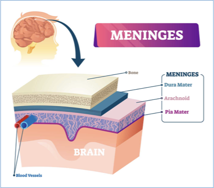
meninges
three protective layers that surround brain and spinal cord
layers:
dura mater:
thick outer layer lining the inner dome of skull (cranium)
arachnoid:
thin middle layer, below is cerebrospinal fluid (CSF)
doesn’t contain nerves or blood vessels
pla mater:
thin inner layer lining surface of brain
rich w/ veins and arteries

falx cerebri and tentorium cerebelli
thick folds of dura mater that anchor brain in place
provides stability to the lt. and rt. hemispheres of the cerebrum and cerebellum
falx cerebri:
separates rt. and lt. hemispheres » ez to see on u/s
midline, echogenic linear structure separating cerebral hemispheres on u/s
tentorium cerebelli:
separates occipital lobe of cerebrum and cerebellum » hard to see on u/s
more posterior and inferior in the brain


brain
the CNS is composed of grey and white matter
grey matter: composed of neuron somas that process and interpret information
white matter: axons and myelin that transmits info to other parts of the CNS
—
brain can be divides into 3 segments:
cerebrum
brainstem
cerebellum
—
weighs ~3 pounds in avg. adult
60% fat, 40% water/protein/carbohydrates/salts
darker outer portion of brain = grey matter
lighter inner section of brain = white matter
—
in spinal cord..
inner = grey matter
outer = white matter

cerebrum
located within bony cranium » extends from frontal bone anteriorly to occipital bone posteriorly
right and left hemispheres
covered in gyri (ridges) and sulci (folds) » they inc. surface area of brain which allows them to take on more complex functions
each hemispheres contains 4 lobes:
frontal (personality, decision-making, and movement)
parietal (identify objects, spatial relationships)
temporal (short-term memory, rhythm)
occipital (vision)

cerebrum sono app
cerebral hemispheres should be symmetrical
gyri (ridges) and sulci (folds) develop over time
18-20 wks: absence of gyri and sulci, brin appears smooth
22-26 wks: gyri and sulci begin forming
28-30 wks: period of greatest brain development, gyri and sulci are detectable
>32wks: gyri and sulci are obvious

brainstem
command center for vital life functions
stalk-like structure connecting cerebrum to spinal cord
contains:
mesencephalon/midbrain
eye movements, reflexed to visual and auditory stimuli
pons
bridge structure, regulates breathing, facial movement, hearing, balance
medulla oblangata
HR, BP, breathing
sono app:
not routinely assessed w/ u/s
can be seen in the middle, posterior aspect of the brain
best seen in SAG


cerebellum
large, bilobed structure w/ symmetrical connected rt. and lt. hemispheres
below the temporal and occipital lobes and above brainstem
vermis = middle of cerebellum
the fourth ventricle and thalamus is anterior to cerebellum
cisterna magna and nuchal fold posterior to cerebellum
usually correlated roughly to GA
sono app:
butterfly shaped brain structure in posterior brain
best seen TRV oblique view (cerebellar view) of fetal head
routinely assessed


posterior fossa
deepest and most posterior part of cranial cavity
contains:
cerebellum
brainstem
fourth ventricle
—
in u/s we often count the cisterna magna and nuchal fold as a part of posterior fossa as they’re in its area » but it’s not technically a part of it

cisterna magna (CM)
largest subarachnoid cistern (pocket) of CSF
space surrounding the posterior and inferior aspects of cerebellum
sono app:
anechoic as it’s filled with CSF
normal = <10mm
within cranium/skull
echoes seen could be artifact or connective tissue


nuchal fold (NF)
skin and subcutaneous tissue at back of fetal neck
transient structure whose measurement provides insight into fetal well-being
normal <6mm


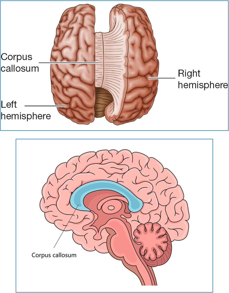
corpus callosum (CC)
thick bundle of fibres that connects the rt. and lt. hemispheres of brain
enables communication b/w both hemispheres
midline, superior to thalamus
not routinely assessed w/ u/s
sono app:
hypo, curved structure midline brain
best seen SAG view
CSP inferior/posterior to CC

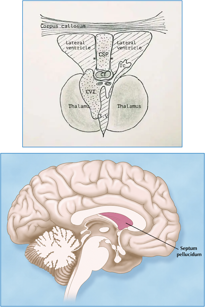
cavum septum pellucidum (CSP)
septum pellucidum:
small structure that lies just inferior to corpus callosum, and in b/w the anterior aspects of lateral ventricles
cavum septum pellucidum (CSP):
small, fluid filled space within septum pellucidum
we focus more on CSP on u/s because it’s visualized more clearly
—
sono app:
anechoic, box-shaped structure in middle of brain, anterior to thalamus
seen in TRV thalamic and oblique cerebellar views
CC anterior, superior to CSP
often there’s a fine hypoechoic ‘U’ shaped structure which represents the anterior leaflets of the CC
if not visualized, there’s a risk of a brain abnormalities

thalamus
inferior to septum pellucidum, superior to brainstem
consists of two ovoid structures (paired thalami) located on either side of the 3rd ventricle
sono app:
thalamic and cerebellar views
heart shaped/ovoid/diamond shaped structure consisting of two lobes


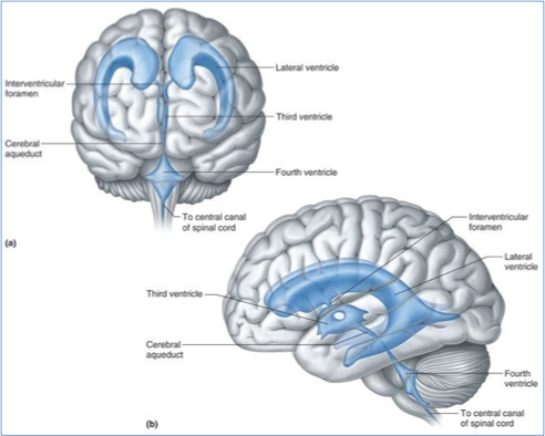
ventricular system
four ventricles of the brain are interconnected chambers that produce and contain cerebrospinal fluid (CSF)
right and left lateral ventricles:
largest
contains the most choroid plexus which produces CSF (ventricle w/ the most CSF)
atrium is the area near posterior aspect of lateral ventricle
seen bilaterally in TRV ventricular view
third ventricle:
small, midline, central in the brain
connects all ventricles together
seen in central brain (near CSP) in thalamic/cerebellar views
not routinely assessed
fourth ventricle:
midline and lower in brain
close to spinal cord
can be seen in posterior brain, near cerebellum, in cerebellar view
not routinely assessed


ventricular system - foramina
the foramina (foramen/aqueduct) connect the ventricles together so CSF can flow b/w them
interventricular foramen:
aka foramen of Monro
connects lateral ventricles to third ventricle
cerebral aqueduct:
aka aqueduct of Sylvius
connects third and fourth ventricles
median aperture:
aka foramen of Magendie
connects fourth ventricle to cisterna magna and subarachnoid space
lateral apertures:
aka foramen of Luschka
connects fourth ventricle to subarachnoid space
ventricular system cont.
develops alongside and within the tube
ventricles within the telencephalon becomes the lateral ventricles
ventricles within the diencephalon become the third ventricle
ventricles within the rhombencephalon become the fourth ventricle
ventricles within the mesencephalon becomes the aqueduct of sylvius

cerebrospinal fluid (CSF)
produced in choroid plexus
travels thru ventricular system to subarachnoid space (b/w arachnoid mater and pia mater layers)
coats the brain, providing lubrication, protection, delivers nutrients and removes waste
typically only see CSF in the ventricles n u/s

vascular system of brain - arterial
arterial supply to brain is from vertebral and carotid arteries
circle of willis is a loop of arteries in the base of the brain that helps keep oxygenated blood flowing throughout entire brain
middle cerebral artery (MCA) is found within circle of willis and can be assessed w/ PW
sono:
MCA assessment of arterial system of fetal brain is most performed
normal = high resistance


vascular system - venous
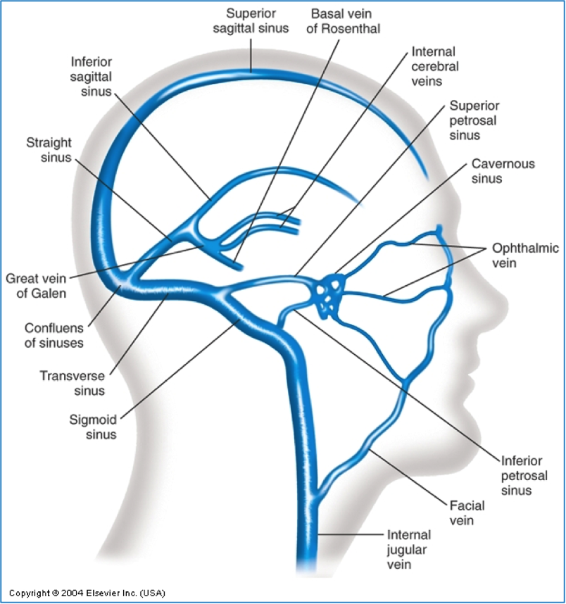
venous drainage of the brain is from dural venous sinuses (large channels b/w the layer of the dura mater) that collects blood and drains into the internal jugular veins
vein of galen (aka “great cerebral vein”) is a prominent vein in posterior aspect of brain
it’s malformation can cause srs congenital vascular problems
sono/doppler:
rarely performed, but can be achieved w/ colour and PW
most often, abnormalities of the venous system causes changes in vein of galen
2nd trimester routine anatomy ultrasound
normal findings:
oval head shape
bone density (echogenic bone structures)
BPD and HC » appropriate for GA
atrium of lateral ventricle = <10mm
CSP, thalamus, falx (midline structures)
cerebellum (measurement appropriate for GA)
cisterna magna = <10mm
nuchal fold =<6mm

fetal head: 8-11 wks GA (1st tri)
anechoic spaces (ventricles) seen within fetal head
will eventially shrink to accomodate growing brain
largest = rhombencephalon at posterior aspect of fetal head
major abnormalities such as acrania, anencephaly, and encephaloceles may be observed at this stage
skull hasn’t ossified yet
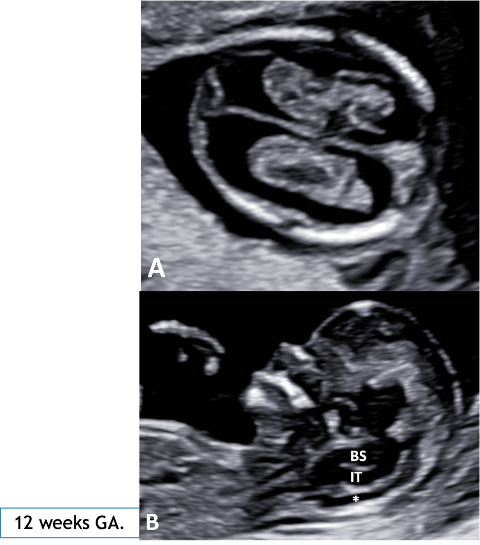
fetal head: 11-14 wks GA (1st tri)
TRV view of fetal head demonstrates:
two lateral ventricles (LV, anechoic spaces)
choroid plexus (CP, hyperechoic structures)
falx (F, echogenix, midline line)
SAG view of fetal head demonstrates:
brainstem (BS)
intracranial translucency (IT, fourth ventricle)
cisterna magna (*)
skull is starting to ossify, becoming mor echogenic

fetal head: 18-20 wks GA (2nd tri) - ventricular view
ventricular view:
aka transventricular view
most superior of the axial views
TRV plane at lvl of..
lateral ventricles
atria » posterior aspect of ventricles
choroid plexus » sits within the lateral ventricles
routinely acquired
measurement acquired..
atria width at lvl of glomus of choroid plexus

measurement of atria width at level of glomus of choroid plexus
ventricular view
inner to inner linear measurement of ventricle at location of atria and glomus of choroid plexus
posterior ventricle is selected bcuz an anterior one is often shadowed by skull
normal = <10mm

fetal head: 18-20 wks GA (2nd tri) - thalamic view
aka “transthalamic view”
found directly inferior to ventricular view
TRV plane at lvl of:
thalamus » midbrain
CSP
falx
routinely acquired
measurements acquired:
BPD, HC

al head: 18-20 wks GA (2nd tri) - cerebellar/posterior fossa view
aka “transcerebellar view”
oblique plane at level of:
cerebellum
cisterna magna and nuchal fold
thalamus and CSP
routinely acquired
measurements acquired:
cerebellum width, cisterna height, nuchal fold thickness
—
MEASUREMENTS:
cerebellum:
linear measurement of max. width
equals roughly GA (ie. 20mm = 20 wks GA)
cisterna magna (CM):
linear measurement of fluid height
normal <10mm
nuchal fold (NF):
linear measurement of skin thickness/height
normal <6mm


brain appearance throughout pregnancy
appearance changes with growth
lateral ventricles and choroid plexus remains a similar size while the rest of brain matter grows
head sonography
routinely assessed during 2nd trimester anatomy scan:
cranium
cerebrum
falx cerebri
lateral ventricles, atria, choroid plexus
CSP
cerebellum
cisterna magna
nuchal fold
not routinely assessed:
meninges
tentorium cerebelli
brainstem
corpus callosum
thalamus
3rd and 4th ventricles
vascular system
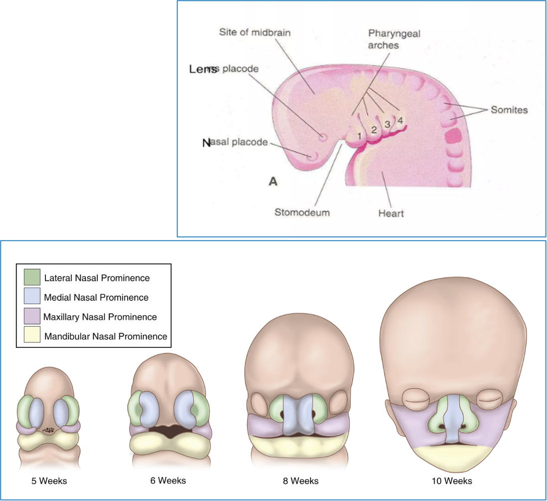
face and neck embryology
face and neck development occurs b/w 4-10 wks GA
complex process involving cells from ectoderm, mesoderm, endoderm, and crest w/ the involvement of 6 pairs of pharyngeal arches
pharyngeal arches play a dominant role in building face and neck
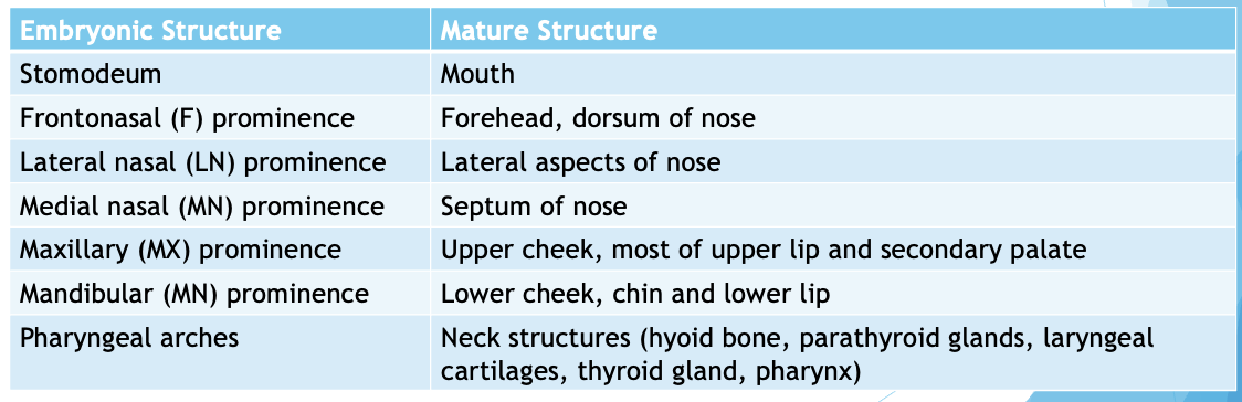
fetal face is made up of 5 main tissue buds (prominences)
failure of development or fusion of prominences contributes to the majority of facial abnormalities

development of orbits
development of orbits begin at 4wks GA
optical grooves emerge on the folds, evolving into optic vesicles
vesicles become most of the structures of the eye
eyelids form at wk 5, fuse closed at wk 8, and remain closed until 26-28 wks GA to protect eye as it develops
—
orbits begin on the sides of the head and move medially over time
orbital angle of 180° at 8 weeks GA, 105° at 12 weeks GA, and 71° at birth (adult is 68°)

face anatomy
face is the anterior portion of head, from forehead to chin and ear to ear
facial shape is determines by skeleton, facial muscles, and subcutaneous tissue
functions:
communication
expression of emotions
external identity
anatomy consists of:
bones
muscles
skin
region
nerves
blood vessels

facial skeleton - face anatomy
aka “viscerocranium”
composed of:
two nasal bones » routinely assessed w u/s
two maxillae » routinely assessed w u/s
two inferior nasal conchae
two palatine bones
two zygomatic bones
two lacrimal bones
mandle (jaw) » routinely assessed w u/s
vomer
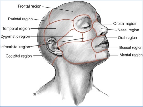
facial regions - face anatomy
superior region:
frontal region (forehead)
orbital region (eyes, orbits)
temporal region
middle region:
nasal region (nose)
infraorbital & zygomatic regions (upper cheek)
auricular region (ear)
inferior region:
oral region (lips)
mental region (chin
buccal region (lower cheek)
parotideomasseteric region (parotid gland)

palate - head anatomy
palate is a structure that forms the roof of the oral cavity and the floor of the nasal cavity
separates the oral and nasal cavities » essential for preventing pressure changes within these cavities
enables important processes (ie. suckling, breathing)
important role in articulation (formation of speech), digestion, and swallowing
hard palate:
anterior, bony portion
formed by fusion of maxilla and palatine bones
soft palate:
posterior
muscular portion
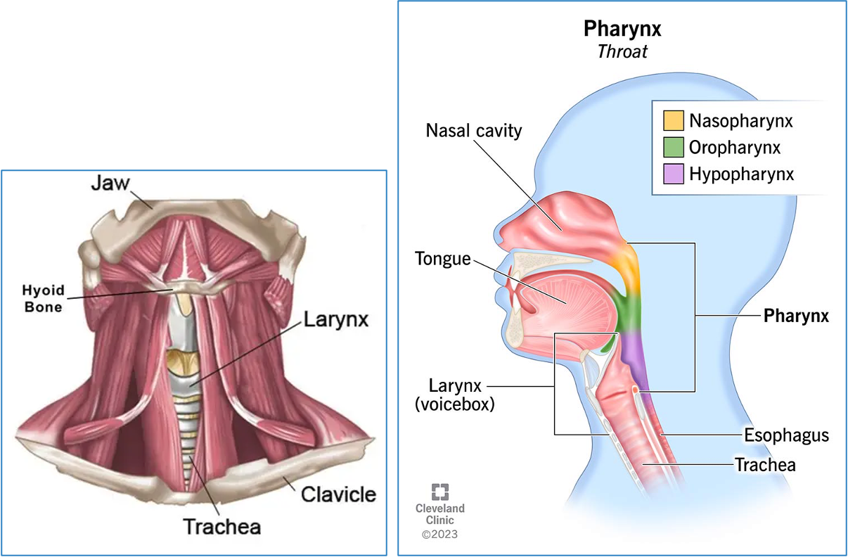
neck anatomy
anchors head
provides stability and allows for rotational movement
contains:
hyoid bone (anchor point, at lvl of “Adam’s apple” in males)
thyroid gland
parathyroid glands
pharynx
larynx (voice box)

face and neck sonography
structures routinely assessed:
slope of forehead
orbits, lenses
nasal bone, tip of bone, nostrils
lips, mouth
chin
maxilla
mandible

face and neck sonography - SAG face/profile
SAG face/profile:
slope of forehead
nasal bone
tip of nose
lips
chin
maxilla
mandible


face and neck sonography - coronal nose/lips
coronal nose/lips:
tip of nose
nostrils
upper lip
lower lip
mouth opening
chin
—
necessary in screening for fetal cleft lip

face and neck sonography - coronal orbits
coronal orbits:
orbits
lenses
maxilla
mandible
chin

face and neck sonography - TRV orbits
TRV orbits:
orbits
lenses
tip of nose
—
orbits are present, normal size, and normal distance apart
lenses are present in each globe
OOD, IOD, OD measurements
face and neck sonography - additional views
optional/additional views:
neck images
TRV maxilla/mandible
3D images of face

TRV maxilla/mandible - optional view
fetal head, maxilla, tooth buds, and palate can be identified
helpful in cleft palate assessment

3D imaging
not required, but can be helpful in visualizing the face if some standard views can’t be acquired due to fetal position
can be performed manually w/ regular curved transducer or automatically w/ a 3D transducer