Anatomy: Lecture 10 / Lab 11 & 12
 Call Kai
 Learn
 Practice Test
 Spaced Repetition
 Match
 Flashcards
 Knowt Play
1/287

There's no tags or description
Looks like no tags are added yet.
288 Terms
What are the cranial limits of the thoracic cavity?
thoracic inlet
What are the dorsal limits of the thoracic cavity?
vertebral bodies
What is the thoracic inlet?
the first pair of ribs and their costal cartilage
What are the lateral limits of the thoracic cavity?
ribs and intercostal muscles
What are the ventral limits of the thoracic cavity?
sternebrae
What are the caudal limits of the thoracic cavity?
diaphragm
What are the three cavities inside the thoracic cavity?
pericardial cavity and 2 pleural cavities
What is pleura?
simple squamous cell layer located within the thoracic cavity
Differentiate between the thoracic cavity and pleural cavity
the pleural cavity is space between pleural and is inside the thoracic cavity
What is mesothelium?
simple squamous epithelial layer that secretes serous fluid and lines cavities
What is serous fluid?
watery fluid that lubricates structures with more pressure and keeps junk out of the body
the mesothelium that lines the thoracic cavity is?
pleura
What is Visceral pleura?
pleura that surrounds the lungs
What is parietal pleura?
pleura that lines the thoracic cavity
What are the different types of parietal pleura?
costal, mediastinal, and diaphragmatic
What is costal pleura?
parietal pleura that lines the ribs and intercostal muscles
What is mediastinal pleura?
parietal pleura that reflects on the midline covering the structures there
which 2 pieces of mesoderm come together to form the diaphragm
septum transversum and pleuroperitoneal folds
What is diaphragmatic pleura?
parietal pleura that lines the diaphragm
Where does the spinal nerve originate from?
intervertebral foramina and innervates the neck trunk and limbs
What is the line of pleural reflection?
imagined line along the horizontal parts of the 8th/9th costal cartilages, across the 10th/11th costal cartilages at costochondral junction and across the middle of last rib
if you stick a needle behind the line of pleural reflection you will be where?
in the abdominal cavity
What is an example of connecting pleura?
pulmonary ligament (not a real ligament but a fold of pleura)
What does the central nervous system include?
brain and spinal cord
what does the peripheral nervous system include?
cranial nerves and spinal nerves
How many pairs of cranial nerves are there?
12
What is cranial nerve 1?
olfactory - smell
What is cranial nerve 2?
optic - sight
what is cranial nerve 3?
oculomotor - eye muscles
What is cranial nerve 10?
vagus - parasympathetic motor and sensory fibers
How many pairs of spinal nerves are present?
35; 8 cervical pairs, 13 thoracic pairs, 7 lumbar pairs, and 3 sacral pairs
How do the spinal nerves arise?
from the dorsal and ventral roots
What type of fibers does the dorsal root carry?
sensory (afferent)
What type of fibers does the ventral root carry?
motor (efferent)
Where do the dorsal and ventral roots join to form a mixed spinal nerve?
within the vertebral foramen
What fiber types does a mixed spinal nerve contain?
both sensory and motor fibers
What are the three immediate branches of a spinal nerve?
dorsal, ventral, and communicating branch (ramis communications)
What muscles does the dorsal branch supply?
epaxial muscles
What muscles does the ventral branch supply?
hypaxial muscles
What is the function of the communicating branch (ramus communicans)?
Connects with the sympathetic trunk (autonomic fibers)
How do spinal nerves exit from C1?
lateral foramen in atlas
How do spinal nerves C2-7 emerge from the axis to C7?
through the intervertebral foramen
How does Cn8 emerge?
Caudal to numerically responding vertebrae because there is no C8
In the thorax, what do the ventral branches of thoracic spinal nerves become?
ventral branches of thoracic spinal nerves become intercostal nerves
What is the source of the dorsal intercostal arteries?
aorta
What superficial vessels and nerves are present in the thoracic wall?
dorsal intercostal arteries, ventral intercostal branches of internal thoracic artery and vein, intercostal nerves, and the axilla
Where do dorsal intercostal arteries run?
along caudal surface of rib
What is the superficial vein of the neck that we’ve covered?
external jugular
What is cranial nerve 11?
accessory nerve
What does the accessory nerve supply?
extrinsic muscles of thoracic limb (omotransversarius and trapezius)
What is the only motor nerve in the trapezius?
accessory nerve
What would happen if the accessory nerve is damaged?
other extrinsic muscles are weakened but trapezius loses motor supply resulting in atrophy and impaired scapular movement
Where are the superficial cervical lymph nodes located?
they lie on the serratus ventralis and scalenus cranial to the supraspinatus
What is the superficial cervical lymph node covered by?
omotransversarius and brachiocephalicus
Where are the axillary lymph nodes located?
latissimus dorsi muscle and deep pectorals (axilla)
Where are the tracheobronchial lymph nodes located?
tracheal bifurcation where windpipe splits
What organs are in the thoracic cavity?
esophagus, trachea, lungs, heart and thymus
Where is the aorta in the thorax?
dorsally (from left ventricle)
What is the cardiac notch?
V-shaped gap in ventral border of R lung (bw cranial and middle lobes)
Where is the cardiac notch located?
low in third and fifth intercostal space and not present on the left

What side and view is this lung and what lobe is located on the left?
left lung, ventral view, caudal lobe

What side and view is this lung and what lobe is located on the middle?
left lung, ventral view, and cranial caudal lobe
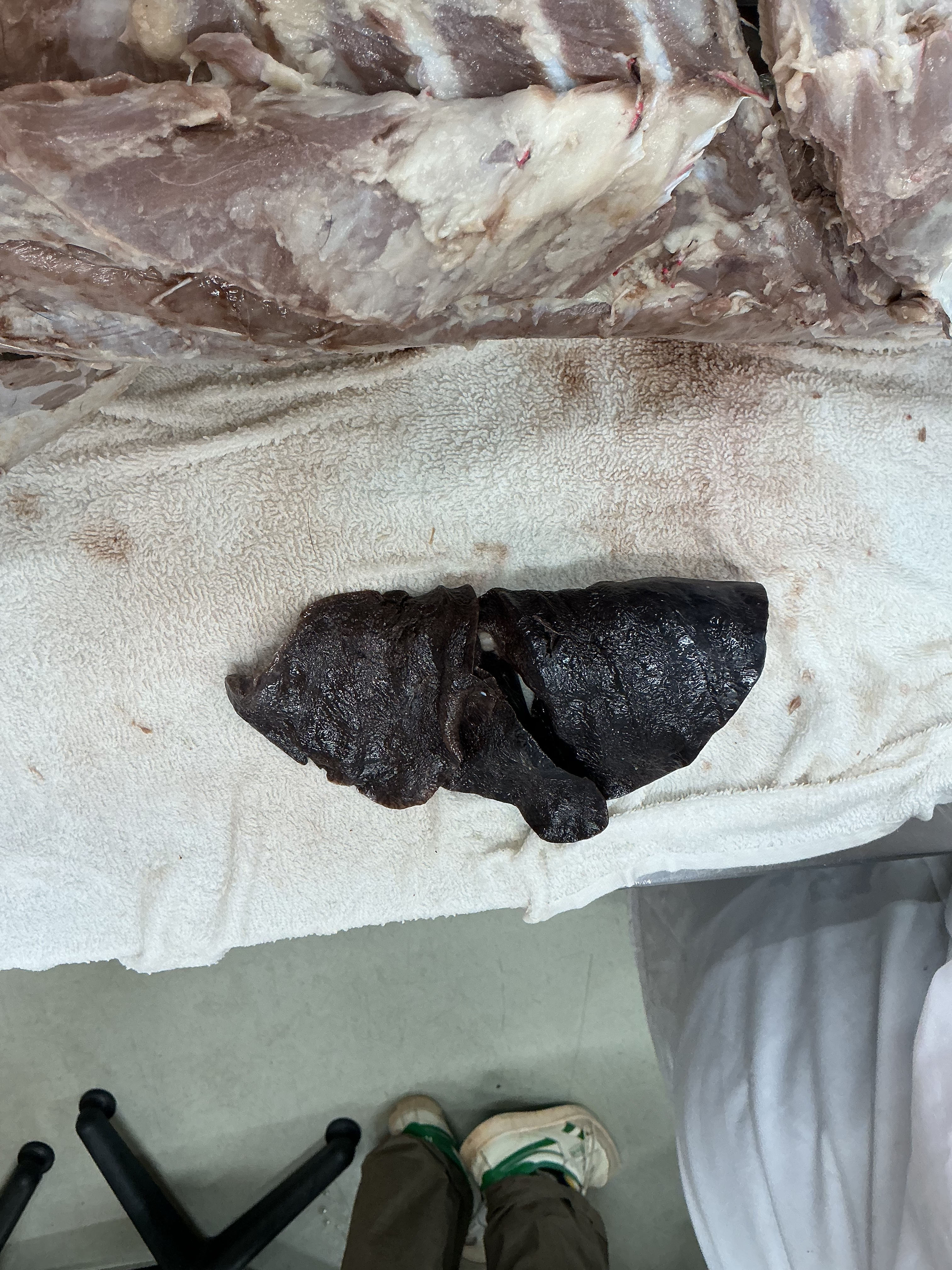
What side and view is this lung and what lobe is located on the right?
left lung, ventral view, and cranial lobe

What side and view is this lung and what is the red lobe?
right lung, dorsal view, cranial lobe

What side and view is this lung and what is the green lobe?
right lung, dorsal view, and middle lobe

What side and view is this lung and what is the blue lobe?
right lung, dorsal view, and caudal lobe

What side and view is this lung and what is the yellow lobe?
right lung, dorsal view, and accessory lobe
What are the first 2 branches off the ascending aorta?
L + R coronary arteries
What two structures does the aorta branch into?
brachiocephalic trunk and left subclavian artery
What does the brachiocephalic trunk branch into?
left common carotid, right common carotid, and right subclavian arteries
What does the left subclavian branch into?
4 branches: vertebral artery, costocervical artery, internal thoracic artery, and superficial cervical artery
What does the left subclavian artery continue as?
axillary artery
What structures form the cranial vena cava?
L + R brachiocephalic veins
What forms from the brachiocephalic vein?
side-specific subclavian and side-specific external jugular
What does the cranial vena cava drain?
front of animal
What does the right brachiocephalic vein drain?
Right head/neck/thoracic limb
What does the left brachiocephalic vein drain
left head/neck/thoracic limb
What does the external jugular vein drain?
head
What do the subclavian veins drain?
thoracic limb on each side
Where does the brachiocephalic trunk supply blood?
to the head, neck, and R forelimb
Where does the right subclavian artery supply blood?
thoracic limb
Where do the common carotid arteries supply blood?
head
Where does the vertebral artery take blood?
brain
Where does the costocervical artery supply blood?
deep neck (first few intercostals)
Where does the internal thoracic artery supply blood?
ventral thoracic and mammaries
Where does the superficial cervical artery supply blood?
base of neck
What do the right and left coronary arteries supply?
myocardium of heart
what lines the thoracic cavity beneath the intercostal muscles?
parietal pleura
what muscles are found within a given intercostal space?
external and internal intercostal muscles
the intercostal artery, intercostal vein, and intercostal nerve run along what side of each rib?
caudal
if you need to make a longitudinal incision in the intercostal space, where should you aim to cut?
cranial area of intercostal space
What is the origin of the phrenic nerve?
C5,C6,C7
How does the phrenic nerve enter the thorax?
thoracic inlet
What is the course of the phrenic nerve?
runs caudally along pericardium
What is the function of the phrenic nerve?
supply the diaphragm for inspiration
Where do the right and left crura of the diaphragm attach?
lumbar vertebrae
Where are the lateral attachments of the diaphragm?
inner surfaces of the rib
What are the three passageways of the diaphragm?
aortic hiatus, esophageal hiatus, and caval foramen
What is the aortic hiatus?
dorsal passageway between the crura for the aorta, the azygous vein, and the thoracic duct
What is the esophageal hiatus?
musclar part of the right crus that transmists the sophagus, vagal nerve trunks, and esophageal vessels