Cell structure
 Call Kai
 Learn
 Practice Test
 Spaced Repetition
 Match
 Flashcards
 Knowt Play
1/30

There's no tags or description
Looks like no tags are added yet.
31 Terms
Mitochondrian
S: -double membrane bound
-inner membrane folds into cristae and matrix
F: site of aerobic respiration
Nucleus
S: double membrane bound, contains nucleolus, nucleoplasm and nucleus pores
F: contains DNA, controls cell activities and is the site of transcription.
Nucleolus
S: dense region inside the nucleus
F: site of ribosome production, synthesises rRNA
Nuclear envelope + nuclear pores
S: double membrane containing pores
F: regulates movement of rRNA and separates nucleus from cytoplasm
Vesicles
S: small sacks surrounded by a single membrane
F: transport substances to and from cell plasma membrane via exocytosis or endocytosis
Ribosome
S: made of proteins, 80S in eukaryotes and 70S in prokaryotes
F: site of protein synthesis, site of translation
Cytoskeleton
S; network of protein fibres
F: maintains cell shape, transports organelles, and separates chromosomes during cell division
Centrioles
S: contains microtubules
F: organises spindle fibres during cell division
Rough endoplasmic reticulum (RER)
S: network of flattened, membrane sacs with ribosomes attached
F: folds, processes and transports proteins to golgi
Smooth endoplasmic reticulum (SER)
S: network of membrane bound flattened sacs, without ribosomes
F: synthesises lipids, detoxification.
Golgi apparatus
S: stack of flattened membrane- bound sacs
F: modifies, packages and secretes proteins into vesicles
Cytoplasm
Where metabolic reactions occur
Lysosome
S: membrane bound vesicle containing hydrolytic enzymes
F: digests pathogens, waste products and worn out organelles
Cell plasma membrane
S: phospholipid bilayer with embedded proteins
F: controls movement of substances into and out of the cell and cell signalling
Cell wall (plants only)
S: made of cellulose
F: supports the cell, maintains shape and prevents it bursting
Chloroplast
S: double membrane bound, contains thylakoids arranged in grana and strong
F: site of photosynthesis
Plasmid
S: small circular DNA found in prokaryotes
F: carries extra genes such as antibiotic resistance
Flagella (bacterial cell only)
S: long membrane bound projections
F: propels cell through liquid
Cilia (bacterial only)
S: short membrane bound projections
F: best to move substances across cell surface

Key similarities and differences between prokaryotic and eukaryotic cells
Differences:
-cells are smaller
-circular DNA
-no membrane bound organelles
-smaller 70S ribosomes
-no nucleus (plasmids: singular, circular DNA)
-cell wall made of peptidoglycan
-flagella (rotates allowing movement)
Similarities:
-both have cell membrane
-cytoplasm
-ribosomes

Protein synthesis
1) nucleolus manufactures ribosomes for protein synthesis in RER
2) nucleolus manufactures mRNA- needed to make proteins
3) ribosomes in RER make protein
4) RER processes proteins, sent in vesicles to Golgi apparatus
5) Golgi further processes proteins, modifies and packages the proteins into vesicles and secrets to cell plasma membrane
6) vesicle fuses with cell plasma membrane, contents released by exocytosis
Magnification
Magnification=actual sizeimage size
F: used to calculate how much larger an image is than the real object
Light microscope
-Uses visible light to form an image of the specimen.
-2D image in colour
Pro: cheap and easy to use
Con: poor resolution and lower magnification
Transmission electron microscope (TEM)
-Passes a beam of electrons through an ultra thin specimen.
-produces a 2D image in black and white
Pro: high resolution and magnification + can view internal structures
Con: expensive + specimen dead
Scanning electron microscope (SEM)
-Scans a beam of electrons across the surface of a specimen.
-produces a 3D image
Pro: high magnification and resolution
Con: can’t see internal structures
Resolution
The ability to distinguish the minimum distance between two separate points, where still seen seprrately
Stage micrometer
A glass slide with a scale of known length, such as 1 mm divided into 100 divisions. It is used to calibrate the eyepiece graticule.
Eyepiece graticule
A scale fitted inside the eyepiece that has no fixed length. It must be calibrated against a stage micrometer for each objective lens because one division's length changes with magnification.
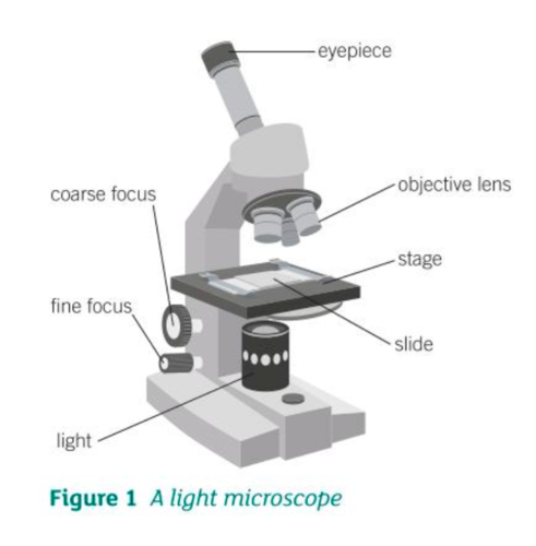
Slide preparation
1) obtain a thin piece/layer of the specimen
2) add a drop of water to specimen
3) staining: stain the specimen, to make the structures visible
4) push coverslip down to squash sample, ensure specimen is thin so light can pass through
5) sample placed at a 45 degree angle, using mounted needle, to avoid trapping air bubbles. Coverslip then placed on top of the slide.
Differential staining
-crystal violet/ methylene blue: positively charged, attracted to negatively charged parts
-Congo red: negatively charged, stains baground as custodial repels
Scientific drawings (rules)
-draw in pencil
-title diagram
-state magnification
-do not shade
-annotate key features
-horizontal label lines without arrowheads
-do not sketch
-continuous lines