Anatomy of the Urogenital Systems
 Call Kai
 Learn
Learn Practice Test
 Spaced Repetition
 Match
Match Flashcards
 Knowt Play
1/74

There's no tags or description
Looks like no tags are added yet.
Name | Mastery | Learn | Test | Matching | Spaced | Call with Kai | Chat |
|---|
No analytics yet
Send a link to your students to track their progress
75 Terms
Kidney Location
retroperitoneal
behind periotenal cavity
-houses GI system organsposterior abdominal region
lateral to vertebral column
t12-l3

Kidney Fat Layers
surrounded+protected by layers of fat+facsia
superficial→deep
paranephric fat
renal fascia
perinephric fat
fibrous renal capsule

Kidney Function
removes
toxins
drugs
metabolic wastes
regulates total water volume
regulates total solute concentration of blood
regulates ECF ion concentrations
produces urine (2-stage process)
glomerular filtration
-non-selectivewater+salt+sugar reabsorbed into venuous system
-further fine-tuning→via secretion
Nephron
functional unit of kidney
2 components
renal corpuscule
-glomerulus
-glomerular capsulerenal tubule
-PCR
-nephron loop
-DCT
Renal Cortex
outer margin of kidney
site of urine production
contains
capillaires
tubes
majority of nephrons
Renal Pyramids
contains numerous urine collecting ducts
-nephrons drain into these
ducts open→at papilla of renal pyramid
Renal Medulla
region containing renal pyramids
Renal Column
pillars between adjacent renal pyramids
contains A+Vs
Urine Drainage Pathway
renal cortex→nephrons→renal pyramid papilla→minor calyx→major calyx→renal pelvis→ureter→bladder
Hilum
slit in kidney
for entrance+exit of vessels
entering kidney
renal A
exiting kidney
ureter
renal V
Renal Artery
branches off abdominal aorta A
enters kidney via hilum
right renal A longer than left renal A
Renal Vein
drains blood from kidney
carries blood back to inferior vena cava V
left renal V longer than right renal V
Ureter
muscular tubes
from kidneys to bladder
Bladder Structure
situated within pelvic cavity
posterior to pubis bones
posterior to pubic symphysis
made of smooth muscle→contraction aids in voiding/emptying bladder
internal surface→contains rugae
Bladder Function
fxn→temporary storage of urine
full→expands superiorly into abdomen
adults→1½ pints urine
Ureteric Orifice
prevents backflow of urine up ureters
Internal Urethral Orifice
tube where urine exits bladder
Bladder Urine Pathway
ureter
ureteric orifice
bladder
internal urethral orifice
urethra
Trigone
smooth triangle surface
between ureteric orifices+urethral orifice
Urethra
drains the bladder to the exterior
internal urethral orifice
external urethral orifice
differences between males+females
Male vs Female Urethra
female
short→4 cm
-clinical→women more susceptible to bladder infxnscarries only urine
external urtheral orifice→anterior to vagina
males
longer
3 distinct parts
-prostatic urethra
-membranous urethra
-spongy urethracarries urine+semen
external urethral orifice→on glans penis
Adrenal/Suprarenal Glands
“chewed bubble gum on top of the kidneys”
above kidneys
responsible for production of over 30 different hormones
estrogen
progesterone
steroids
cortisol
dopamine
adrenaline
True vs False Pelvis
true pelvis
inferior parts of pelvic bones
sacrum
coccyx
has inlet+outlet
pelvic cavity
false pelvis
superior region→upper parts of pelvic bones
lower lumbar vertebrae
considered part of abdomen
Pelvic Cavity Structure
bowl-shaped cavity→in true pelvis
continuous with abdomninal cavity
formed by
pelvic inlet
pelvic walls
pelvic floor
Pelvic Cavity Location
fxn→house organs
bladder
rectum
sigmoid colon
male/female reproductive tract
Male Pelvic Cavity
contains
bladder
rectum
connection bewteen urinary+reproductive tracts
seminal vesicles
vas deferens (portion)
Female Pelvic Cavity
contains
anterior→bladder
intermediate→uterus
posterior→rectum
fallopian tube
ovary
sigmoid colon
connection between vagina+uterus
Pelvic Inlet (General)
hole in pelvic bone
posterior components
body of S1
TP of S1
rim on pelvic bones to pubic symphysis
Female Pelvic Inlet Pathway
pregnancy→uterus expands into abdomen
fetus passes through pelvic inlet
fetus passes through pelvic outlet
Pelvic Wall Components
bones
sacrum
coccyx
inferior half of os coxae
muscles
obturator internus
piriformis
ligaments
Sacrotuberous Ligament
links os coxae to sacrum
from sacrum→ischial tuberosity
closes greater sciatic notch
Sacrospinous Ligament
from sacrum→ischial spine
closes lesser sciatic notch
Pelvic Outlet
diamond-shaped
formed by
bone
-ischiopubic ramus
-ischial tuberosity
-coccyxsacrotuberous ligament
pubic symphysis
Pelvic Floor
separates pelvic cavity from perineum
contains
fascia
muscles
levator ani muscles
coccygeous muscles
Levator Ani Muscles
walls of pelvis→midline
form pelvic diaphragm
Coccygeous Muscles
sacrum+coccyx→ischial spine
posterior portion of pelvic diaphragm
Pelvic Floor
muscles→form pelvic diaphragm
supported by
perineal membrane
-thick facsial sheetdeep perineal pouch
-narrow space
-deep to perineal membrane
pierced by
vagina
urethra
anus (on way to perineum)
Perineum Location
inferior to pelvic floor
between lower ribs
margins→formed by pelvic outlet
divided into
urogenital triangle
anal triangle
-”imaginary” line
-between ischial tuberosities
Perineum: Urogenital Triangle
part of perineum
urogenital hiatus→houses openings of
vagina
urethra (distal end enclosed in erectile tissue of penis)
Perineum: Anal Triangle
houses opening of anus→external ana sphincter
-circular muscle fiber orientation
Male vs Female Bony Pelvis
females→increased diameter→obsterical demands
greater sciatic notch→wider in females
body of pubis→taller in males
subpubic angle
female→80-85°
male→50-60°
pelvic inlet
female→oval shaped+wider
male→heart shaped
auricular surface
males→cover first 3 sacral elements
females→cover first 2 sacral elements
Primary Sex Organs vs Accessory Reproductive Organs
primary sex organs→gonads
produce gametes
-ova
-spermtestes vs ovaries
secrete sex hormones
-male→androgen
-female→estrogen+progesterone
accessory reproductive organs
ducts
glands
external genitalia
Vulva
composed of
mons pubis
-skin+adipose tissue
-cover pubic bones+symphysislabia majora
-outer borderslabia minora
-medial to labia majoraglans clitoris
-partially covered by prepuce
-homologous to penis
Vestibule
space between labia minora
contains
external urethral orifice
-opening of urethravaginal orifice
Uterus
pear-shaped
thick muscular walls
hollow
anterior to rectum
posterior to bladder
3 sections
fundus
-domedbody
-uterine canalcervix
-extends into vagina
Features of the Cervix (Uterus)
cervical canal
internal os
external os
fornix
Vagina+Vaginal Canal
smooth muscle
superior→cervix
inferior→external vaginal opening
Ovary of Uterus
produces eggs
releases egg every month
only connected to uterus via ligaments

Fallopian/Uterine Tube of Uterus
site of fertiliation
components
ostium
-opening of fallopian tubeinfundibulum
-widened portion of fallopian tubefimbriae
-finger-like extension of infundibulum
-”sweeps” egg from ovary→fallopian tube

Uterus
site of implantation of the egg
3 parts
fundus
body
cervix

Cervix of Uterus
inferior portion of uterus
extends into vaginal
cervical canal
canal through cervix
openings
internal os
external os

Fornix of Uterus
the surrounding space of the cervix (on the sides of the opening)
components
anterior fornix
posterior fornix
lateral fornix x2

Broad Ligament
sheet-like ligament
fold in peritoneum
connects uterus+fallopian tubes→pelvic walls

Suspensory Ligament
suspends ovary from lateral wall of pelvis
runs with ovarian A+V
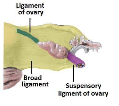
Ovarian Ligament
connects ovary to uterus
found within broad ligament

Round Ligament
exits pelvis via inguinal canal→ends in connective tissue of labia majora
found within broad ligament→until inguinal canal

Male Reproductive Structures (General)
primary sex organs→external→sperm production needs to occur below body temperature
many structures→truly urogenial in fxn
External Genitalia
scrotum
houses testes
muscles+fascia
penis
root
body
glans
prepuce
Root of Penis
anchors penis to body
crura x2
proximal parts of corpus cavernosa
bulb
proximal part of corpus spongiosum

Body of Penis
entirely covered in skin
terminates in glans penis
-contains external urethral orifice
contains erectile tissue
corpus cavernosa
corpus spongiosum

Corpus Cavernosum of Penis
paired
erectile tissue
each contains deep artery of penis

Corpus Spongiosum of Penis
single
erectile tissue
spongy urethra runs through
proximal→bulb

Spongy Urethra of Penis
runs through corpus spongiosum
caries urine/semen

Glans Penis
distal
expanded end of corpus spongiosum

Testes
produces sperm+testosterone
before birth→abdominal→descend before birth
external→sperm can’t be at body temperature
-housed in scrotum
components
straight tubules
rete testis
epididymydis

Seminiferous Tubules of Testes
site of sperm production

Epididymydis of Testes
storage+maturation of sperm
head
body
tail

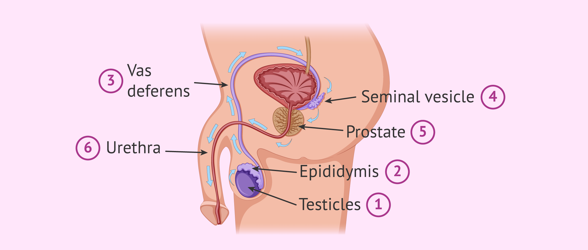
Vas/Ductus Deferens
carries sperm from testes→into pelvic cavity
runs with testicular A+V→form spermatic cord in scrotum
located in inguinal canal
within pelvis
runs along bladder
combines with seminal vesicle duct
forms ejaculatory duct
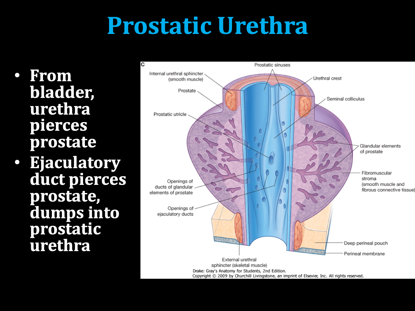
pierces prostate
dumps into prostatic urethra

Male Bladder Location
inferior to bladder→prostate gland
posterolateral to bladder→seminal vesicles
Seminal Vesicles
lie on posterior surface of bladder
produce components of seminal fluid

Prostate Gland
inferior to bladder
produces prostatic secretions→mix with seminal fluid+sperm→semen
pierced by prostatic urethra

Prostatic Urethra Pathway
bladder
urethra
prostate+ejaculatory ducts
prostatic urethra
Membranous Urethra of Penis
small portion of urethra
from prostatic urethra→through deep perineal pouch+pelvic floor→spongy urethra

Spermatic Cord
runs through inguinal canal
contains
cremaster muscle+fascia
cremasteric A+V
testicular A
testicular V (in pampiniform plexus)
vas deferens
A to vas deferens
branch of genitofemoral N
sympathetic+visceral N fibers
exits via
deep inguinal ring
inguinal canal
superficial inguinal ring

Sperm Pathway
sperm produced in testes
sperm stored in epididymis
vas deferens
inguinal canal
pelvic cavity
seminal vesicles+vas deferens combine together
ejaculatory duct
-carry sperm+seminal fluid togetherprostatic urethra
prostate→secretes prostatic secretions
sperm+seminal fluid+prostatic secretions→semen
prostatic urethra
membranous urethra
spongy urethra
-in corpus spongiosum