Micro 10
 Call Kai
 Learn
 Practice Test
 Spaced Repetition
 Match
 Flashcards
 Knowt Play
1/168

There's no tags or description
Looks like no tags are added yet.
169 Terms
why when embryo begins to develop does the cardiovascular system develop first?
the early embryo can be sustained by diffusion but as get larger cant be supported by diffusion as distance nutrient sneed to cross get larger so a circulatory system become snecessary before organs mature so they can be supplied
is the cardiovascular system the first functional system in an embryo?
yes
what does the mesoderm give rise to for cardiovascular?
vascular and cardiac mesenchymal precursors
what do angioblastic cells do?
contribute to blood vessel formation
what call process of blood vessel formation?
angiogenisis
what does early vessel formation develop?
a network that later expands and remodels
what is the cardiogenic field?
horse shoe shaped cardiogenic region in embryonic disc that lies cranial and lateral to neural plate that has blood forming cavities in the visceral mesoderm that will coalesce and form the primitive heart, blood vessels and blood cells
what does embryonic folding change?
the position of the cranial field
what are the folding and steps that the cranial cardiogenic field undergoes in order?
the field is brought caudo-ventrally then the developing aorta approaches the cardiac tube which is then brought to a ventral position to the dorsal aorta and the vitelline veins. The caudal part of the cardiogenic tube fuses with the vitelline veins and the two sides of the cardiac tube fuse as well. The paired cardiac primordia move towards the midline and the dorsal and ventral aorta connect.
what do dorsal and ventral aorta connect?
the early circulation
what does fusion of the dorsal and ventral aorta establish?
a primitive heart tube
what occurs to the primitive heart tube?
the tube is initially straight but then undergoes elongation and looping resulting in the bulbus cordia, primitive ventricle and atrium acquire new spatial relationship and the establishment of the arrangement needed for subsequent septation to form heart chambers
what is a primitive atrium?
anterior/trabeculated regions of the right and left atria
what is primitive ventricle?
much of the left ventricular trabeculated region
what is the bulbus cordis?
the right ventricle and ventricular outflow region in embryo
what is the truncus arteriosus?
the aorta and pulmonary trunk after partitioning
what is the sinus venous?
the smooth part of the right atrium and associated venous structures
what do endocardial cushions partition?
the common atrioventricular canal
what does cushion tissue contribute to?
the cardiac septa and atrioventricular junction
what must partitioning preserve?
suitable fetal blood flow during development
what is the steps of partitioning to form heart?
the primary atrial septum/septum primum and intraventricular septum begins to develop and the septum primum grows towards the endocardial cushions forming ostium primum that is a foramen that premits interatrial blood flow
the ostium primium narrows and closes and the ostium secundum forms and preserves blood flow allowing right to laeft atrial passage
the second atrial septum/septum secundum grows to the right of septum primum creating the fetal foramen ovale connecting the right and left atria
the interventricular septum growing upwards from the ventricular floor and fuses with the endocardial cushion seperating right and left ventricles
what does ostium primum initially permit?
interarterial blood flow
when does the ostium primum narrow?
narrows as the septum primum approaches the cushions
what does the septum primum form adjacent to the foramen ovale?
a mobile flap
what allows blood to flow from right atrium to left atrium through foramen oval in fetus?
the fetal right atrial pressure
what closes the formen ovale after birth?
firs breaths after birth lower pulmonary vascular resistance and the left atria pressure rises switching the sides of the pressure gradient which forces the mobile flap formed by septum primum closed
does functional closure of the foramen ovale precede anatomical fusion?
yes
what does the truncus arteriosus divide into?
the aorta and pulmonary trunk
what establishes the normal crossing pattern of thevessels of the heart?
the spiral development of the aorticopulmonary septum
where is right ventricular blood directed to?
pulmonary trunk
where is the left ventricular blood directed to?
aorta
what can alter aorta and pulmonary trunk alignmnet?
abnormal outflow sepation
what happens if complete transposition of the great vessels occurs?
the aorta arises from the right ventricle and then the deoxygenated blood will flow to body while oxygenated flows to lungs
what do embryonic aortic arch arteries form paired connections with?
dorsal aortae
what occurs to selected arch segments?
they persit, regress or remodel
what can abnormal remodeling of arch segments do?
alter the arrangement of the great arteries which can have bad outcomes
prior to birth what is fetal circulation designed for?
the in utero aqueous enviroment where the placenta oxygenates fetal blood
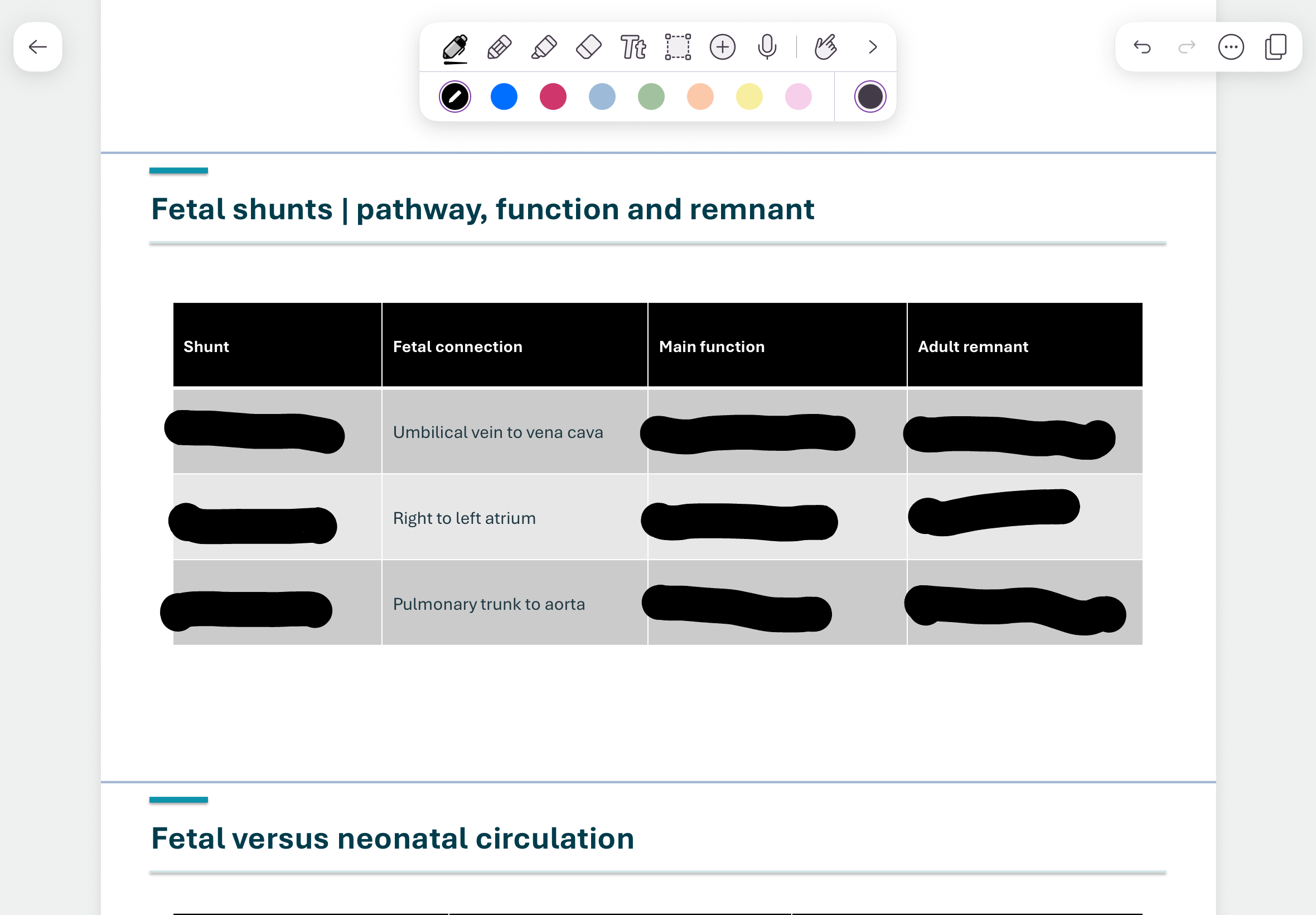
what are the 3 essential fetal shunts? what does each do and where located?
ductus venosus: connects the umbilical vein to inferior vena allowing oxygenated blood to bypass liver
foramen ovale: located between atria allows blood to flow from right atrium to left atrium so it doesnt have to go to lungs since cant get oxygen from lungs in utero
ductus arteriosus: connects pulmonary arteries and aorta allowing blood to flow from pulmonary artery into aorta so doesnt have to go to lung since dont get oxygen in lungs while in utero
what oxygenates fetal blood?
the placenta
where does oxygenated umbilical venous blood return and what occurs following this?
blood returns to the fetus and reaches the caudal vena cava where goes into right atrium and then a substantial portion of blood enters the left atrium through the foramen ovale
what does the left fetal heart supply?
the systemic arterial circulation
where does gas exchange occur in fetus?
placenta not alveoli
what is pulmonary resistance like in fetus? what does this do to blood flow?
high pulmonary resistance so blood flow to lungs is limited since no gas exchange occurs there
what do the fetal shunts allow blood to bypass?
the liver and pulmonary circulation
do different streams of fetal blood have different oxygen content?
yes
what steps occur in order when transition from fetal to neonatal circulation?
lung expansion decreases pulmonary vascular resistance
placental circulation is lost when umbilical vessels close
left atrial pressure rises which functionally closes the foramen ovale
flow through ductus arteriosus declines as it constricts
whata re the 3 postnatal remnants of the fetal shunt?
ligamentum venosum- remnant of ductus venosus
fossa ovalis- remnant of foramen ovale closure
ligamentum arteriosum- remnant of ductus arteriosus
which closure precedes the other in foramen ovale closure?
functional closure precedes fibrous anatomical closure
what are the 6 steps of the essential sequence of cardiac development?
1-cardiogenic field and cranial mesodermal precursors
2-heart tube from the midline fusion of primordia
3-looping for reorientation of early chambers
4-septation into atria and ventricles
5-form the great vessels (aorta and pulmonary trunk)
6-close shunt and perfuse lungs at birth













what does the heart do?
create pressure and circulate blood
what do arteries do?
distribute blood
what do veins do?
return blood to heart
what recover tissue fluid?
lymphatics
what do capillaries do?
exchange nutrients and waste
whata re 4 main functions of cardiovascular system?
-maintain adequate blood flow and cardiac output
-deliver oxygen, nutrients, hormones, electrolytes and water to tissues
-remove carbon dioxide and other metabolic waste products
-support thermoregulation and normal renal perfusion
what is pulmonary circulation?
from heart to lungs and back to heart
what is systemic circulation?
from heart to body/tissue back to heart
what is the first organ to form in embryo?
heart
who have 4 chambers of their heart?
mammals and birds
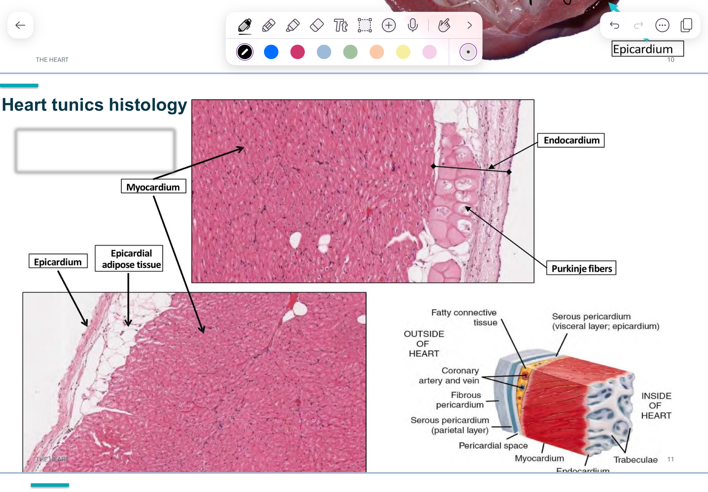
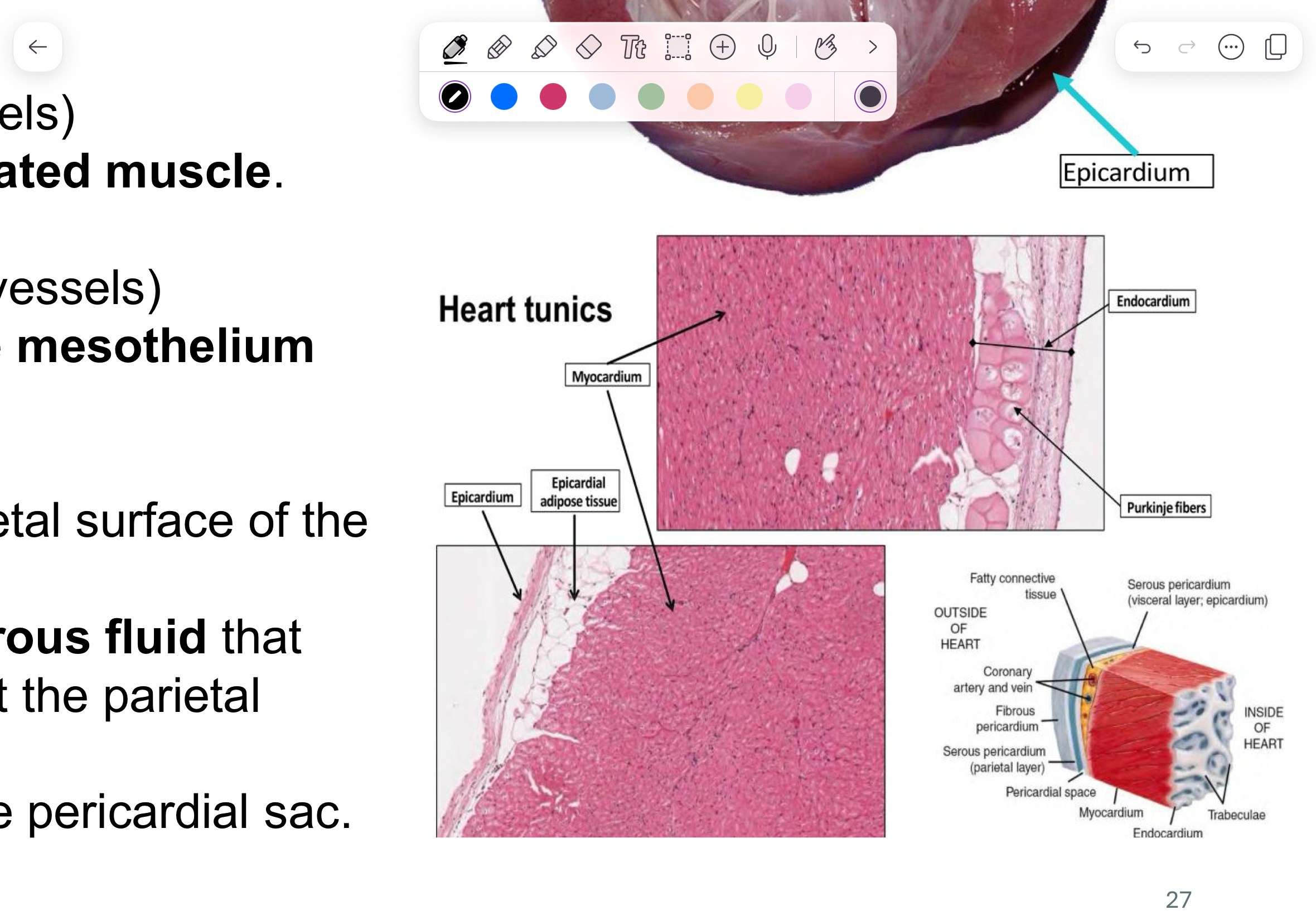
what 3 layers compose the heart in order from inside to outside?
endocardium
myocardium
epicardium
what is another way can call epicardium?
visceral pericardium
what layer/tunic find purkinje fibers?
endocardium
what does the endocardium form?
inner lining and valves
what layer of heart is in direct contact with blood?
endocardium
what is the innermost surface of endocardium?
endothelial cells (stratified squamous)
what is the endocardium equivalent to in blood vessels?
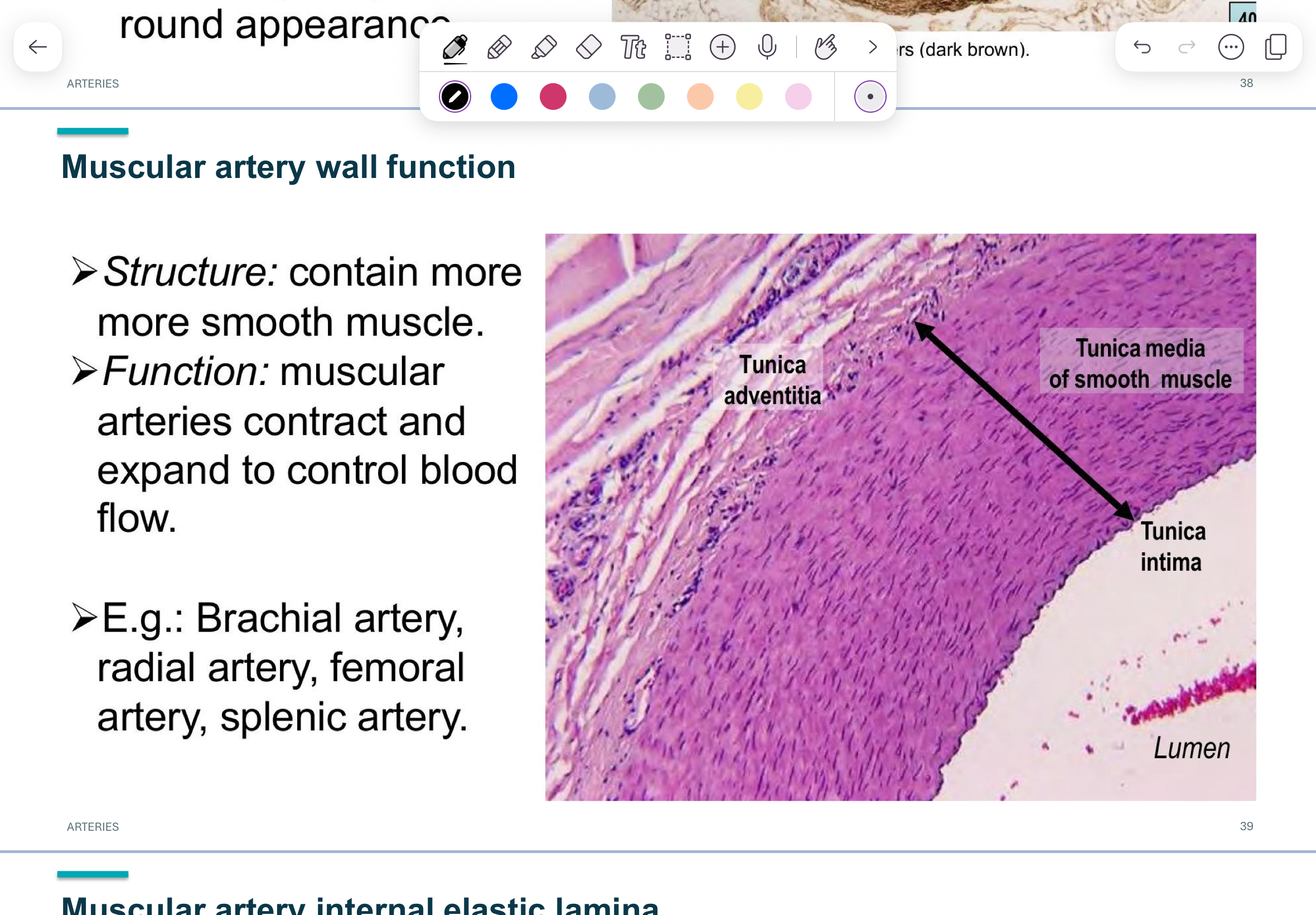
tunica intima
what layer of heart is important in hemostasis?
endocardium
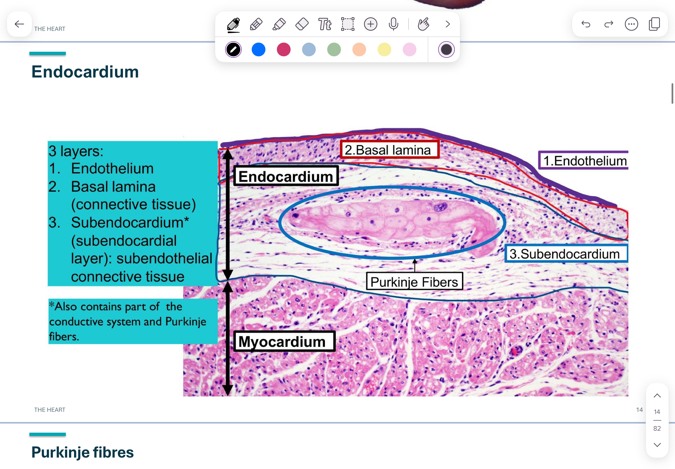
what are the 3 layers of endocardium?
endothelium
basal lamina (connective tissue)
subendocardium
what is subendocardium?
subendothelial connective tissue that also contains purkinje fibers and some of the conductive system
what are purkinje fibers?
fast conducting cardiac muscle cells that rapidly transmit electrical impulses to coordinate heart contractions
what are myofibrils like of purkinje fibers?
few myofibrils and are located on periphery
do purkinje fibers have lots of glycogen and mitochondria?
yes
do purkinje fibers have t tubules?
no