CNS Disease meurodegeneration & tumours part 1
 Call Kai
 Learn
 Practice Test
 Spaced Repetition
 Match
 Flashcards
 Knowt Play
1/84

There's no tags or description
Looks like no tags are added yet.
85 Terms
What is epilepsy and seizure
epilepsy is a group of disease which exhibit unprovoked seizures
seizure is transient occurence of signs ± sx due to abnormal excessive or synchronius neuronal activity in brain
Seizure classification and example
focal onset : tonic, myoclonuc, sensory emotional
generalised onset : tonic (stretch) - clonic (rythmic jerking, typical
unknown onset
mb all have motor and nonmotor
Stages of seizure
Prodromal : Days to hours before, anxiety, irritability, mood changes
Early ictal : auras like dejavu fear (amygdala activated)
ictal : aura to seizure stage
post ictal : recovery
What form of epilepsy most common in adults + risk factors
temporal lobe epilepsy
brain injuries
Dravet syndrome
seizures develop in 1st year of lofe
febrile seizures, usually heat induced
denovo variants causing scn1a defective containing na+ → inhibitory neurone cant act
CHD 2 encephalopathy
chd 2 regulates gene expression in brain development
mutation may lead to hyperexcitable brain
How inflammation cause epilepsy
Disrupted gene expression from injury, stroke, infection etc
microglial dysfunction cause alteration in synaptic structure
Astrocyte lose ability to control, causing dysregulated synaptic reorganisation
glutamate and gaba processing also dysregulated (more glutamate receptor while gaba a receptors internalised)
Dx of epilepsy
EEG
MRI, Blood test (to rule out others)
Hx, genetic testing
Note high rate of misdiagnosis (20%)
GABA Potentiating drugs eg and moa
benzodiazepines (diazepam, acute) , barbiturates
Positive allosteric modulator for GAVAa receptor
chloride ion channels open more → cl influx → hyperpolarisation
Gaba transmission targeting drugs
Vigabatrin (1st line tx for infants): inhibits GABA- T causing less GABA metabolism
Tiagabine : Blocks GATs causing less GABA reuptake → more GABA in synaptic cleft
Gaba transmission targeting drugs
Vigabatrin (1st line tx for infants): inhibits GABA- T causing less GABA metabolism
Tiagabine : Blocks GATs causing less GABA reuptake → more GABA in synaptic cleft
Ca signalling target drugs for epilepsy eg and mia
gabapentin, pregabalin
block vg ca ion channels → less calcium ion influx at presynaptic terminal → less exocytosis
Felbamate (nmda, 1st for infantile partial), topiramate (ampa)
block nmda or ampa at postsynaptic terminal → less glutamate → less excitability
Valporate MAC
M : monocarboxylic adid for tx of epilepsy
A : GABA T inhibitor, VG Na channel blocker, T type calcium channel blocker (histone deacetylase inhibitor)
C : reduce hyperexcitability (+ neurodevelopmental disprder)
Levetiracetam MAC
focal onset Epilepsy medication
binds to sv2a on vesicles (mainly with glutamate) prevent exocytosis
reduce excitation
Epilepsy side effects
sedation, fatigue
dizziness, less coordination
anxiety
retinal nerve atrophy (vigabatrin)
nausea
excitement
other non pharmacological tx for epilepsy
vagus nerve stimulation
keto diet
surgery
gene replacemnt
stem cell transplant
Cerebrovascular accidents classification and subclassification
arterial : subdivided into ischaemia or haemorrhage
venous : subdivided into ischaemia
What is a stroke
blood flow to brain is interrupted either from blockage or bleeding
defined clinically as :-
1. Acute functional neurological deficit
2. localised within cns w vascular cause
3. lasting more than 24 hours (if death does nit occur)
WHAT IS TIA
Resolutioj of all neurollgical sign and symptoms within 24hrs wo brain infarct on imaging
What happens if ACA + MCA Supply blocked
All 3 of TACS (total anterior circulation stroke) criteria
unilateral weakness / sensory
homonymous hemianopia
higher cerebral dysfunction
What happen if either aca or mca supply knicked PACS
2 of TACS (total anterior circulation stroke) criteria
unilateral weakness / sensory
homonymous hemianopia
higher cerebral dysfunction
What happen if lacunar stroke (deep penetrating branches)
one of :-
pure motor/sensory stroke
sensori-motor stroke
ataxic haemparesis (weakness + poor movement coordinatiom in one side)
Posterior circukation stroke sx
One of :-
CN palsy and comtra motor/sensory deficit
bilateral motor/sensory deficit
ocular motor / sensory deficit
cerebellar dysfunctiom
What cause blocked vessel
embolism: artherosclerosis, AF, Carotid dissection
Thrombosis.dehydration, atherothrombosis, lipohyalinosis, hypercoaguable disorder
Vasculitis: autoimmune or infectious
Fibromuscular dysplasia: abnormal cell growth (string of beads)
Causes of bleeding vessel
Trauma
HTN
Aneurysm
amyloid
drugs

What are a to e

Mechanism of neuronal injury
Neurones have less fuel and experience excitotoxicity
Inappropriate release of excitatory neurotransmitter (glutamate)
excessive calcium influx from nmda glutamate receptor
mitochondrial and ~dna Damage → necrosis and apoptosis
permanent damage
Cellular reaction of astrocyte to cns injury
gliosis
condensation of thick fibre ( rosenthal fibre)
Glial cells multiply/swell to form scar like structure to protect neurone
Microglia response to cns injury
proliferation
nodular aggregates
neuronophagia
Ischaemic stroke vs haemorrhagic strole infarct gross appearance
ischaemic : pale, softened, liquefied brain
haemorrhagic : blood clot (haematoma)

find

What is the distribution of peripheral neuropathy
stocking glove neuropathy
affects hands or feet first
Causes of raised icp + eg
space occupying lesion. haemorrhage, infection, tumour
cerebral oedema : cytotoxic, interstitial, vasogenic
Expanded csf : reduced csf resorption, increase csf production
What is vasogenic oedema + imaging
bbb disruption w increased vascular permeability
fluid shift from intravascular space to parenchyma, intercellular spaces
due to inflammation/tumout or generelosed (ischaemia)
gray-white matter differentiation maintained
what is cytotoxic oedema + imaging
increased intercellular fluid following neuronal/glial/endothelial injury
caused by hypoxia or metabolic derangement
gray-white matter differentiation lost in ct
What is hydrocephalus and types
Hydrocephalus is accumulation if excessive csf in ventricular system
communicating hydriceohalus
obstructed csf :subarachnoid haemorrhage, infection
non obstructed : normal pressure hydrocephalus
non communicating (obstructive)
colloid cyst in foramen of monro
aqueduct sylvius stenosis
compression of fourt ventricle
tumours
clinical consequences of raised icp (5)
early morning headache/cephalgia
cushings sign (brady, widened pulse pressure, irregular respiration)
papilloedma
n&v
seizures, behaviour changes
What is brain herniation and type
increased icp displaces brain tissue
subfalcine herniation : involves cingulate gyri
transtentorial herniation : medial aspect of temporal lobe compress cniii
tonsillar herniation : cerebellar tonsil compress brainstem → compromise resp/cardiac centre
What happens if there is panda eyes and bruising begind ear
panda eyes/ raccoon eyes : basilar skull fracture
bruising behind ear : battle sign due to bruise around mastoid
Intraparenchymal haemorrhage location causes, source, shape on imaging
brain paremchyma
htn, avm, trauma
atrial or venous
rounded lobes
intraventricular haemorrhage location causes, source, shape on imaging
in the ventricle
htn, avm, trauma, tumour
arterial/ venous
conforms to ventricular shape
subarachnoid haemorrhage location causes, source, shape on imaging
between arachnoid and pia mater
berry aneurysm, rupture, avm , trauma
mainly arterial
‘star sign’
subdural haemorrhage location causes, source, shape on imaging
between dura and arachnoid
trauma to bridging veins
venous source
crescent shape
extradural haemorrhage location causes, source, shape on imaging
between dura and skull
trauma (pterion fracture hit mma)
arterial mostly
lentiform shape
Extradural haemorrhage clinical features
brief period of unconciousness
then lucid interval of minutes to hours
as blood accumulates, haematoma compress brain causing worse sx
Subdural haemorrhage clinical features
subacute w step wise deterioration + bleed
non accidental injury (sdh _ retunal haemorrhage + encephalopathy)
more common in older people as bridging veins stretch more
Subarachnoid haemorrhage clinical features
thunderclap headache
blood in csf during lumbar puncture (later xanthochromia)
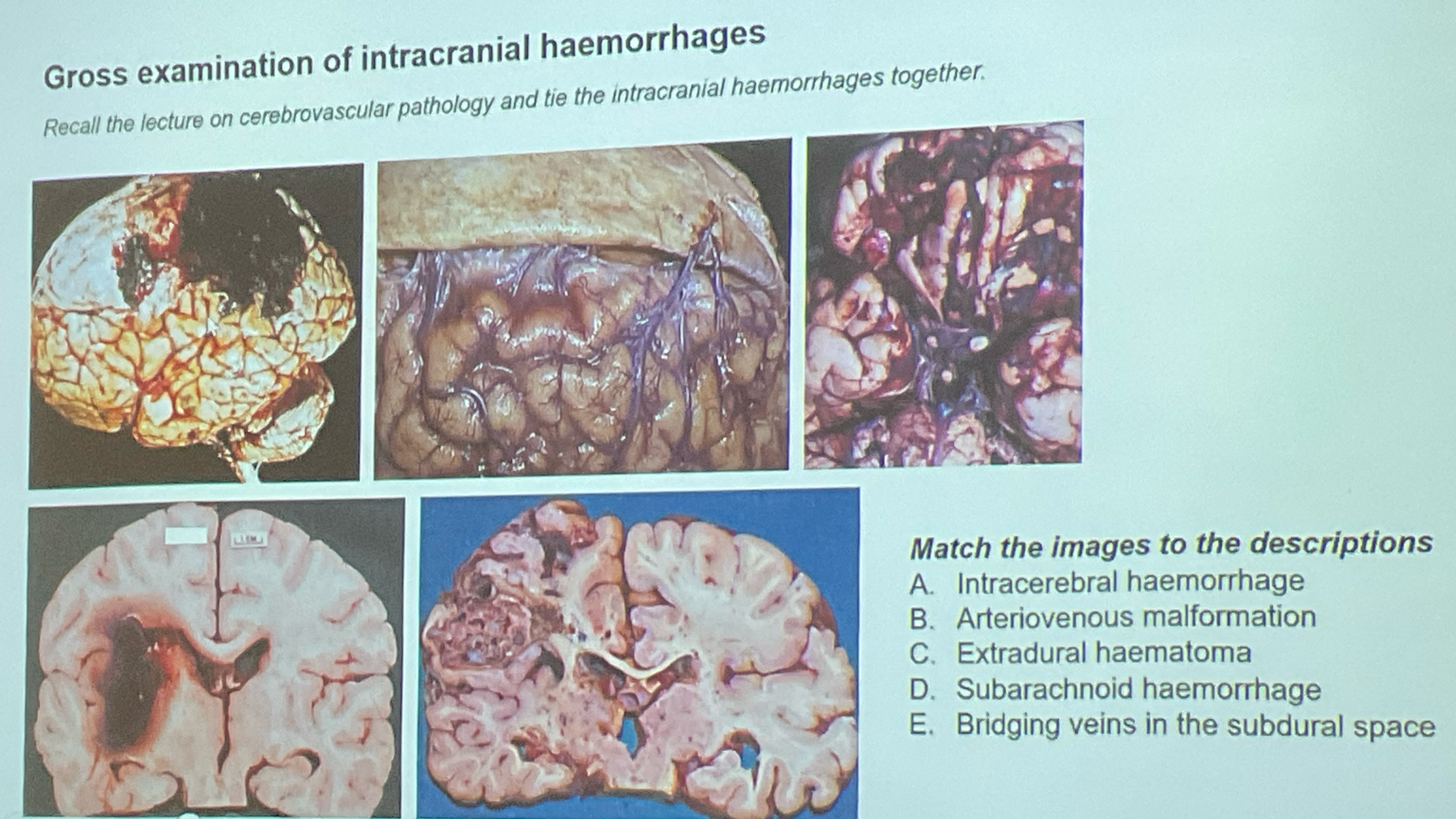
match please

Hydrocephalus ex vacuo
compensatory enlargement of brain ventricle due to loss of brain tissye
brain volume shrink
icp normal, no blockage nor fluid imbalance
Histological evolution of brain infarcts
acute (6-12 hrs ) : red neurones surround oedema and neutrophils, endothelial and glial cells swell, myelinated fibre disintegrate
subacute (48-72) : phagocytic cells clean up myelin breakdown
healed infarct : dense network lf glial fibres + capillary and ct
Axonal loss vs demyelination difference electrophysiologically
Action potential amplitude reduced but velocity normal for axonal loss and vv
fibrillations present in axonal loss but absent in demyelination
Axonopathy vs myelinopathy progression
axonopathy usually with myelinopathy
myelinopathy +- axonopathy
peripheral innervation and patterns of injury in a muscle
when reinnervation, each axon will be associated with more motor units
one axon will innervate more myofibres
Paraproteinaemic polyneuropathy what and manifestations
nerve damaging disorder due to production of paraproteins/mproteins(MAb) from plasma proteins
demyelinating or axonopathic
Manifest as POEMS
Polyneuropathy
organimegaly
endocrinopathy
M- Band
skin changes
What is gullainnbarre syndrome (4)
acute inflammatory demyelinating neuropathy
may spread as ascneding paralysis, cause areflexia, fatal respiratory paralysis
preced by acute flulike illness
increased csf protein but inflammatory cells confined to roots
Chronic inflammatory demyleinating neuropathy what
symmetric, mixed sensorimotor polyneuropathy, >= 2 mths
T cells, antibodies targetmolecules at schwann cell-azon junction
complement fixing igg and igm cause recruitement of macrophages
Myasthenia gravis vs LEMS
MG is when autoantibody bloc ACH receptors at post synaptic
LEMS is when autoantibody block ach release at presynaptic terminal
MG worsen when muscle exert while in LEMS, usage w muscle response improved
Genetic neuronal disorder (3)
SCA : AR mutation in fxn gene cause degeneration in spinal cord, pns, cerebellum
CMT :PMP22 duplication commonly causing sensorimotor demyelination → deterioration
LHON : mitochondrial inheritance causing increased ros → apoptosis of retinal ganglion cells
Inflammatory muscle disorders (4)
dermatomyositis : autoantibodies (anti-m2, anti-jo1) causing inflammation of bv causing perifascular atropphy
polymyositis : dermatomyositis wo cutaneous features due to cd8+ infiltration
immune mediated necrotising myopathy : subacute muscle weakness w increased CK assoc w autoantibody agains hmg coa reductase
inclusion body myositis : progressive muscle weakness in older adults involving myofibres contained rimmed vacuoles (blue)
DMD vs myotonic dystrophy
dmd early onset while myotonic dystrophy is adult onset
X linked recessive (dmd) vs mutation in dmpk or cnbp (myotonic dystrophy)
genetic congeintal muscle disorder (-dmd ans MD) (4)
myotonia congemita ; mutation in clcn1 for skeletal muscle
channelopathies : hypokalaemic (prolonged) and hyperkalaemia (brief episodic weakness)
Inborn error of metabolism : mcardles disease involving lack pf g1p
mitochondrial disorder
what are the peripheral nerve sheath tumours (3)
schwannoma : benign, encapsulated w either celullar (antoni A) / hypocelullar (antoni B)
neurofibroma : benign unecapsulated spindle cell tumours
MPNST : malignant sarcoma w aggresive behaviours
PNS muscle tumours (3)
Rhabdomysarcoma » rhabdomyoma : mesenchymal tumours
leiomyomas (fibroids) » leiomyosarcomas : mesenchymal tumours usually in uterus
Global standards in diagnosing tumours (3)
WHO classification of tumour
iccr (RCpath, RCPA, CAP)
TNM staging ( by uicc, ajcc)
Gliomas and astricyte grading system (4)
astrocyte, oligodendroglioma, ependymoma
pillcytic astrocytoma = grade 1
grade 2-4 are known as infiltrating astrocytoma
glioblastoma is grade 4 astrocytoma wo idh mutation
clinical features of gliomas
pilocytic astrocytoma l infratentorial mass, may exhibit rosenthal fibres
infiltrating astrocytoma : well differentiated → anaplastic + mitosis →microvascular proliferation ± necrosis + idh1/idh2 mutant
glioblastoma : diffuse infiltration, malignant
non astrocytoma gliomas (2)
oligodendroglioma : grade 2 or 3, idh mutant mostly in frontal lobe, fried egg appearance
ependymoma : grade 2 or 3 in post fossa/spinal canal, perivascular pseudo-rosette
both have microvascular proliferation, necrosis and brisk mitosis (eho grade 3 )
meningioma(3)
derived from meningothelial cells of arachnoid (who 1-3)
15 subtypes : most common meningithelial, fibrous and transitional
grading based on atypia, mitotic figures and invasion + tert promoter region
Vestibular schwannoma (acoustic neuroma) (4)
who grade 1
sx : tinnitus, unilateral hearing loss, vertigo, disequilibrium
assoc w NF2 (inactivating mutatiin of gene)
histologically, cells kine up to form nuclear palisades
Primary diffuse large b cell lymphoma of cns
solitary lesions, affectingcerebral hemispheres, thalamus and basal ganglia usually
dx : aurora borealis sign on fundoscopy, angiocengric cuffs of neoplastic b cells (cd20+, cd19+)
assoc w myd88 l265p mutation
cns metastatic tumours
haematogenous spread
to brain/spinal cord parenchyma : lung cancer, breast cancer in adults + leukaemias and lymphomas on childrem
to meninges : leptomeninges, metastasis
neural tube defects (2)
anencephaly : absence of brain and cavarium
spina bifida: occulta (small gap in vertebrae), meningocoele (outpouching wo cns) or myelomeningocoele (+ cns extension)
chiari malformation types (2)
downward displacement of cerebellum through enlarged foramen magnum
chiari type i : low lying cerebellar tonsil
chiari type ii : arnold chiari, mishapen midline cerebellum w downward extension of vermis
Entamoeba histolytica pathogenesis
exist in cyst and trophozite forms
ingestion of cyst
cyst in small intestine form trophozoites
trophozoites invade and penetrate colonic mucosa
tissue destruction → increased intestinal secretion
ascends to portal venous system to cause liver abcess
Amoebic dysentry sx and dx
diarrhoea (usually bloody)
abdominal pain. weightloss
dx by stool microscopy (3 samples from seperate days) or antigen
nb serology does not distinguish past and present infection
Amoebic liver acess epi sx dx tx
in returning travellers
ruq pain w fever, sometimes diarrhoea and hepatosplenomegaly
dx by imaging on right lobe, serology or aspirate ‘anchovy paste’
oral metronidazole (+ paromycin for intraluminak cyst)
Dementia definition
acquired persistent loss of intellectual function
with compromise in memory
and at least2 other cognituve domaina such as language, visuspatial skills or executive function
Dementia beside classification and examples
dementia w other medical disease : HIV, immune, paraneoplastic
dementia w other neurological disease : HD,MS,STROKES (always;pften present w clonical signs)
dementia in isolation : alzheimers, fronto-temporal
Alzheimer Amyloid cascade deposition theory
APP gene codes for APP protein
if cut by b secretase then gamma secretase then build up of Amyloid- beta 42protein
Ab oligomerise then make plaques in brain
sx depends on deposition
ultimateky cause neurodegenration
Alzheimers tau theory
in concussion, release of ca2+
activate calpain → activate cdk5
hyperphosphorylation of tau
tau detavhed from microtubules and form clumos called neurofibrillary tangles
Fromti-Temporal dementia
Affects :-
Language : apahasia
Behaviou : socially inappropriate
motor : mnd, parkimsons
Tau related, sx depends on place
Huntington disease pathophysiology
CAG repeats of huntingtin gene (anticipatiin)
dominang toxic gain of function
increase cag repeats → misfolded confirmation
at the end, produce nig insiluble aggregates
elicits neutotoxicitu → caudate atrophy
MND factors
genetic : C9orf72 (RAN-ATG translation aggregates cause neurotoxic)
toxic : expsome, occupational hazards
Metabolic : Oxidative stress, atp depletion
Parkinsons disease motor vs non motor
Motor
Resting tremor : coarse, asymmetric
Gait Impairment : shuffking
Bradykinesia : flexed oosture
Non motor
D : Depression
A : Anxiety
S : Sleep Rem disorder
H : hallucination
Parkinson pathophysiology + tx
a- synuclein metabloism and aggregatiin
make fibril which can become lewy bodies (wo lewy bodies called Multiple system atrophy)
Spread tp different places of brain according to stages
give dopamkne (L DOPA)