Abdominal Imaging I
 Call Kai
 Learn
 Practice Test
 Spaced Repetition
 Match
 Flashcards
 Knowt Play
1/38

There's no tags or description
Looks like no tags are added yet.
39 Terms
Should abdominal rads be taken on peak inspiration or expiration?
Expiration
Abdominal margins that should be included in every rad
- diaphragmatic crura (and whole diaphragm)
- ventral body wall
- coxofemoral joints
- lumbar vertebrae
What is detail?
How well the margin of an organ or structure can be discerned from the adjacent organ/structure
AKA, ability to see organs
Ideal BCS rad detail
Adequate
Mildly to moderately obese BCS rad detail
Excellent - fat and soft tissue are different opacities
Thin/cachectic BCS rad detail
Poor - lack of fat and superimposition of organs
Why do neonates have decreased rad detail?
Higher accumulation of brown fat - higher water content
What is within the peritoneal space?
- liver
- spleen
- GI tract
- urinary bladder
- LNs
- mesentery
What is within the retroperitoneal space?
- kidneys
- perirenal fat
- ureters
- adrenal glands
- abdominal aorta
- abdominal vena cava
- +/- ovaries
- +/- prostate
Causes of decreased detail
- young animal
- decreased BCS
- peritoneal effusion
- large masses
- steatitis/peritonitis
- carcinomatosis/sarcomatosis
Cause of increased detail
- gas in peritoneal space/retroperitoneal cavity
- obesity
Causes of pneumoperitoneum
- hollow viscous rupture/GI perforation
- penetrating abdominal wound
- iatrogenic (procedure within 28 days)
- abscess rupture (gas forming bacteria)
- perforated emphysematous cystisis/cholecystitis
Where are the best places to look for pneumoperitoneum?
In front of the liver and around the falciform fat - stay away from SI
Generally, what is the cause of pneumoretroperitoneum?
Extension of pneumomediastinum
Further evaluation of pneumoperitoneum diagnosis technique
Horizontal beam rad - put patient in left lateral recumbency for 10 min prior to shooting the rad
Causes of peritoneal mineralization
- bate's body (nodular fat necrosis)
- dystrophic mineralization of a structure to prior mineralization
- vessel mineralization (endocrinopathies)
- incidental adrenal mineralization in older cats
Causes for generalized hepatomegaly
- metabolic hepatopathy (hepatic lipidosis, cushings, diabetes)
- toxicity
- viral/inflammatory hepatitis
- infiltrative neoplasia (lymphoma)
- congestion
Causes for focal hepatomegaly
- nodular hyperplasia
- neoplastic mass
- cyst
- abscess
- hematoma
- hepatic lobe torsion
Rad findings of generalized hepatomegaly
- rounded caudoventral margins
- caudally shifted gastric acid
- caudal margins extends beyond costal arches
Rad findings of focal hepatomegaly
- focally rounded margin
- asymmetry on ventrodorsal projection
- mass can be pedunculated and caudal to stomach
Rad findings of microhepatia
- caudoventral margin difficult to visualize
- cranially displaced gastric axis
- cranial displacement of other abdominal organs
2 common differentials for microhepatia
- normal variation
- chronic hepatitis with hepatic cirrhosis
- congenital PSS
2 spots of gas accumulation in the liver
- portal vein gas
- parenchymal gas
Where is the spleen found on VD rads?
Head is found left cranial
Causes of generalized splenomegaly
- breed related
- drug induced congestion (anesthesia, sedation)
- infiltrative neoplasia (lymphoma, MCT)
- extramedullary hematopoiesis
- splenic torsion
- infectious disease
Breed related generalized splenomegaly
- greyhounds
- shepherds
Causes of focal splenomegaly
- malignant neoplasia (hemang, histiocytic sarcoma, MCT)
- hematoma
- benign neoplasia/nodular hyperplasia
- abscess (rare)
Radiographic findings of pancreatitis
- cranial abdominal mass effect
- widening of the pyloroduodenal angle
- reduced detail in the right cranial abdomen
- gastric and duodenal functional ileus
Common pancreatic neoplasias
- carcinoma
- insulinoma2

Diagnosis?
Liver parenchymal gas - hepatic abscess

Diagnosis?
Hepatosplenomegaly - high suspicion for lymphoma
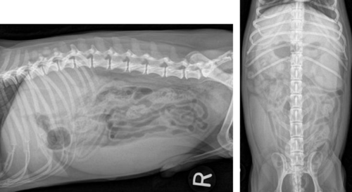
Diagnosis?
Generalized splenomegaly

Diagnosis?
Pneumoretroperitoneum

Diagnosis?
Generalized hepatomegaly

Diagnosis?
Focal splenomegaly

What is this and what is the significance?
Bate's body/nodular fat necrosis
- incidental
- common in cats

Diagnosis?
Pneumoperitoneum

Diagnosis?
Portal vein gas

Diagnosis?
Focal hepatomegaly