Uterine Size, Shape, Position, and Congenital Anamalies
 Call Kai
 Learn
Learn Practice Test
 Spaced Repetition
 Match
Match Flashcards
 Knowt Play
1/58

There's no tags or description
Looks like no tags are added yet.
59 Terms
Following menopause, the uterus typically becomes — than the premenopausal uterus
much smaller

Postmenopausal uterus. Sagittal (A) and transverse (B) views demonstrating an atrophic postmenopausal uterus (between calipers).
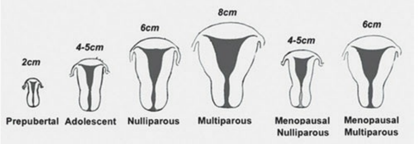
Changes in uterine size with age.
A newborn baby girl may have a thickened endometrium. This is caused by the —
stimulation of the baby’s endometrium by maternal hormones
The normal position of the uterus is considered to be —
anteversion or anteflexion
Anteversion describes the uterine position in which the body —
tilts forward or anteriorly, forming a 90-degree angle with the vagina
Anteflexion of the uterus denotes the position in which the uterine body —
folds forward, possibly coming in contact with the cervix
Retroversion of the uterus is the position in which the uterine body —
tilts backward or posteriorly, without a bend where the cervix and body meet
Retroflexion is the uterine position that results in the uterine body —
tilting backward and actually coming in contact with the cervix
The uterus that is located more on the left is referred to as a —
levoverted uterus
the uterus that is located on the right is referred to as —
dextroverted uterus
A bicornuate uterus, also referred to as bicornis unicollis, is a —
common uterine anomaly that is present when the endometrium divides into two endometrial cavities with one cervix, with a prominent concavity noted in the outline of the uterine fundus
The unicornuate uterus is present when the uterus has only —
one horn
The septate uterus, which is also a common Müllerian duct anomaly, describes a uterus that has —
two complete separate uterine cavities separated by an anteroposterior septum
The subseptate uterus, which is characterized by an incomplete septum, has a normal uterine contour with an —
endometrium that branches into two horns
The arcuate uterus is a subtle variant in which the endometrium has a —
concave contour at the uterine fundus
The uterus didelphys is —
complete duplication of the vagina, cervix, and uterus

anteverted uterus

retroverted uterus

anteflexed uterus
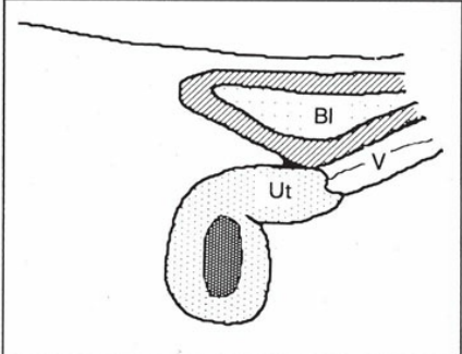
retroflexed uterus
The word part “colli” refers to the —
neck or cervix of the uterus
the word part “cornu” refers to the —
horn of the uterus
bicornis unicollis is interpreted as —
two horns with a cervix
bicornis bicollis is interpreted as —
two horns with a double cervix
Some studies claim that intrauterine — has resulted in the formation of congenital malformation of the uterus
exposure to diethylstilbestrol (DES)
DES was a —
drug administered to pregnant woman from the 1940s to the 1970s to treat threatened abortions and premature labor.
The female fetus exposed to DES in utero had an increased likelihood of developing a —
congenital uterine malformation
Congenital malformations have been linked to
menstrual disorders, infertility, and obstetric complications
Specifically, the septate uterus has an explicit connection with —
spontaneous abortion
hysterosalpingography, which is a radiographic study that utilizes contrast to evaluate the uterine cavity and fallopian tubes, was often performed on women with suggested —
congenital uterine malformations or who are suffering from infertility
Saline infusion sonohysterography, which may also be referred to as simply —, can be used to provide additional helpful information in regard to congenital uterine malformations
sonohysterography
For patients with a septate uterus, sonography can also aid in the resection of the septum during a —
hysteroscopic uterine septoplasty
— is said to be one of the most common Müllerian duct anomalies.
The septate uterus
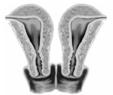
double uterus (didelphys) and double vagina
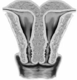
uterus duplex bicornis
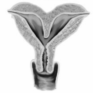
uterus bicornuate

uterus unicornuate
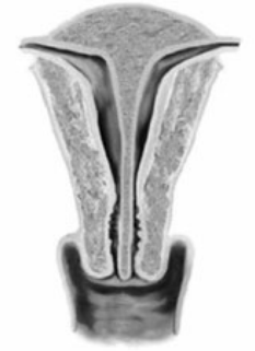
complete septate uterus

subseptate uterus
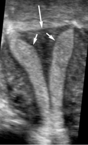
Septate uterus. Three-dimensional reformatting of a septate uterus in the coronal plane demonstrating the presence of two separate endometriums (short arrows) within the fundus (long arrow) of the uterus.
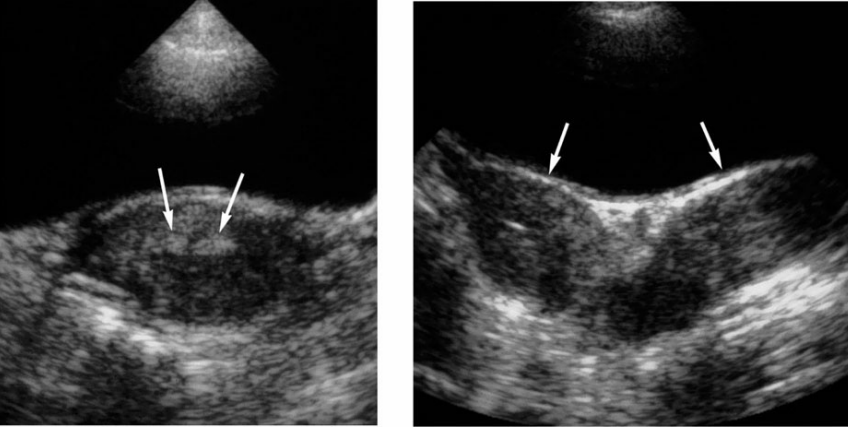
Didelphic uterus. A. Transverse transabdominal image of the cervix revealing two separate endocervical canals (arrows). B. Transverse transabdominal image of the upper uterus demonstrating a separate right and left uterine horn (arrows).
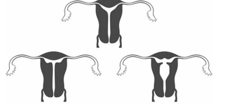
DES anomalies. Uterine anomalies associated with in utero diethylstilbestrol (DES) exposure. The classic anomaly is a T-shaped uterus.
the classic anomaly with DES exposure is
T-shaped uterus

Normal hysterosalpingogram. Radiograph of the female pelvis after the installation of a radiopaque compound into the uterine cavity.
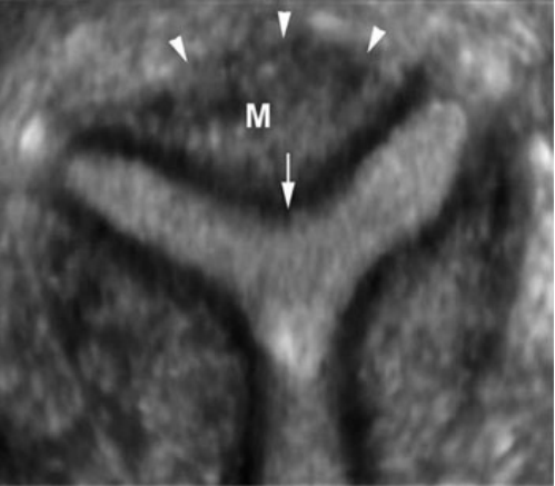
3D of arcuate uterus. True coronal view of the uterus, obtained by reconstruction using 3D sonography, demonstrating the myometrium (M) in the uterine fundus dips down with a rounded configuration (arrow) into the endometrium, indicating an arcuate uterus. The fundus had a normal shape (arrowheads).
Congenital malformations of the vagina can lead to the
accumulation of fluid within the female genital tract secondary to an obstruction
Hydrocolpos describes the condition in which —
the vagina is distended with simple, anechoic fluid, and is seen more often in the neonatal period
As the vagina distends with fluid, excessive amounts may lead to further accumulation of the fluid into the uterus, a condition known as —
hydrometrocolpos
Patients may have blood components from menstruation retained in the uterine cavity or vagina, termed —
hematometra and hematocolpos
hematometrocolpos, a condition when
both the uterine cavity and vagina are filled with blood
Vaginal obstruction is frequently associated with the presence of an —
imperforate hymen in young girls.
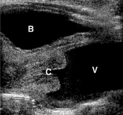
Hydrocolpos. Simple appearing fluid is noted posterior to the urinary bladder (B) and inferior to the cervix (C) within the vagina (V) in this sagittal image of the neonatal pelvis.

Hematometra. The uterus is distended with echogenic fluid (f) representing blood

Hematocolpos. Sagittal image of a 12-year-old girl with cyclic pain demonstrating a blood-filled distended vagina (V).
The word part “colpo” refers to —
the vagina
the word part “metra” refers to —
the uterus
clinical findings of vaginal obstructions
Cyclic pelvic pain (often at the time of menses in adolescent girls)
enlarged uterus
abdominal pain
urinary retention
amenorrhea (adolescent girls)
Sonographic findings of vaginal obstructions
Distension of the uterus, vagina, or both with anechoic or complex fluid