digestive system anatomy lab quiz
 Call Kai
 Learn
 Practice Test
 Spaced Repetition
 Match
 Flashcards
 Knowt Play
1/154

There's no tags or description
Looks like no tags are added yet.
155 Terms
which two anatomical parts is the digestive system divided into
digestive tract
accessory organs
which structures are included in the digestive tract
oral cavity
pharynx
esophagus
stomach
small intestine
large intestine
which structures are accessory organs to the digestive systems
teeth
tongue
salivary glands
liver
gallbladder
pancreas
what are the functions of the digestive system
processes food
breaks down food into small molecules for use in body
extracts nutrients from food
absorbs small molecules into blood to be distributed to tissues
eliminates waste
what are meat and other dietary proteins broken down into
amino acids
what are carbohydrates broken down into
monosaccharides and disaccharides
what are lipids broken down into
glycerol and fatty acids
what are the five stages of digestion
ingestion
mechanical and chemical digestion
absorption
compaction
defecation
what happens during Stage 1 (ingestion) of digestion
takes place wherever food enters (oral cavity, HG tube, ostomy, IV, etc.)
intake of food or drink
what happens during Stage 2 (mechanical and chemical digestion) of digestion
begins in the mouth with chewing and salivary amylase
continues with enzymes in the stomach and small intestine
what happens during Stage 3 (absorption) of digestion
occurs in the small intestine
bile mixes with chyme (food, enzymes, etc.) from stomach
mixture is churned inside the small intestine
what happens during Stage 4 (compaction) of digestion
occurs in the large intestine
gas and H2O are removed from indigestible food residue
what happens during Stage 5 (defecation) of digestion
occurs in rectum and anal canal
indigestible food residue is passed out of the body through feces
what is the enteric system
“brain in the bowel”
almost as many neurons in the digestive system as there are in the brain
nerves that innervate the digestive system are largely parasympathetic (CN X plays a big part)
what is our digestive system influenced by
endocrine system
autonomic system
emotions
what are amino acids and monosaccharides absorbed by
venous blood capillaries in the villi of the small intestine
what are fatty acids and glycerol absorbed by
lymphatic capillaries in the villi of the small intestine
what is the route of absorption for amino acids and glucose
venous capillaries in the lamina propria (of the small intestine)
small veins which converge into larger veins
larger veins empty into the hepatic portal vein
liver removes some of the amino acids and glucose for use and storage
blood leaves the liver
right side of the heart
lungs
left side of the heart
systemic circulation
what is the route of absorption for fatty acids and glycerol
lymphatic capillaries in the lamina propria (of the small intestine)
becomes chyle when lipids are combined with lymph fluid
flows through collecting ducts, lymphatic trunks, lymphatic ducts
flows into the subclavian vein
right side of the heart
lungs
left side of the heart
systemic circulation
what is chyme
stomach contents (food, enzymes, stomach acid, etc.)
what happens at the duodenum
chyme is received and mixed with bile and pancreatic juice
what are the two steps of lipid breakdown in the duodenum
broken down by bile into fat droplets
broken down by pancreatic lipase from fat droplets into fatty acids and glycerol
how is stomach contents (chyme) neutralized in the duodenum
acidity of HCL and pepsinogen is neutralized by the bicarbonate in pancreatic juice
what deactivates pepsin in the duodenum
pepsin is inactivated by the rise in pH
what finishes chemical digestion and what are the end products
pancreatic enzymes
results in amino acids, fatty acids/glycerol, and monosaccharides

which structure is shown
mucosa layer

which structure is shown
simple columnar epithelium in mucosa
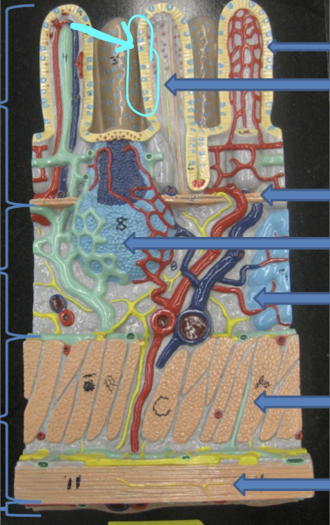
which structure is shown
goblet cells in simple columnar epithelium

which structure is shown
villi (projections)

which structure is shown
lamina propria in mucosa

which structure is shown
muscularis mucosa in mucosa

which structure is shown
submucosa layer
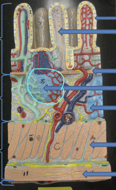
which structure is shown
lymph nodule of submucosa

which structure is shown
submucosal gland in submucosa

which structure is shown
muscularis externa layer

which structure is shown
circular muscle in muscularis externa

which structure is shown
longitudinal muscle in muscularis externa

which structure is shown
serosa layer
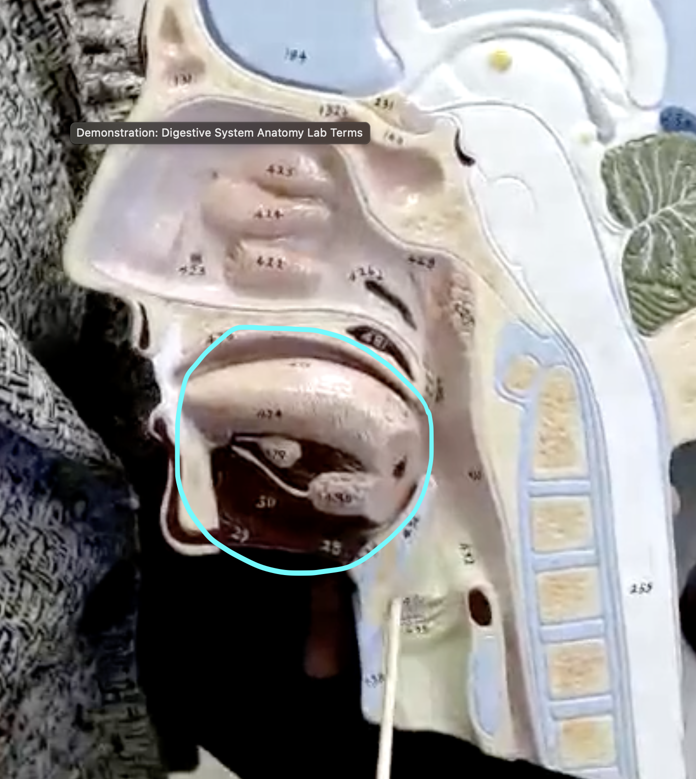
which structure is shown
oral cavity
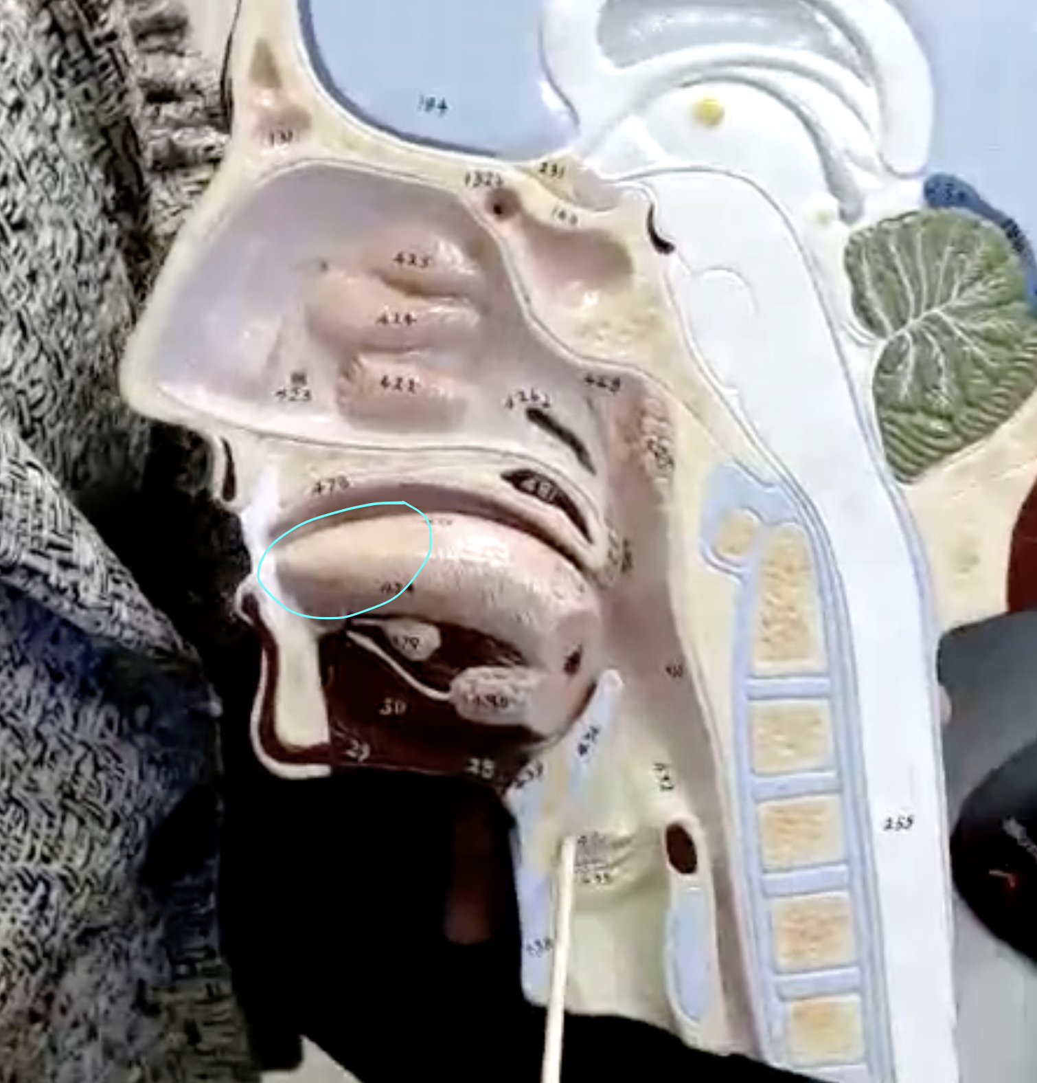
which structure is shown
body of the tongue

which structure is shown
root of the tongue
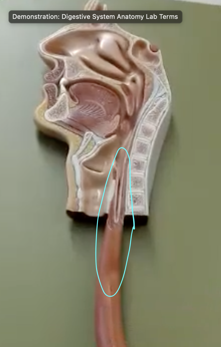
which structure is shown
esophagus

which structure is shown
stomach

which structure is shown
body of stomach

which structure is shown
cardiac region of the stomach

which structure is shown
esophagus

which structure is shown
fundus (outer protrusion/bump on stomach)
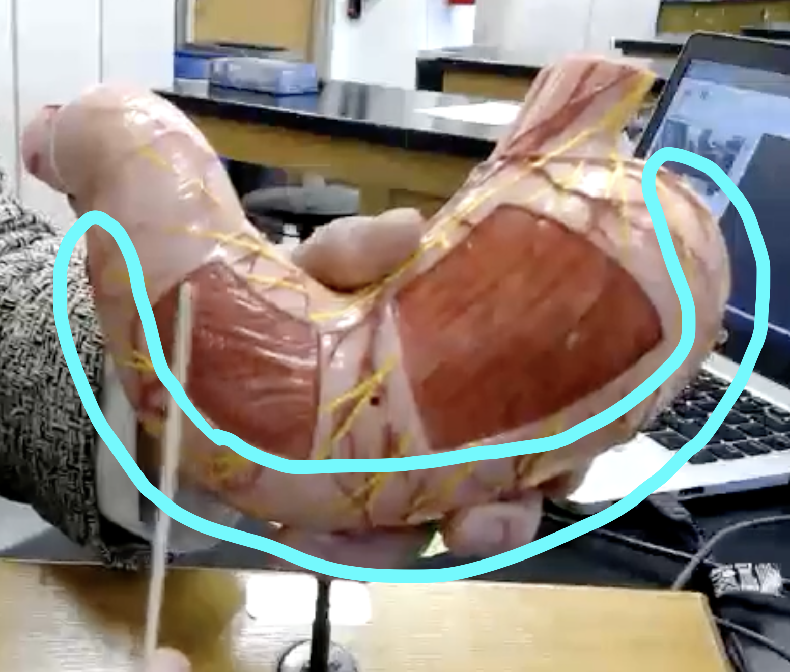
which structure is shown
greater curvature of stomach
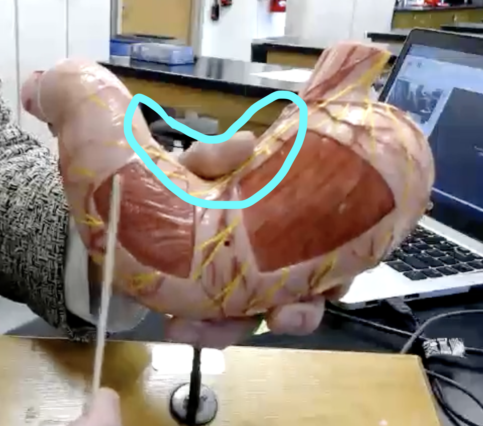
which structure is shown
lesser curvature of stomach
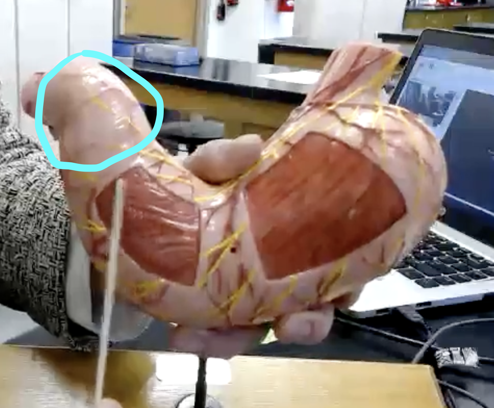
which structure is shown
pyloric region of stomach

which structure is shown
pyloric sphincter

which structure is shown
rugae of stomach (folds/grooves inside stomach)

which structure is shown
small intestine

which structure is shown
mesentery layer on posterior side of small intestine

which structure is shown
duodenum

which structure is shown
jejunum

which structure is shown
ileum

which structure is shown
ileocecal valve

which structure is shown
pancreas

which structure is shown
pancreatic duct

which structure is shown
hepatopancreatic ampulla and sphincter

which structure is shown
major duodenal papilla (bump)
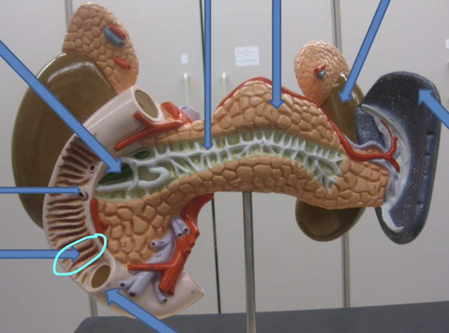
which structure is shown
plicae circularis in small intestine

which structure is shown
transverse colon

which structure is shown
ascending colon
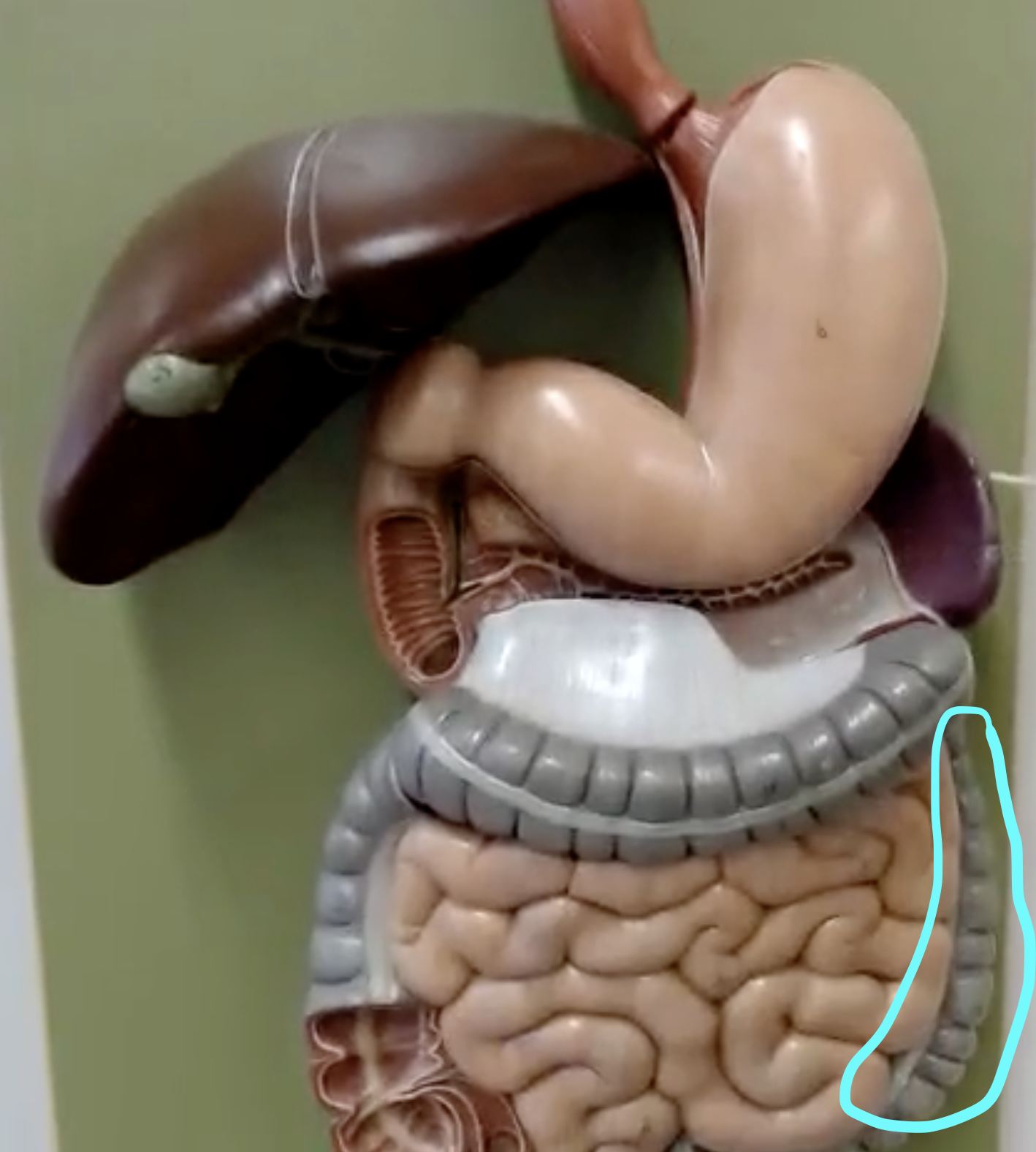
which structure is shown
descending colon

which structure is shown
haustra in colon

which structure is shown
teniae coli

which structure is shown
cecum

which structure is shown
vermiform appendix

which structure is shown
right colic (hepatic) flexure (90 degree turn in colon)

which structure is shown
mesocolon

which structure is shown
left colic (splenic) flexure (90 degree turn in colon)
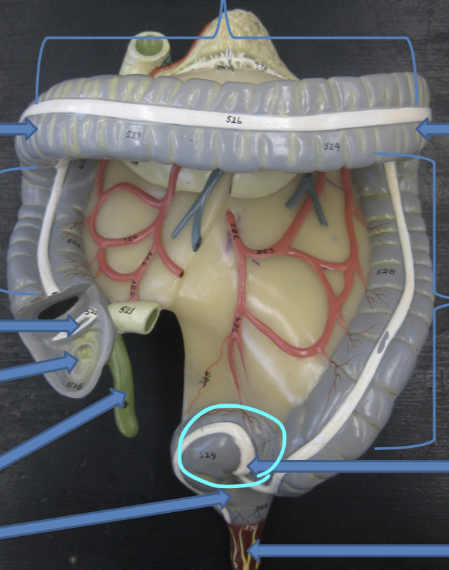
which structure is shown
sigmoid colon
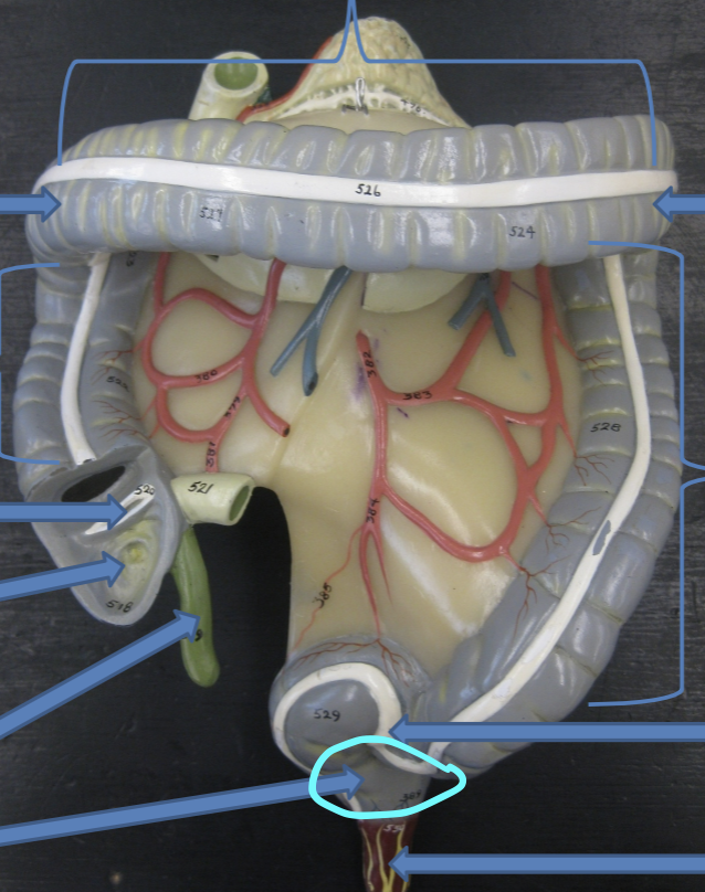
which structure is shown
rectum

which structure is shown
anal canal

which structure is shown
anus

which structure is shown
central incisor tooth

which structure is shown
lateral incisor tooth

which structure is shown
canine tooth

which structure is shown
2nd and 1st premolars

which structure is shown
3rd, 2nd, and 1st molars

which structure is shown
crown of tooth

which structure is shown
neck of tooth

which structure is shown
root of tooth

which structure is shown
gingiva

which structure is shown
gingival sulcus

which structure is shown
enamel of tooth

which structure is shown
cementum of tooth

which structure is shown
periodontal ligament

which structure is shown
dentin of tooth
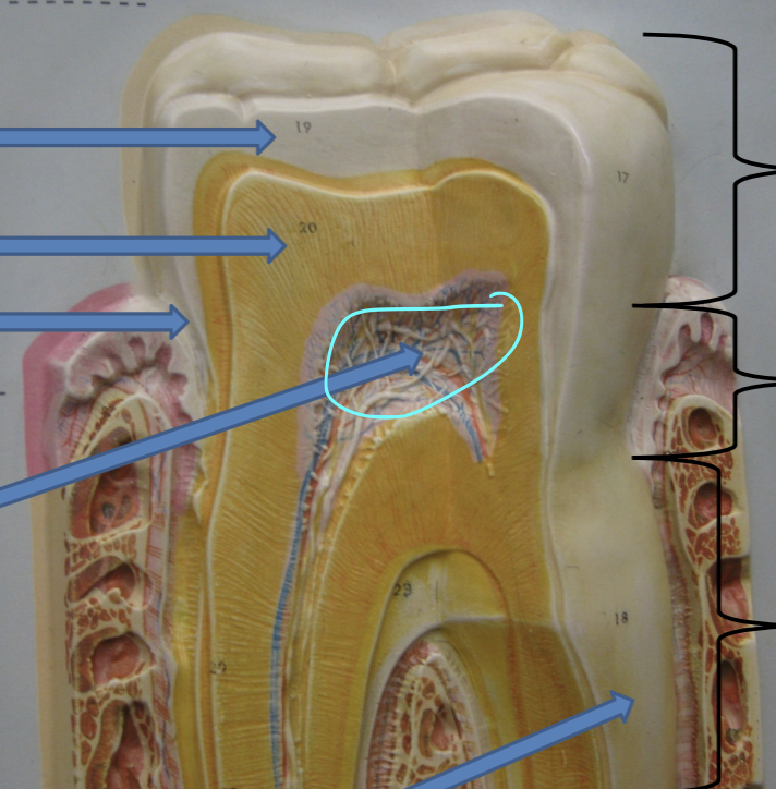
which structure is shown
pulp in pulp cavity

which structure is shown
root canal of tooth
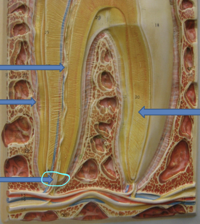
which structure is shown
apical foramen of tooth
which structure is shown
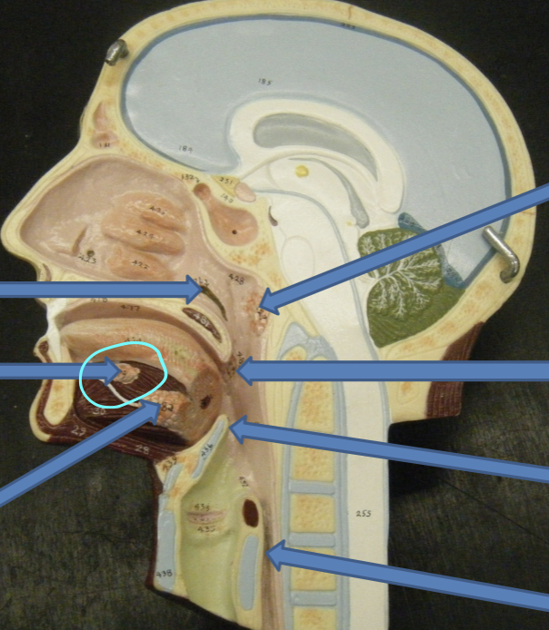
sublingual gland
which structure is shown
submandibular gland

which structure is shown
parotid gland
which structure is shown
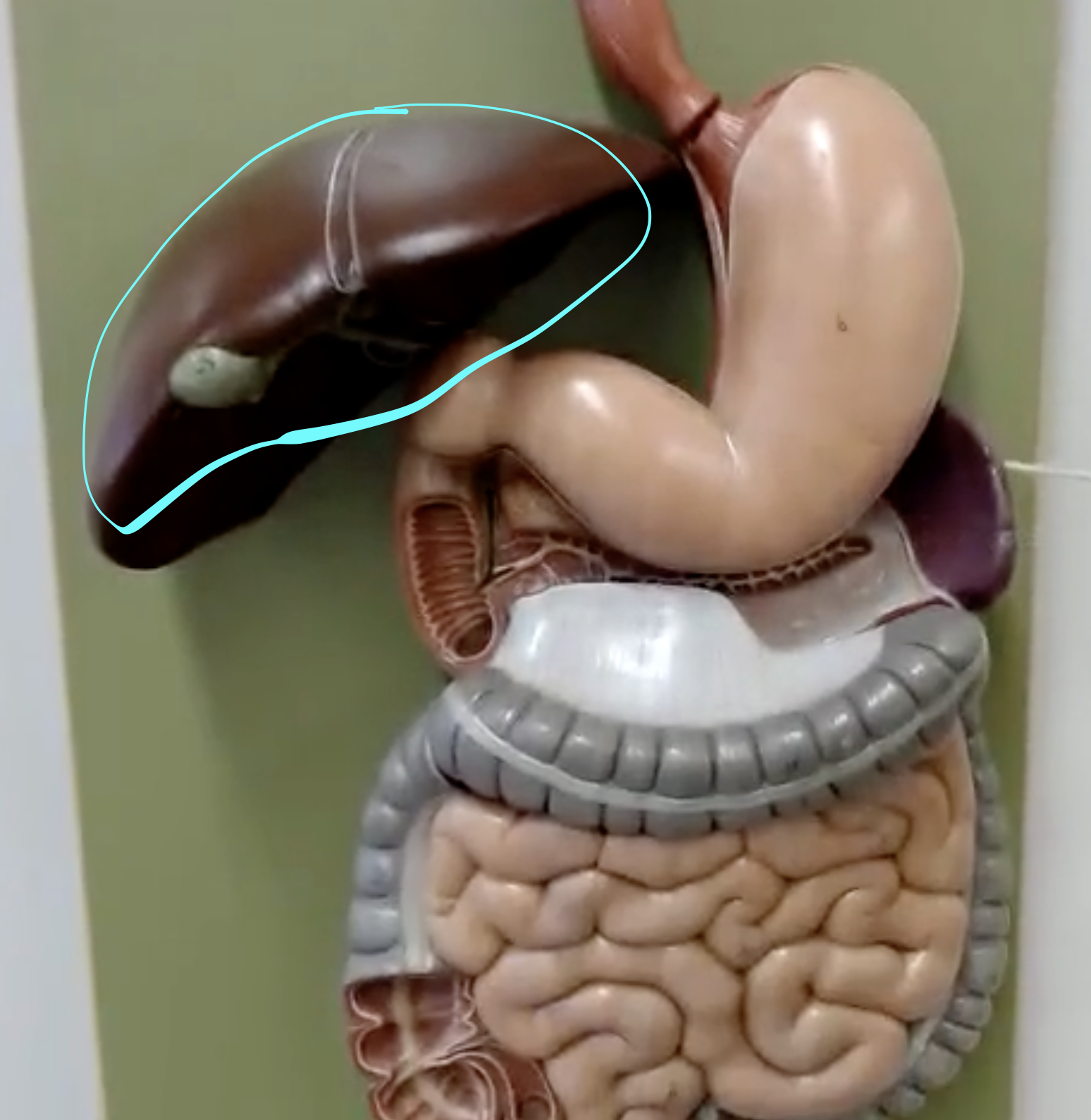
liver

which structure is shown
right lobe of liver (always bigger than left)

which structure is shown
left lobe of liver (always smaller than right)