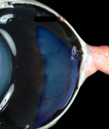
Week #6 Eye - Internal Anatomy
 Call Kai
 Learn
Learn Practice Test
 Spaced Repetition
 Match
Match Flashcards
 Knowt Play
1/5

There's no tags or description
Looks like no tags are added yet.
Name | Mastery | Learn | Test | Matching | Spaced | Call with Kai | Chat |
|---|
No analytics yet
Send a link to your students to track their progress
6 Terms
Iris
• Colored portion of the eye
• Pigmented, muscular diaphragm that controls the amount of light that enters the posterior part of the globe
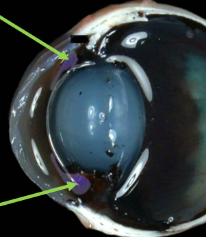
Pupil
• Opening in the center of the iris

Lens
• The lens is a soft, transparent structure made up of microscopic fibers that are arranged like the layers of an onion
• Elastic and biconvex
• Focus
• The process where the shape of the lens is changed by the ciliary body is called accommodation
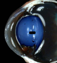
Ciliary Body
• Contains ciliary muscles that adjust the shape of the lens
• Located immediately behind the iris
• Located in the middle, vascular layer of the eye

Retina
• Complex, multilayer structure that lines most of the vitreous compartment
• This is where the visual image is formed, sensed, and converted to nerve impulses that are transmitted up the optic nerve to the occipital lobe
• The rest of the eye exists to support the retina

Tapetum
• Sandwiched between the sclera and retina
• Mostly made of pigment and blood vessels that supply the retina
• Forms the tapetum lucidum, which is a highly reflective area in the rear of the eye
• Acts as a light amplifier to aid in low-light vision