chapter 9: neurons
 Call Kai
 Learn
 Practice Test
 Spaced Repetition
 Match
 Flashcards
 Knowt Play
1/40

There's no tags or description
Looks like no tags are added yet.
41 Terms
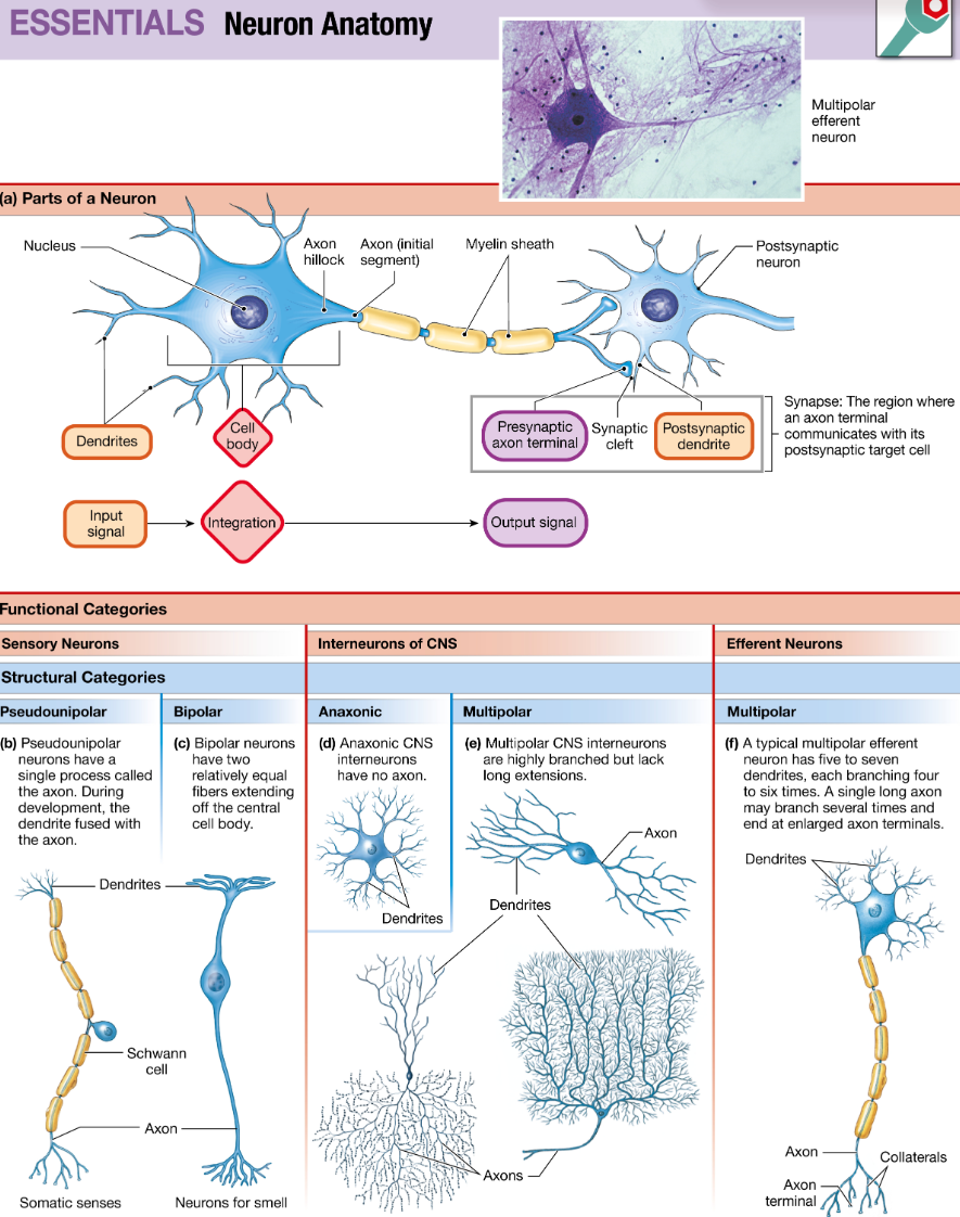
neuron
b
neurotransmitter
b
afferent/efferent
Nerves that carry only afferent signals are called sensory nerves; those that carry only efferent signals are called motor nerves.
The peripheral nervous system (PNS) consists of sensory (afferent) neurons and efferent neurons.
sensory (afferent) neurons, interneurons, and efferent (somatic motor and autonomic) neurons. Sensory neurons carry information about temperature, pressure, light, and other stimuli from sensory receptors to the CNS.
Efferent neurons: A peripheral neuron that carries signals from the central nervous system to the target cells
Efferent neurons are subdivided into the somatic motor division, which controls skeletal muscles, and the autonomic division, which controls smooth and cardiac muscles, exocrine glands, some endocrine glands, and some types of adipose tissue.
dendrite
Dendrites: thin, branched processes that receive and transfer incoming information to an integrating region within the neuron
These processes are classified as dendrites, which usually receive incoming signals, or axons, which usually carry outgoing information.
Dendrite → receives
cell body
cell body (soma) contains the nucleus and most of the organelles. It keeps the neuron alive and processes incoming information.
axon
The axon carries electrical signals away from the cell body toward the axon terminals.
Dendrites → cell body → axon
interneuron
Neurons that lie entirely within the CNS are known as interneurons (short for interconnecting neurons)
Neurons that lie entirely within the CNS are known as interneurons (short for interconnecting neurons)
nerve
The long axons of all peripheral neurons are bundled together with connective tissue into cordlike fibers called nerves that extend outward from the CNS.
sensory/motor/mixed nerve
Nerves that carry only afferent signals are called sensory nerves;
those that carry only efferent signals are called motor nerves.
Nerves that carry signals in both directions are mixed nerves.
cranial/spinal nerve
Cranial nerves: Nerves that connect to the brain.
Spinal nerves: Nerves that connect to the spinal cord.
Both are part of the PNS.
axon hillock
Most peripheral neurons have a single axon that originates from a specialized region of the cell body called the axon hillock
synapse
The region where an axon terminal meets its target cell is called a synapse
presynaptic/postsynaptic cell
The neuron that delivers a signal to the synapse is known as the presynaptic cell, and the cell that receives the signal is called the postsynaptic cell (Fig. 9.2a).
synaptic cleft
the narrow space between two cells is called the synaptic cleft
glial cells(glia) myelin
Glial cells, or neuroglia, are the support cells for neurons.
myelin, a substance composed of multiple concentric layers of phospholipid membrane
myelin acts as insulation around axons and speeds up their signal transmission.
nodes of Ranvier
In the PNS, each Schwann cell wraps around a segment of the axon, leaving tiny gaps, called the nodes of Ranvier, between the myelin-insulated areas
nodes play an important role in the transmission of electrical signals along the axon
depolarization/hyperpolarization
Depolarization
Membrane becomes less negative.
ex. −70 mV → −60 mV
The Na+ channels open very rapidly, but the K+ channels are slower to open. The result is an initial flow of Na+ across the membrane, followed later by K+ flow.
Many channels that open in response to depolarization close only when the cell repolarizes.
Hyperpolarization
Membrane becomes more negative.
ex. −70 mV → −80 mV
voltage-gated ion channel
Voltage-gated ion channels respond to changes in the cell’s membrane potential. Voltage-gated Na+ and K+ channels play an important role in the initiation and conduction of electrical signals along the axon.
graded potential
Graded potentials are variable-strength signals that travel over short distances and lose strength as they travel through the cell. They are used for short-distance communication.
If a depolarizing graded potential is strong enough when it reaches an integrating region within a neuron, the graded potential initiates an action potential.
action potential
Action potentials are very brief, large depolarizations that travel for long distances through a neuron without losing strength. Their function is rapid signaling over long distances, such as from your toe to your brain.
If a depolarizing graded potential is strong enough when it reaches an integrating region within a neuron, the graded potential initiates an action potential.
refractory period
The double gating of Na+ channels plays a major role in the phenomenon known as the refractory period
excitatory/inhibitory postsynaptic potential (EPSP/IPSP)
If the synaptic potential is depolarizing, it is called an excitatory postsynaptic potential (EPSP) because it makes the cell more likely to fire an action potential.
If the synaptic potential is hyperpolarizing, it is called an inhibitory postsynaptic potential (IPSP) because hyperpolarization moves the membrane potential away from threshold and makes the cell less likely to fire an action potential.
Differentiate structurally between the central and peripheral nervous systems
The central nervous system (CNS) consists of the brain and the spinal cord.
The peripheral nervous system (PNS) consists of sensory (afferent) neurons and efferent neurons.
Reproduce Figure 9.1 essentially, at least orange and purple sections and CNS (e.g. understanding that efferent neurons come in two types, autonomic and somatic motor, and so on for the rest of the figure)
ok

Differentiate functionally between the somatic motor and autonomic divisions of the efferent division of the PNS
Efferent neurons are subdivided into the somatic motor division, which controls skeletal muscles,
and
the autonomic division, which controls smooth and cardiac muscles, exocrine glands, some endocrine glands, and some types of adipose tissue. T
Differentiate functionally between the sympathetic and parasympathetic nervous systems
Sympathetic | Parasympathetic |
|---|
Fight or flight | Rest and digest |
Active/stress | Rest/maintenance |
↑ heart rate | ↓ heart rate |
↓ digestion | ↑ digestion |
Describe or identify the contribution of the Na+/K+ pump, K+ leak channel, and Na+ and K+ voltage-gated channels to the resting membrane potential* and to the action potential
Explain why the sodium equilibrium potential (ENa) and the potassium equilibrium potential (EK) are different
Explain or identify what creates the resting membrane potential (what it “is”)
Explain what is happening at each stage of the action potential and the relationship between the ion movement and voltage change across the membrane
Explain what keeps action potentials from adding up/ piggybacking on each
other
Explain how action potentials propagate down the axon in the absence of
myelin, and in its presence
Calculate the equilibrium potential for an ion using the simplified Nernst
equation
Calculate the membrane potential using the GHK equation; more importantly, be
able to use understanding of this equation to qualitatively think through the
effect of changes in these variables on the membrane potential, such as making
the action potential harder or easier to initiate, and the resulting effects on the person
Predict qualitatively how changes in permeability to an ion or to ion
concentration gradients would change the equilibrium potential for that ion or
the membrane potential
Predict qualitatively how changes in the characteristics of the Na+ and K+
channels (e.g., what voltage they open at, or how many of them there are the
cell membrane) and starting concentration gradients would affect the action
potential
Recognize that a single action potential doesn’t significantly change ion
concentrations across the membrane
Differentiate structurally and functionally between fast (ionotropic) and slow
(metabotropic) postsynaptic responses
Name several major neurotransmitters
Provide or identify the correct sequence of steps in synaptic communication at a chemical synapse
Explain why a depolarizing PSP is excitatory (an EPSP) and a hyperpolarizing PSP is inhibitory (an IPSP)