1.5 BODY CAVITIES, MEMBRANES AND FLUIDS
 Call Kai
 Learn
Learn Practice Test
 Spaced Repetition
 Match
Match Flashcards
 Knowt Play
1/80

Earn XP
Description and Tags
Body cavities and their fcns, serous membrane and its fcns, different types of fluids involved with body cavities and their fcns.
Name | Mastery | Learn | Test | Matching | Spaced | Call with Kai | Chat |
|---|
No analytics yet
Send a link to your students to track their progress
81 Terms
What are body cavities?
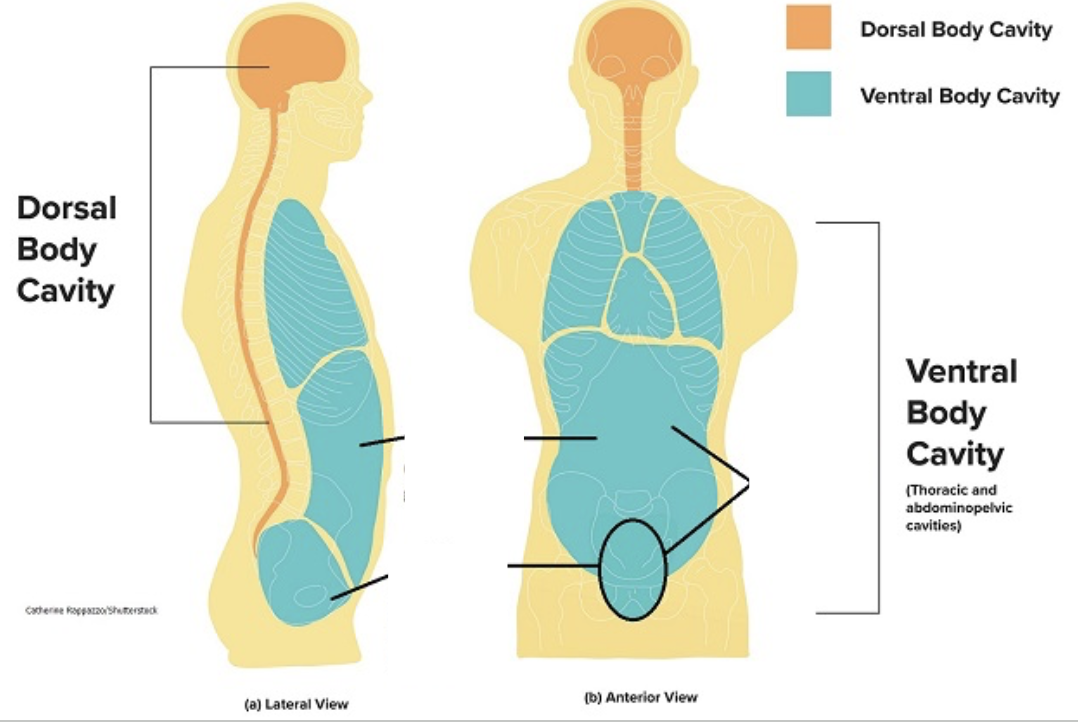
Inside the human body are hollowed out areas, or cavities, where the organs reside.
What are the functions of the body cavities?
Protection, Compartmentalization, Support and Allowance for Movement, and Organ Expansion and Contraction.
Protection
Hard outer structures, like the skull and vertebral column for the dorsal cavity, and the rib cage for the thoracic cavity, shield delicate internal organs from trauma.
Compartmentalization
Membranes and physical dividers separate organs into distinct spaces. This organization prevents infections or injuries from easily spreading between organ systems.
Support and Allowance for Movement
Fluid inside these cavities cushions organs and reduces friction when they move.
Organ Expansion and Contraction
The ventral cavity allows organs like the lungs, heart, stomach, and bladder to change in size and shape during normal function (such as breathing or digesting) without disrupting neighboring tissues.
The body is divided into two major cavities:
Dorsal Cavity and the Ventral Cavity

Dorsal Cavity
Located in the posterior side of the body and contains the cranial and vertebral cavities.

Cranial Cavity
Houses the brain
Vertebral Cavity
Houses the spinal cord
Ventral Cavity
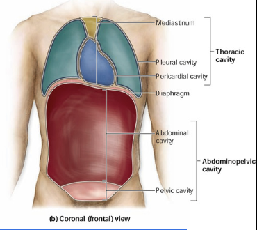
On the anterior side of the body. Divided into the thoracic cavity and abdominopelvic cavities.
Thoracic Cavity
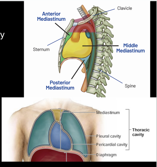
Contains the Mediastinum and the Pleural cavities.

Mediastinum Cavity
Contain the pericardial cavity
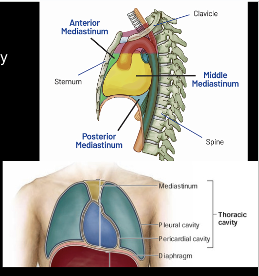
Pericardial Cavity
Contains the heart
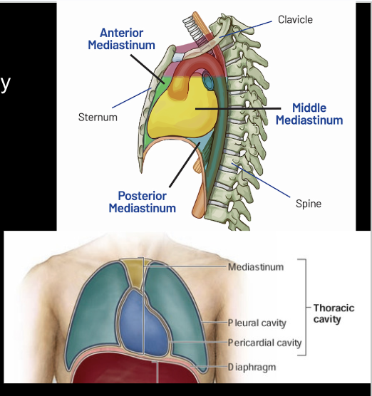
Pleural Cavities
Houses the lungs

Abdominopelvic cavities
Below the diaphragm. Subdivided into a superior abdominal cavity and a inferior pelvic cavity. There is no membrane or wall dividing these two regions.

Superior abdominal cavity
Contains the liver, gallbladder, stomach, pancreas, intestines, kidneys and the spleen.

Inferior pelvic cavity
Contains the bladder and reproductive organs
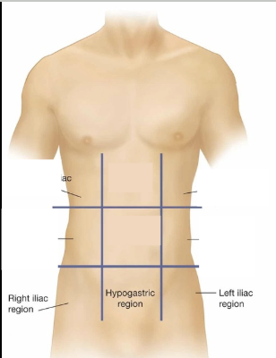
What are the regions of the abdominopelvic cavity?
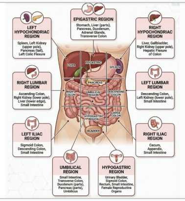
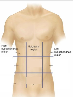
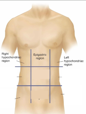
The abdominopelvic cavity is divided into nine specific regions using two vertical (parasagittal) and two horizontal imaginary lines.

Top Row (Superior) of the abdominopelvic cavity
Right Hypochondriac Region, Epigastric Region, and Left Hypochondriac Region.

Middle Row (Central) of the abdominopelvic cavity
Right Lumbar Region, Umbilical Region, and Left Lumbar Region

Bottom Row (Inferior) of the abdominopelvic cavity
Right Iliac (Inguinal) Region, Hypogastric (Pubic) Region, and Left Iliac (Inguinal) Region

Right Hypochondriac Region
Contains the right part of the liver, gallbladder, right kidney, and small intestine parts
Epigastric Region
Contains most of the stomach, part of the liver, pancreas, duodenum, and spleen.

Left Hypochondriac Region
Contains part of the spleen, left kidney, part of the stomach, and pancreas.
Right Lumbar Region
Contains the gallbladder, right kidney, liver part, and ascending colon.

Umbilical Region
Contains the belly button (navel), parts of the small intestine, and transverse colon.

Left Lumbar Region
Contains the descending colon, left kidney, and spleen part.

Right Iliac (Inguinal) Region
Contains the appendix, cecum, and right iliac fossa.

Hypogastric (Pubic) Region
Contains the urinary bladder, part of the sigmoid colon, and reproductive organs.

Left Iliac (Inguinal) Region
Contains part of the descending colon and the sigmoid colon.
What are Serous Membranes?
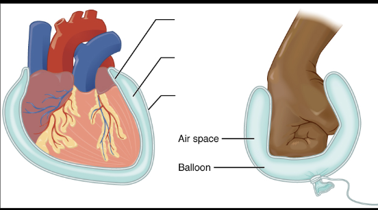
A serous membrane (also called a serosa) is a thin, double-layered sheet of tissue that lines closed body cavities and covers the organs inside them.

Visceral Layer
The internal side of the serous membrane

Parietal Layer
The external side of the serous membrane

Pericardium
Serous membrane that lies on top of the heart.
Pleura
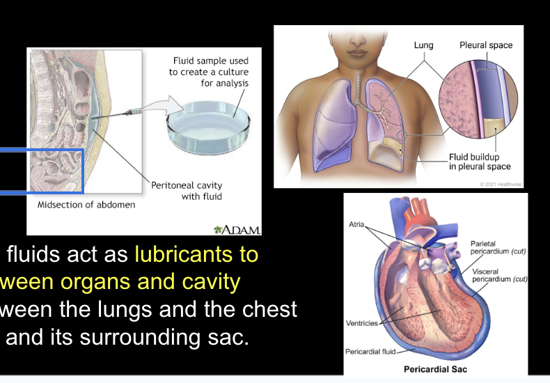
Thin, slippery membrane that covers the outside of your lungs and lines the inside of your chest wall. The pleura forms a continuous, two-layered sac around each lung:

Pleural cavity
The narrow space between these the visceral pleura and the parietal pleura. It holds a tiny amount of slippery liquid called pleural fluid.

Visceral Pleura
The inner layer that wraps tightly around the surface of the lungs.

Parietal Pleura
The outer layer that attaches to the chest wall and diaphragm.

The brain and the spinal cord are surrounded by a series of 3 protective membranes called
meninges

Dura Mater
The tough, thick outer layer closest to the skull and bones of the spine.

Arachnoid Mater
The delicate, web-like middle layer.

Pia Mater
The thin, soft inner layer that hugs the surface of the brain and spinal cord

Peritoneum
Lines the abdominal cavity and covers organs like the stomach and intestines

Main Layers of Peritoneum
Parietal peritoneum, Visceral peritoneum, Peritoneal cavity, Folds and Connections (a. mesentery, b. omentum)
1.Parietal peritoneum
The outer layer that attaches to and lines the walls of the abdominal and pelvic cavities.

2.Visceral peritoneum
The inner layer that wraps around and covers the abdominal organs, such as the stomach, liver, and intestines.

3.Peritoneal cavity
The narrow, fluid-filled space between the parietal and visceral layers that reduces friction when organs move.

4.Folds and Connection
Specialized double layers of this membrane, such as the mesentery and the omentum.

The mesentery
a continuous double fold of peritoneal tissue that anchors the intestines to the back wall of the abdomen while letting them move during digestion

The omentum
a large, apron-like fold of fatty tissue that hangs from the stomach and drapes over the abdominal organs. It suspend organs, store fat, and act as pathways for blood vessels, nerves, and lymphatics.

What are Serous Fluids?
A pale yellow, transparent, watery fluid that fills the inside of closed body cavities (such as the pleura around the lungs, the pericardium around the heart, and the peritoneum in the abdomen). Between the visceral and parietal layers.

Serous Fluid is also a term used for
clear wound exudate which helps heal wounds.
What are the different types of wound exudates?
Serous, Serosanguineous, Sanguineous, Seropurulent, Purulent
Serous
Clear, thin, and watery fluid (or light straw colored). This is normal during the early inflammatory healing stage. Made mostly of plasma. Beneficial to remove foreign substances and keep the wound moist for healing.Too much may indicate infection .

Serosanguineous
Thin, pink, or light red fluid that mixes blood and plasma. This is the most common type seen during normal healing.

Sanguineous
Fresh, bright red, bloody drainage. This is normal for deep or fresh wounds, but ongoing heavy bleeding can mean new trauma

Seropurulent
Thin, cloudy, yellow, or tan fluid. This can be an early sign that bacteria are colonizing the wound.

Purulent
Thick, opaque, milky pus that appears yellow, green, brown, or gray. This often has a foul odor and is a clear sign of a severe infection.

Are all cavity serous fluids the same?
While all serous fluids share a general watery or plasma-like consistency, fluids in different cavities, have have different origins and interact with different organs, producing several key differences:
Composition Variations
Composition Variations
The precise levels of proteins, electrolytes, and cellular components (like lymphocytes or mesothelial cells) depends on where the fluid is found and whether a disease is preset.
How do serous fluids changes depending on health?
Transudate and Exudate.

Transudate
Clear and watery fluid caused by systemic changes like high blood pressure or low blood protein; has low protein and cell counts.


Exudate
Cloudy or turbid fluid caused by local inflammation, infection, or cancer; has high protein levels, cellular debris, and white blood cells

What are the serous fluids of the thoracic cavity?
Pleural fluid, Pericardial fluid, Peritoneal fluid
These specialized fluids act as lubricants to reduce friction between organs and cavity walls, such as between the lungs and the chest cavity or the heart and its surrounding sac.
What is the CSF?
In the brain and spinal cord, the fluid is called cerebrospinal fluid (CSF).
It floats the brain and has large daily production.

Floats the Brain
CSF provides physical buoyancy that reduces the effective weight of a 1,400 g human brain down to about 25 g preventing the lower parts of the brain from being crushed under their own heavy mass.

Large Daily Production
The body produces about 400 to 600 ml of CSF every single day (about a gallon /week). Since only about 125 to 150 ml are present inside the central nervous system at any one time, the entire fluid supply is completely absorbed and refreshed four to five times every 24 hours
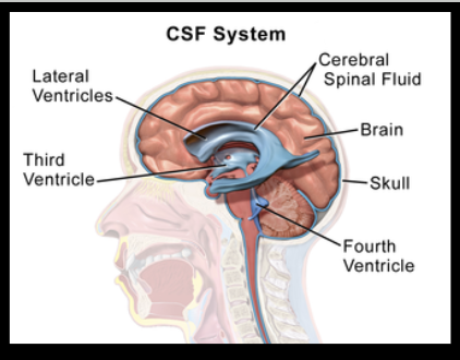
Lateral Ventricles
are the starting point for the brain’s “irrigation” system.These ventricles is were the primary production of CSF takes place.
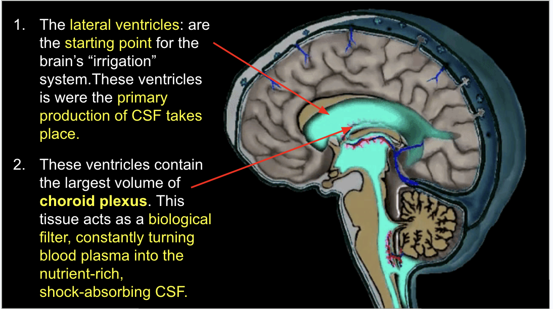
The lateral ventricles contain
the largest volume of choroid plexus. This tissue acts as a biological filter, constantly turning blood plasma into the nutrient-rich, shock-absorbing CSF.

Flow Dynamics
As CSF is produced, it creates a pressure gradient that pushes the fluid through the Interventricular Foramen, also known as Foramen of Monro (the “doorway”) into the Third Ventricle

Waste Clearance
The flow of CSF from the the lateral ventricles into the third ventricle acts like a constant “flush,” clearing metabolic byproducts from deep brain structures.

The Cerebral Aqueduct
also known as the Aqueduct of Sylvius, is a narrow channel located within the midbrain that connects the third and fourth ventricles.

Fourth Ventricle
the “grand terminal” of the brain’s internal plumbing.The fourth ventricle is the only part of the system that allows fluid to escape the “internal” brain and reach the “external” surface.

The Foramina of Luschka (Lateral Apertures)
Two openings on the sides that direct fluid around the brain.

Foramen of Magendie (Median Aperture)
A single opening in the middle that directs fluid toward the spinal cord and brainstem.

What is synovial fluid?
Synovial fluid is derived from blood plasma filtered through the synovial membrane. It contains very few cells and no clotting factors.

The joints contain
synovial fluid
Synovial fluid contains
specialized cells that secrete large amounts of hyaluronic acid, which gives the fluid its signature slickness, and Lubricin which provides boundary lubrication when cartilage surfaces physically touch.
Synovial fluid is a Non-Newtonian liquid.
Its viscosity changes depending on the force applied. Under high shear stress (fast movement), it thins to allow smooth gliding; under sudden heavy impact (shocks), it thickens temporarily to absorb the force before returning to its normal state.