Intro to Neuro anatomy ,
 Call Kai
 Learn
 Practice Test
 Spaced Repetition
 Match
 Flashcards
 Knowt Play
1/33

There's no tags or description
Looks like no tags are added yet.
34 Terms

Cranium (skull)

Viscero cranium (Mandible, Maxilla, Nasal, Zygomatic bones)

Foramen of Skull

Neurocranium

Sutures

Cranial Fossa

Internal Surface of cranial base

3 meninges (protective membranes)
1. Dura mater — outermost layer
The dura mater is the tough, thick outer covering around the brain.
It has TWO layers:
Periosteal layer: attached to the inner surface of the cranium (skull).
Meningeal layer: lies underneath the periosteal layer.
2. Arachnoid mater — middle layer
It sits underneath the dura mater.
It looks web-like.
It does NOT closely follow the grooves and folds of the brain.
Important: Subarachnoid space = space between the arachnoid mater and pia mater. (has CSF to cushion brain)
3. Pia mater — innermost layer
The pia mater is the thin, delicate innermost covering.
It directly touches the brain.
It closely follows the brain's surface & the grooves and folds.

3 meninges (protective membranes)
• Falx cerebri (cerebral falx)
➢ the largest dural infolding; sickle-shaped
➢ lies in the longitudinal cerebral fissure
• Dural venous sinuses
➢ endothelial-lined spaces between the periosteal and meningeal layers of the dura
• Arachnoid granulations
➢ collections of arachnoid villi
➢ tufted prolongations of the arachnoid that protrude through the meningeal layer and dural venous sinuses
➢ transfer CSF to the venous system

Dural Septa (dura mater folds that separate parts of brain)
Falx cerebri
A fold that dips into the longitudinal fissure between the cerebral hemispheres.
Function: Separates the right and left cerebral hemispheres.
2. Falx cerebelli
Keeps going inferiorly from the posterior falx cerebri; runs along the vermis (middle) of the cerebellum.
Function: Separates the right and left cerebellar hemispheres and helps stabilize the cerebellum.
3. Tentorium cerebelli
Nearly horizontal; extends into the transverse fissure between the cerebral bad cerebellum hemispheres.
Function: Separates the cerebrum (above) from the cerebellum (below) .

Dural Venous Sinus (brings deoxygenated blood from brain to heart)
Pathway A (starts from superior sagittal sinus or top of falx cerebri):
Superior sagittal sinus
Confluence of sinuses
Transverse sinus
Sigmoid sinus
Pathway B (starts from inferior Sagittal sinus or bottom of falx cerebri):
Great cerebral vein (vein of Galen) + Inferior sagittal sinus
Straight sinus
Confluence of sinuses
Transverse sinus
Sigmoid sinus
After all the blood goes to the internal jugular vein then to the SVC


Sense, Integrate & Response

Organization of the nervous system
The nervous system has two main divisions:
A. Central nervous system (CNS)
Consists of the brain and spinal cord.
Acts as the body's control and processing centre.
Receives information, processes it, and decides how the body should respond.
B. Peripheral nervous system (PNS)
Consists of cranial nerves and spinal nerves.
Connects the CNS to the rest of the body.
The PNS has two major divisions:
Sensory/afferent division (Info goes to CNS)
Includes somatic and visceral sensory nerve fibres.
Somatic sensory
Information from skin, muscles, tendons and joints. (Ex. Touch, pain)
Visceral sensory
Information from internal organs. (Ex. Hunger, headache)
Examples: information your organs or intestines
Motor (efferent) division (Info goes from CNS to body)
Sends instructions to effectors (muscles and glands).
Has two divisions:
A. Somatic nervous system
Controls voluntary movements. Sends signals to skeletal muscles.
Examples: Walking, Lifting your arm & Writing
B. Autonomic nervous system (ANS)
Controls involuntary functions.
Sends signals to: Cardiac muscle (heart), Smooth muscle (organs and blood vessels) & Glands
Examples: Heart rate, Digestion, Sweating, Changes in blood vessel diameter
The autonomic nervous system has TWO divisions:
Sympathetic vs. parasympathetic
Sympathetic division (flight or fight)
Mobilizes body systems during activity.
Prepares the body for stressful or demanding situations.
Parasympathetic division (relaxation)
Conserves energy.
Promotes housekeeping functions during rest.
Ganglions
The autonomic nervous system uses TWO neurons to send a signal from the CNS to an organ.
Steps:
CNS
Preganglionic fibre
Ganglion (main connection spot)
(First neuron: CNS → ganglion)
Postganglionic fibre
Target organ
(Second neuron: ganglion → target organ)
Parasympathetic vs sympathetic
Parasympathetic
Originates from the brain stem and sacral spinal cord.
Preganglionic fibres are LONG.
Postganglionic fibres are SHORT.
Ganglia are near visceral organs.
Sympathetic
Originates from the thoracic and lumbar spinal cord.
Preganglionic fibres are SHORT.
Postganglionic fibres are LONG.
Ganglia are close to the spinal cord.
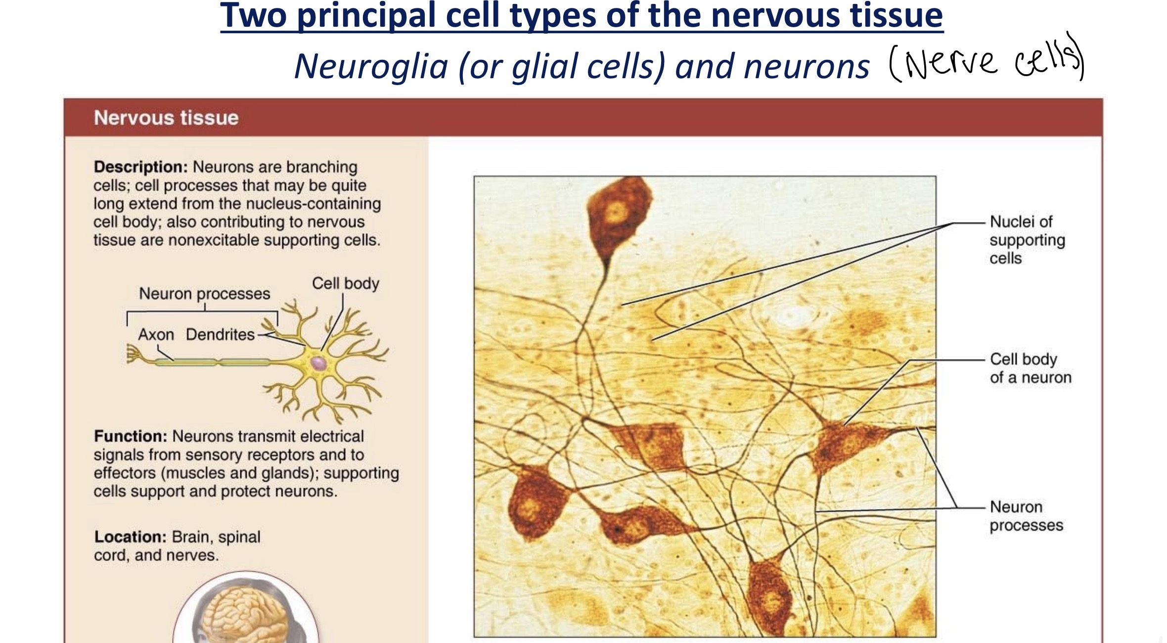
Glial cells (supporting cells) and nerves cells (neurons)
Neurons:
Specialized nerve cells of the nervous system.
Transmit electrical signals between sensory receptors, the CNS and effectors (muscles and glands).
Have a cell body, dendrites and an axon. (Neuron processes contain axon and dendrites extending from cell body containing neuron)
Supporting cells (glial cells):
Nonexcitable cells, but Supports and protect neurons.
Location: brain, spinal cord & nerves
Neuroglia (or glial cells)
Six types: four in the CNS and two in the PNS
➢ In the CNS: astrocytes, microglial cells, ependymal cells, and oligodendrocytes
➢ In the PNS: Satellite cells and Schwann cells
Functions of neuroglia cells
1. Glial cells in the CNS (4 types)
Astrocytes: Support neurons and help maintain their ECF environment.
Microglial cells: Act as the immune defence cells by removing debris and harmful substances.
Ependymal cells: Line the cavities of the brain and spinal cord that contain CSF.
Oligodendrocytes: Form myelin sheaths around axons.
2. Glial cells in the PNS (2 types)
Satellite cells: Support neurons and help maintain their ECF environment.
Schwann cells: Form myelin sheaths around axons.
Neurons (Cell Body)
Neuron cell body
also called perikaryon or soma
consists of a spherical nucleus (with a conspicuous nucleolus) surrounded by cytoplasm
acts as part of the receptive region (helps receive signals from other neurons)
Groups of cell body Location:
Nuclei: Clusters of cell bodies in the CNS (spinal cord & brain)
Ganglia: Cluster of cell bodies along the nerves in the PNS (spinal cord and nerves)
What the PNS and CNS consist of:
1. CNS (brain and spinal cord)
Neuron cell bodies & Neuron processes
2. PNS (nerves outside the CNS)
Consists mainly of neuron processes

Dendrites
• Short, tapering extensions
• The main receptive or input regions that carry messages toward the cell body
> Provide an enormous surface area for receiving signals from other neurons
> Bristle with dendritic spines which represent points of close contact (synapses)

Axon
Axons
• one axon per neuron
• cone shape area of cell body = axon hillock
• The initial segment narrows to form a slender process of axon (stays the same thinness)
• may have occasional branches along its length, called axon collaterals
Distal part of terminal branch = axon terminal
Functions of axon
Generate nerve impulses and transmit them away from the cell body.
In motor neurons:
The nerve impulse begins at the initial segment (trigger zone) of the axon.
The impulse travels along the axon toward the axon terminals.
Axon terminals are the secretory region, where the neuron releases chemical messengers to communicate with another cell.
Tracts & Nerves (bundles of axons) Location:
Tracts: Bundles of axons in the CNS
Nerves: Bundles of axons in the PNS
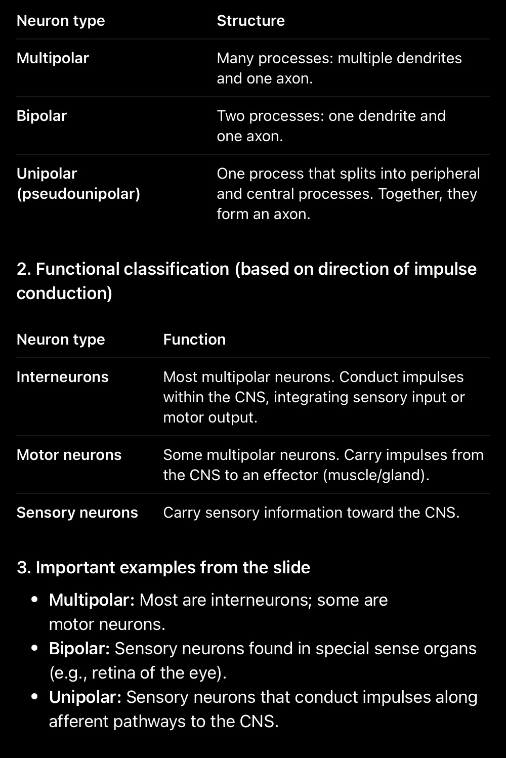
Structural Classes of Neurons

Myelin Sheath & Myelination
Myelin Function:
• Covers many nerve fibers, particularly those with large diameters
• Protects and electrically insulates fibers
• Myelinated fibres increases the transmission speed of nerve impulses
Myelination in the PNS
• Formed by Schwann cells, gaps in between are called nodes of ranvier
Myelination in the CNS
• Formed by oligodendrocytes (can coil around axon 60 times), gaps in between are called nodes of ranvier

Lobes of the brain
Surface & poles of the brain

Diencephalon
Cerebrum
Location: upper part of the brain
> Function: thinking, memory, interpreting senses, and voluntary movement
• Cerebellum
Location: back and lower part of the brain
Function: balance and coordination
Diencephalon
Thalamus: the relay station for information coming into the cerebral cortex
Hypothalamus: main visceral control center of the body
Epithalamus: includes the pineal gland, which produces melatonin

Brain Stem
Midbrain: located between the diencephalon and pons
Pons: the bulging brain stem between the midbrain and the medulla
Medulla oblongata: the most inferior part of the brain stem; blends into the spinal cord
Grey & White Matter
Gray Matter
Contains: neuron cell bodies and nonmyelinated neuron processes.
Has two main regions:
A. Cortex
- Outer layer of the cerebral hemispheres.
B. Deep nuclei
- Groups of neuron cell bodies with similar functions.
1) Basal ganglia:
Caudate
Lentiform nucleus: Putamen & Globus pallidus
2) Diencephalon
Thalamus
Hypothalamus
White Matter:
Contains: myelinated axons. Myelin is fatty so it’s white
White fibres (bundles of myelinated axons) directions they run in:
Regions within the brain (same or opposite sides).
The brain to the spinal cord through long pathways.
Transmit information to and from the body.

Sulcus & Gyrus
Lateral sulcus (or lateral fissure): separates the temporal lobe from the parietal and frontal lobes
Precentral sulcus (in frontal lobe): anterior from precentral gyrus
Precentral gyrus (in frontal lobe): borders the central sulcus anteriorly
Central sulcus: separates the frontal lobe from the parietal lobe
Postcentral sulcus (in parietal lobe) : posterior from postcentral gyrus in the parietal lobe
Postcentral gyrus (in parietal lobe): borders the central sulcus posteriorly

Functional areas of cerebral cortex

Vertebral & Internal Carotid Artery
Vertebral arteries (posterior route up the neck):
Comes from the subclavian artery
Two vertebral arteries from the left & right side of neck enters the skull through the foramen magnum.
Combination of the 2 arteries = basilar artery.
Internal carotid arteries (anterior route up the neck):
Comes from the common carotid artery
Two internal carotid arteries from the left & right side of neck enters the skull through the carotid canal.
Where the arteries branch off to
Internal Carotid Artery
Left & Right Anterior cerebral artery (ACA)
(connected by Anterior communicating artery)
Left & Right Middle cerebral artery (MCA)
Left & Right Posterior communicating artery (this also connects the posterior cerebral artery)
Basilar Artery
Left & right posterior cerebral artery
What each branch supplies
Arteries bring oxygenated blood to the brain from the subclavian and common carotid arteries
Vascular Supply:
ACA: medial surface, frontal pole
MCA: superolateral surface, temporal pole
PCA: inferior surface, occipital pole
