FPP - Lower Limb Muscles
 Call Kai
 Learn
 Practice Test
 Spaced Repetition
 Match
 Flashcards
 Knowt Play
1/18

Earn XP
Description and Tags
19 Terms
Psoas Major
Origin:
Vertebral body of T1 - L4
Transverse process of L1 - L5
Lateral surfaces of intervertebral discs
Insertion:
Lesser trochanter
Movement:
Hip flexion
External rotation of thigh
Trunk flexion
Lateral flexion of trunk
Postural stability

Iliacus
Origin:
Iliac fossa
Insertion:
Lesser trochanter
Movement:
Hip flexion
External rotation of thigh
Trunk flexion /anterior pelvic tilt

Gluteus Medius
Origin:
Posterior surface of Ilium
Insertion:
Greater trochanter
Movement:
Anterior fibers -
Hip flexion
Internal rotation
Middle fibers:
Hip abduction
Assisting pelvic tilt
Posterior fibers:
Assists with extension
Assists with external rotation

Gluteus Maximus
Origin:
Gluteal surface of ilium
Posterior superior iliac crest
Sacrum
Coccyx
Insertion:
Iliotibial tract (IT Band)
Gluteal tuberosity
Movement:
Hip extension

Adductor Longus
Origin:
Anterior surface of pubis
Insertion:
Middle third of the linea aspera of the femur
Movement:
Hip adduction

Sartorius
Origin:
Anterior superior iliac spine
Insertion:
Medial aspect of proximal tibia
Movement:
Hip flexion
Hip abduction
External rotation

Vastus Medialis (Quads)
Origin:
Lower, medial, half of the intertrochanteric line
Medial lip of linea aspera of femur
Insertion:
Patella tendon into the tibial tuberosity
Movement:
Knee extension

Vastus Intermedius (Quads)
Origin:
Anterior and lateral surfaces of body of femur
Insertion:
Patella tendon into the tibial tuberosity
Movement:
Knee extension

Vastus Lateralis (Quads)
Origin:
Intertrochanteric line
Greater trochanter
Gluteal tuberosity
Lateral lip of linea aspera of the femur
Insertion:
Patella tendon into the tibial tuberosity
Movement:
Knee extension
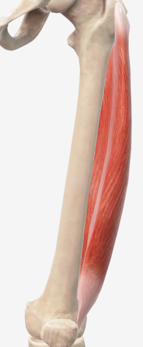
Rectus Femoris (Quads)
Origin:
Anterior inferior iliac spine
Insertion:
Patella tendon into the tibial tuberosity
Movement:
Knee extension

Biceps Femoris (Hamstrings)
Origin:
Lower medial ischial tuberosity (Long head)
Lateral linea aspera (Short head)
Insertion:
Head of fibula
Lateral tibial condyle
Movement:
Knee flexion
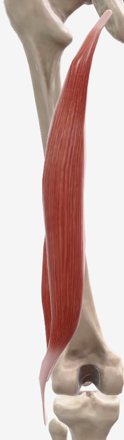
Semitendinosus (Hamstrings)
Origin:
Ischial tuberosity
Insertion:
Medial aspect of proximal tibia
Movement:
Knee flexion

Semimembranosus (Hamstrings)
Origin:
Ischial tuberosity
Insertion:
Medial tibial condyle
Movement:
Knee flexion

Peroneus (Fibularis) Longus
Origin:
Head and lateral surface of fibula
Insertion:
Plantar surface of medial cuneiform bone
Base of first metatarsal bone
Movement:
Eversion of the foot
Plantar flexion
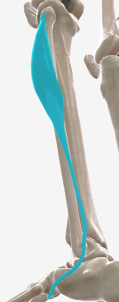
Peroneus (Fibularis) Brevis
Origin:
Lateral surface of fibula
Insertion:
Tuberosity of fifth metatarsal bone
Movement:
Eversion of the foot

Gastrocnemius
Origin:
Lateral and medial condyles of femur
Insertion:
Posterior surface of calcaneus, via Achilles tendon
Movement:
Plantar flexion

Soleus
Origin:
Head and upper third of the posterior surface of the fibula
Medial border of the tibia
Soleal line
Tendinous arch spanning between the tibia and fibula
Insertion:
Posterior surface of the calcaneus, via Achilles tendon
Movement:
Plantar flexion
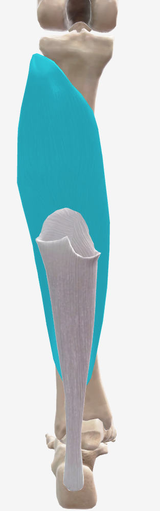
Tibialis Posterior
Origin:
Upper 2/3 of the posterior surface of tibia
Superior 2/3 of the medial posterior surface of the fibula
Insertion:
Tuberosity of the navicular bone and the medial cuneiform
Intermediate cuneiform, lateral cuneiform, cuboid, and bases of 2nd, 3rd, and 4th metatarsals
Movement:
Foot inversion
Plantar flexion

Tibialis Anterior
Origin:
Upper 2/3 of the lateral surface of the tibia and adjacent interosseous membrane
Insertion:
Medial cuneiform and base of the first metatarsal bone of foot
Movement:
Dorsiflexion
Foot inversion
