0450 Agnosia
 Call Kai
 Learn
Learn Practice Test
 Spaced Repetition
 Match
Match Flashcards
 Knowt Play
1/45

There's no tags or description
Looks like no tags are added yet.
Name | Mastery | Learn | Test | Matching | Spaced | Call with Kai | Chat |
|---|
No analytics yet
Send a link to your students to track their progress
46 Terms
What is object recognition?
The ability to recognize everyday objects easily over a wide range of distances, orientations, and lighting conditions.
What is object constancy?
Recognizing an object despite changes in viewing conditions (distance, orientation, lighting) — the percept remains stable.
Why must descriptions of object structure be stored in the brain?
So we can recognize objects even when seen from a new angle; patients with agnosia have helped reveal the multiple routes to object constancy.
What does agnosia mean?
"Not knowing" (Freud).
What did Munk (1878) find?
Dogs with specific bilateral occipital lobe lesions apparently could see okay but lost the ability to appreciate the meaning of visual stimuli — he called it "mindblindness."
Who was Patient GL (Lissauer, 1889)?
An 80 y/o salesman with a closed head injury: normal visual acuity, but he no longer recognized things around him — thought pictures in his room were boxes, mistook his jacket for trousers, couldn't recognize cutlery.
What was striking about what Patient GL could still do?
He could draw objects that he could not recognize; he retained knowledge of objects (referred to them appropriately in conversation) and recognized them from touch and sound.
What two types of agnosia did Lissauer (1890) distinguish?
Apperceptive agnosia (object perception) and associative agnosia (object meaning).
What is apperceptive agnosia?
Unable to differentiate between visually similar items; unable to construct an accurate 3-D percept (object constancy fails).
What is associative agnosia?
Able to differentiate between visually similar items and able to construct an accurate 3-D percept — but the object has lost its significance or meaning.
What is the key bedside distinction between the two agnosias?
Associative agnosics can copy (draw) but can't identify.
What did associative agnosic patients say when shown objects?
"I still don't know." / "Could be a dog or any other animal." / "Could be a beach stump." / "A wagon or a car of some kind. The large vehicle is being pulled by the smaller one."
What was the Warrington & Taylor (1973) Unconventional Views Test?
Examined object constancy by testing identification of objects in unusual orientations — perceptually challenging conditions.
What did the Unconventional Views Test show about RH patients?
Right-hemisphere patients were impaired at identifying objects in unconventional views, and the impairment was not at the conceptual level.
What were De Renzi et al.'s (1969) two object recognition tasks?
a) Perceptually challenging: Ghent Overlapping Figures Test. b) Conceptually challenging: Picture-Object Matching Test — match each of 10 colored photos to each of 10 objects on a desk, where the matching object was the same conceptually but different physically.
What did De Renzi et al. (1969) find?
A double dissociation between object perception and object meaning in posterior RH and LH lesion patients, using the perceptually challenging (overlapping figures) vs. conceptually challenging (object matching) tasks.
What is Warrington & Taylor's (1978) 3-stage theory of object recognition?
Three stages: perceptual processing → object constancy → "post-sensory" → object meaning. Apperceptive agnosia, associative agnosia, and pseudo-agnosia map onto damage (X) at different stages of this model.
In Figure 2.15, what are lesion loci A, B, and C?
A = visual sensory impairment (occipital pole); B = apperceptive agnosia (posterior cortex); C = visual associative agnosia (anterior/temporal cortex).
Who was Patient JB (Riddoch & Humphreys, 1986)?
A patient who was intact on the unusual views test and intact at discriminating real objects from plausible nonobjects, but impaired at naming and making conceptual judgments about visual objects — impaired access to conceptual knowledge (an associative pattern).
What is Marr's (1982) computational theory of visual object recognition?
Stages: primal sketch → 2½-D sketch → 3-D representation; processing is viewer-centered (data-driven) early and object-centered (stored structural descriptions) late.
What are stored structural descriptions (Marr)?
Object-centered representations of an object's 3-D structure, stored in the brain and matched against viewer-centered input.
Who was Patient MS (Ratcliff & Newcombe, 1982)?
A police cadet with a febrile illness: poor object recognition, but could name from verbal description of function (intact semantic system); could copy line drawings with a line-by-line strategy (intact primal sketch); could match identical stimuli and match visual to felt stimuli (intact 2½-D sketch); was almost at chance on the Unconventional Views Test (impaired 3-D representation).
What does Patient MS's line-by-line copying strategy reveal?
An intact primal sketch with an impaired 3-D representation — he copies local lines without grasping the whole.
What is the principal axis / canonical view (Marr)?
The brain stores objects in a single viewpoint — the canonical viewpoint that contains the principal axis. An unconventional view foreshortens or obscures the principal axis.
What is Warrington's distinctive-features model?
Features are mapped onto stored descriptions of objects; you need sufficient features to specify the stimulus and differentiate it from perceptually similar stimuli.
What is "degradation of visual vocabulary" (Warrington)?
RH patients need more distinctive features than normal to recognize an object — their stored visual vocabulary is degraded.
When can deficits be elicited in the distinctive-features model?
When distinctive features are obscured, degraded, distorted, or foreshortened.
What did Humphreys & Riddoch (1984) test with two unconventional views?
Whether foreshortened-principal-axis views and intact-principal-axis views minimize the salience of distinctive features equally — both minimize distinctive features.
How did Humphreys & Riddoch (1987) fractionate apperceptive agnosia?
Into three subtypes: form agnosia, integrative agnosia, and transformational agnosia.
What are the four broad stages of object recognition (Riddoch & Humphreys, 2001)?
1) Early visual processing. 2) Grouping of visual elements. 3) Matching grouped visual elements to a representation of the object's structural description. 4) Attaching meaning to the accessed structural description (Warrington).
How do the agnosias map onto the four stages?
Form agnosia → early visual processing; integrative agnosia (HJA) → grouping of visual elements; associative agnosia → attaching meaning.
Who was Patient HJA, and what is integrative agnosia?
"Seeing parts but not wholes": HJA said "I can see the details but I don't get an impression of how they relate."
What were HJA's spared abilities?
Able to copy drawings of objects he cannot recognize (he can "see" them at some level); able to draw objects from memory — he can access structural descriptions from memory but not from vision.
What were HJA's impaired abilities?
Unable to do an object decision task with "novel" objects made by recombining parts of real objects; unable to recognize pictures but gives reasonable descriptions of their parts ("The bottom point seems solid and the other bits are feathery. It does not seem logical unless it is some sort of brush.").
What was HJA's surprising silhouette advantage?
He was better at the object decision task with silhouettes — he has coarse (fast) coding of shape but is impaired at integrating parts into more complex representations.
What are the two proposed routes to object constancy?
Warrington's route: feature matching — features matched to stored structural descriptions; disruption → posterior RH. Marr's route: the brain stores a single canonical viewpoint; recognition involves view normalization from the seen viewpoint to the stored viewpoint via mental rotation; disruption → parietal cortex.
Who was Patient EL (Harris et al., 2001), and what is object orientation agnosia?
EL could not choose the correct orientation for an object (shown in four orientations) — but could still recognize objects in all viewpoints (unusual and canonical), relying on distinctive features; associated with parietal cortex.
What did Patient EL show about view normalization?
View normalization through mental rotation does not appear to be used as a route to object recognition — it may instead be used as a route to guide actions.
What is the primary route to object recognition, and where?
Feature matching to stored structural descriptions within the ventral stream.
What is the RH vs. LH division in feature representation (Vuilleumier et al., 2002)?
RH may represent complex features in a view-specific holistic manner; LH may code for a view-independent conjunction of characteristic features.
In the dorsal/ventral pathway diagram, what is the M-cell route?
M cells → M layers of LGN → V1 layer 4B → V2 thick stripes → V5 (MT) → parietal cortex (dorsal pathway).
In the dorsal/ventral pathway diagram, what is the P-cell route?
P cells → P layers of LGN → V1 interblob and blob → V2 interstripe and thin stripes → V4 → temporal cortex (ventral pathway).
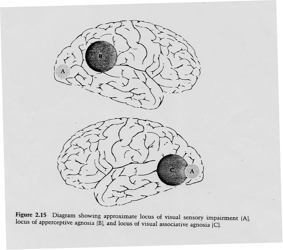
Lesion loci in visual agnosia

Unconventional-views stimuli

Dorsal and ventral visual pathways
In Warrington & Taylor's (1978) 3-stage theory, which hemisphere handles perceptual vs. semantic categorization?
Perceptual categorization (object constancy across viewpoints) → posterior right hemisphere. Semantic categorization (accessing stored conceptual knowledge / object meaning) → posterior left hemisphere.