Anatomy 337 Exam #3, Brain-UW Madison
 Call Kai
 Learn
 Practice Test
 Spaced Repetition
 Match
 Flashcards
 Knowt Play
1/68

There's no tags or description
Looks like no tags are added yet.
69 Terms
Telenchephalon
cerebral hemispheres

Diencephalon
thalamus, hypothalamus, epithalamus

Mesencephalon
the midbrain; a region of the brain that surrounds the cerebral aqueduct; includes the tectum and the tegmentum

Metencephalon
pons and cerebellum

Myelencephalon
medulla oblongata

gray matter
cell bodies, dendrites, unmyelinated axons
gray matter cortex
covers the surface of the brain and forms discrete internal clusters called cerebral nuclei
White matter
myelinated axons, towards the middle
Cerebral cortex
-awareness, knowledge, memory, perception, information processing, and thinking
Motor areas
control voluntary movement
sensory areas
conscious awareness of sensation
multimodal association areas
integration and interpretation of sensory and motor information
cytoarchitecture
The arrangement of neuronal cell bodies in various parts of the brain (ie. cerebral cortex has 6 layers, each layer has different function)
Brodmann's Areas
regions of cortex defined by the relative distribution of cell types across cortical layers (cytoarchitecture), location varies from person-to-person
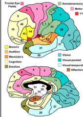
Cerebral hemispheres
-mirror images, different function
-connected by white matter pathways (corpus callosum, anterior commissure, and posterior commissure)
Median longitudinal fissure
separates left and right hemispheres

Corpus callosum
the large band of neural fibers connecting the two brain hemispheres and carrying messages between them, white matter

Anterior commissure
connects medial temporal lobes

Posterior commissure
white matter

Corpus callosum and TBI
-rotational injuries are usually worse
-almost always damaged in TBI/repetitive trauma

Parietal Lobe
general sensory functions, tactile sensation, proprioception, taste, language, spatial orientation, directing attention
-primary somatosensory cortex, somatosensory association cortex (where DCL pathway ends)
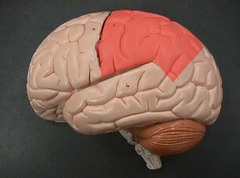
Central sulcus
separates frontal and parietal lobes

Lateral sulcus
Separates temporal lobe from parietal and frontal lobes

parieto-occipital sulcus
separates parietal and occipital lobes

Primary somatosensory cortex
receives general somatic sensory information from touch, pressure, pain, and temperature receptors; located within the postcentral gyrus

Somatosensory association cortex
-posterior to primary somatosensory cortex
-integrates and interprets sensory information

Prefrontal cortex
Dorsolateral, orbitofrontal, anterior cingulate
Dorsolateral Prefrontal Cortex
-organization and planning
-managing behavior
-high-level decision making
-multitasking
-working memory

Orbitofrontal Cortex
modulating emotions, inhibition, adaptive learning, rewards, and emotion

Anterior Cingulate Cortex
-Motivational behavior
-Reward-based learning: error detection, outcome monitoring
-Pain processing

Phineas Gage
-understanding of the localization of brain function
-prefrontal cortex is not vital to being alive
Primary motor cortex
-voluntary motor activity
-beginning of corticospinal tract
-innervation similar to sensory homunculus

sensory homunculus
Demonstrates that the area of the cortex dedicated to the sensations of various body parts is proportional to how sensitive that part of the body is.

Motor homunculus
broad areas of primary motor cortex devoted to controlling movements of different body regions

Prefrontal/supplementary motor cortices
-process motor information
-plans and coordinates learned, skilled motor activity
-"making the plan"
Broca's area
-left side of brain
-controls muscles of speech

Broca's aphasia
can understand spoken language, but can't speak it
Temporal Lobe
-hearing and smell
-medial associated with limbic system (memory, learning, aggression, emotion)

Wernicke's area
-recognizing and comprehending written and spoken language
-left side
-overlaps the parietal and temporal lobes

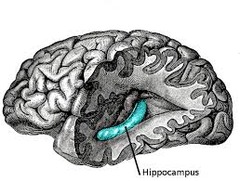
Hippocampus
-medial temporal lobe
-essential in learning, storing memories, and forming long-term memory
Patient HM
Fell off his bike in the 1950's which caused him to have seizures. As a solution, doctors took out his medial temporal lobe which led to him not being able to form any new memories

Amygdala
-anterior to hippocampus (medial temporal lobe)
-associations between sensory input and emotions
-sort and code memories based on emotions
-fear

Insula
-primary gustatory cortex (taste)
-"old area", primal, not entirely sure function

Limbic system structure
form ring around diencephalon
Limbic system
-establishes and processes emotional states and behavioral drives
-affects memories (formation, storage, retrieval) through integration, emotional states, physical sensations
-links consciousness, intellectual cortex with unconscious ANS functions
cerebral nuclei (basal ganglia)
-control of posture and voluntary motor movements
-adjust rhythm/pattern (+ learned movement)
-muscle tone
-process, integrate, and relay info from cortex to thalamus
Cerebral nuclei structure
-paired gray matter nuclei deep within the white matter
-caudate nucleus
-putamen
-globus pallidus
Hypothalamus
-controls release of regulatory hormones from anterior pituitary gland
-specific hormones from posterior pituitary gland
-controls adrenal medulla

Pituitary gland
-inferior to hypothalamus
-attached to hypothalamus by infundibulum
-anterior: adenohypophysis
-posterior: neurohypophysis

Adenohypophysis (anterior pituitary)
controlled by releasing and inhibiting hormones via the hypothalamus, travels through hypothalamohypophyeal tract
-uses a portal system

Neurohypophysis (posterior pituitary)
neural connection between the hypothalamus and the posterior pituitary (axons make the hypothalmohypophyseal tract
-does not make the hormones, the hypothalamus does (stores and releases hormone when needed)

Cerebellum
-voluntary motor activity (precision, timing, error-correction)
-muscle tone and posture
-eye blink, visceromotor, vestibuloocular reflex
-higher cognitive functions
-emotional processing
-ipsilateral
Midbrain
Tectum and reticular formation
Tectum
-midbrain
-contains 2 pairs of sensory nuclei call corpora quadrigemina
-visual reflexes: superior colliculi
-auditory reflexes: inferior colliculi

Reticular Formation
arousal and consciousness

cerebral peduncles (midbrain)
-primarily motor axons
-corticospinal tract

substantia nigra (midbrain)
-neurons secrete dopamine
-voluntary movements through connections to basal ganglia

red nucleus (midbrain)
involuntary motor control, maintaining muscle tone and limb position

Substantia Nigra and Parkinson's

Pons
-contains corticospinal tract fibers
-somatosensation (DCML pathway)
-process and relay cerebellar commands
-origin of four cranial nerves
-sleep regulation, breathing, hearing, balance
Pons breathing
-autonomic nuclei in the pontine respiratory center
-regulate activity of respiratory rhythmicity center of the medulla oblongata
Pons Hearing
superior olivary nuclei receive auditory input and help localize sound
Pons Balance
nuclei that relay info from the cerebellum
Medulla Oblongata
-connects brain and spinal cord
-white matter tracts through it
-origin of 5 cranial nerves
-sensory nuclei from DCML
-motor: pyramids
Cardiovascular Center
-medulla
-autonomic nuclei, adjust HR, strength of contraction, vasoconstriction/dilation
Medullary Respiratory Center
sets the pace of breathing
Other Medullary Nuclei
coughing, sneezing, salivation, swallowing, gagging, and vomiting
Subdural Hematoma
-rapid acceleration or deceleration of head
-tearing of a cerebral vein as it pierces the arachnoid mater
-typically slower

Epidural Hematoma
-can occur with skull fracture
-damages the meningeal artery
-often fatal
