Electrical and Mechanical Events of the Heart Cycle
 Call Kai
 Learn
 Practice Test
 Spaced Repetition
 Match
 Flashcards
 Knowt Play
1/28

Earn XP
Description and Tags
Comprehensive vocabulary flashcards covering the electrical conduction system, electrocardiogram waves, valve dynamics, and mechanical phases of the cardiac cycle.
29 Terms

Sinoatrial (SA) node
The natural pacemaker of the heart situated in the right atrium that exhibits an intrinsic firing rate of approximately 100 beats per minute and initiates cardiac contractions without external nerve or hormonal input. made up by pacemaker cells
Pacemaker potential
An unstable resting membrane potential characteristic of autorhythmic cells in the SA node that continuously drifts upward toward threshold (this means that the potential will always be reached it just depends on how fast it will be reached).

Sympathetic stimulation of the SA node
Autonomic input that increases the heart rate by steepening the slope of the pacemaker drift toward threshold through increased permeability of the Na(F)+ (funny) inward current. (releases norepinephrine and epinephrine into the atrium and ventricles from the thoric spinal nerves)
Parasympathetic stimulation of the SA node
Autonomic input via the vagus nerves that lowers the heart rate by decreasing the inward current slope and hyperpolarising the cell membrane through increased K+ permeability. (from vagus nerves release acetylcholine (ach), into atria)
Atrioventricular (AV) node delay
A brief pause in the transmission of electrical impulses through the AV node, ensuring the atria complete their contraction and empty blood into the ventricles prior to ventricular systole.
Purkinje fibres
Specialised myocardial conducting fibres branching throughout the ventricular walls that rapidly distribute electrical depolarisation upward through the ventricular myocardium.
P wave
The deflection on an electrocardiogram representing atrial depolarisation/contraction and excitation spreading across both atria from the SA node.

QRS complex
The rapid waveform on an electrocardiogram reflecting ventricular depolarisation/contraction and excitation, which electrically initiates ventricular contraction and conceals atrial repolarisation.

T wave
The electrocardiogram deflection corresponding to ventricular repolarisation/relaxation, marking electrical recovery during ventricular diastole.

Tricuspid valve
The right atrioventricular valve that separates the right atrium and right ventricle; it opens when right atrial pressure exceeds right ventricular pressure and closes when right ventricular pressure exceeds right atrial pressure.

Mitral (bicuspid) valve
The left atrioventricular valve that separates the left atrium and left ventricle; it opens when left atrial pressure exceeds left ventricular pressure and closes when left ventricular pressure exceeds left atrial pressure.

Aortic valve
The left semilunar valve separating the left ventricle from the aorta; it opens when left ventricular pressure exceeds aortic pressure and closes when left ventricular pressure falls below aortic pressure.

Pulmonary valve
The right semilunar valve separating the right ventricle from the pulmonary artery; it opens when right ventricular pressure exceeds pulmonary artery pressure and closes when right ventricular pressure drops below pulmonary artery pressure.

Isovolumetric ventricular contraction
The early phase of systole where the ventricles contract with both AV and semilunar valves closed, causing ventricular pressure to rise rapidly while ventricular volume remains unchanged.

Ventricular ejection
The second phase of systole wherein ventricular pressure surpasses arterial pressure, forcing open the aortic and pulmonary valves to expel blood into circulation.

Isovolumetric ventricular relaxation
The initial phase of diastole wherein the ventricles relax and their internal pressure drops below arterial pressure, closing the semilunar valves while the AV valves remain shut, keeping volume constant.

Ventricular filling
The diastolic phase occurring when ventricular pressure falls below atrial pressure, causing the AV valves to open and blood to fill the ventricles, concluding with atrial contraction.
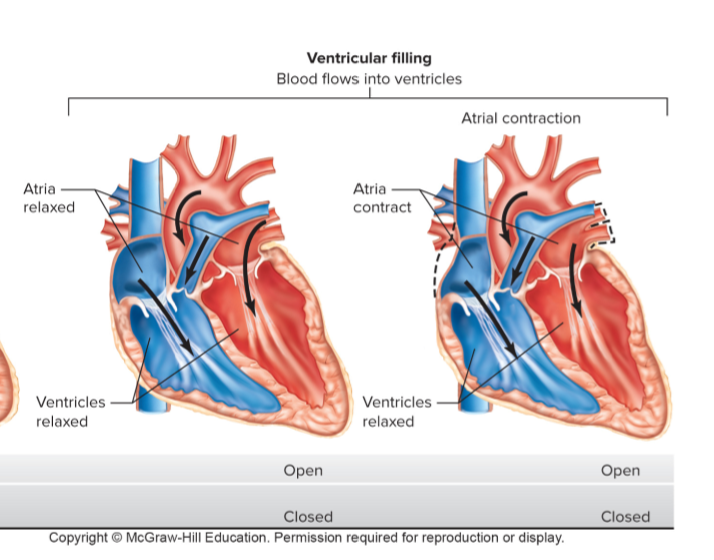
End-diastolic volume (EDV)
The volume of blood contained within a ventricle at the conclusion of diastole, typically around 135mL.
End-systolic volume (ESV)
The residual volume of blood remaining in a ventricle following ejection at the end of systole, typically around 65mL.
Stroke volume
The volume of blood pumped out of the ventricle per contraction, equal to the difference between end-diastolic volume and end-systolic volume (EDV−ESV, approximately 70mL at rest).
Dicrotic notch
A brief rebound spike observed on the aortic pressure tracing caused by blood rebounding against the closed aortic valve leaflets at the onset of ventricular diastole.
First heart sound (lub)
The heart sound produced by the sudden closure of the atrioventricular (mitral and tricuspid) valves at the onset of ventricular systole.
Second heart sound (dub)
The heart sound produced by the closure of the semilunar (aortic and pulmonary) valves at the beginning of ventricular diastole.
heart beat phases shown on pressure graph and left ventricle vol?

pressure volume curve
it shows pressure changes
left → right the heart is filling up with blood
down → up pressure increases
atria contraction adds a bit more blood and stretches the ventricles so the pressure begins to rise

what does it mean if the peak on the ecg is bigger?
the heart muscle is bigger and stronger
the electrical events of a heart beat

what 2 things does the rate of firing from the cells of the sa node depend on?
on the initial value of the membrane potential
on the slope of the drift towards threshold
threshold potential for firing rate
a = control
b = during sympathetic stimulation
c = during parasympathetic stimulation
