L5 and L6- Morphology of Chromosomes and Karyotyping
 Call Kai
 Learn
 Practice Test
 Spaced Repetition
 Match
 Flashcards
 Knowt Play
1/39

There's no tags or description
Looks like no tags are added yet.
40 Terms
Chromosomes
rod-shaped, filamentous bodies
present in the nucleus, which become visible during
cell division
Number of chromosomes
46 (23 pairs)
Chromosomes can be identified
• Their size
• Their shape (the position of the centromere)
• Banding patterns produced by specific stains (Giemsa)
Karyotype
A display of the chromosome pairs of a cell arranged by size and shape.
Karyotype is during
chromosome measurements are generally taken during mitotic metaphase.
Chromosome 1
largest chromosome
Chromosome 21
smallest chromosome
X chromosome
is relatively large (~156 Mb)
Y chromosome
is much smaller (~57 Mb)
p arm
short arm of chromosome

q arm
long arm of chromosome

Centromere
Area where the chromatids of a chromosome are attached

telomeres
ends of chromosomes

Centromeric index
ength of p arm ÷ total chromosome length × 100
metacentric chromosome
Chromosome in which the two chromosome arms are approximately the same length.

Submetacentric
centromere slightly off center

Acrocentric
centromere close to end , has satellite

satellite chromosome
small chromosomal segment

Satellite stalks
contain nucleolar organizer regions (NORs), the sites of ribosomal RNA genes and production of rRNA

Telocentric
the centromere is at or very near the end of the chromosome

chromosome by groups
Group A: 1-3, largest metacentric and submetacentric chromosomes
Group B: 4 & 5, large submetacentric
Group C 6-12 & X ; Very similar, medium-sized (arranged in order of decreasing size sub-metacentric)
Group D: 13-15, medium-sized acrocentric plus satellites
Group E 16-18 Smaller, metacentric and submetacentric
Group F, 19-20; even smaller, metacentric and submetacentric
Group G 21, 22 & Y; Smallest acrocentric with satellites. Y submetacentric
Banding pattern
result from dies showing a pattern of light and dark bands from karyotyping

dark stain
densely packed heterochromatin. (AT rich).
usually not transcribed; late s-pahse.
centromer, telomere, stalite dna regions.

light stain
less dense euchromatin (GC rich).
activity transcribe; replicated in eraly s phase

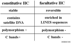
Facultative heterochromatin
exhibits variable levels of condensation, related to levels of transcription of resident genes.
Lyonization/X-inactivation
When one of the X chromosomes present is rendered inactive forever to ensure that the female does not have twice as many X chromosomes as the male.
by Xist non-coding RNA produced by XIC
constitutive heterochromatin
chromosomal regions that remain condensed in heterochromatin at most times in all cells

centromeres
the point on a chromosome by which it is attached to a spindle fiber during cell division.
of highly repetitive DNA and specific proteins that form the kinetochore,
Telomeres
Repeated DNA sequences at the ends of eukaryotic chromosomes
provide terminal stability to the chromosome and ensure its survival .
T-loop and D-loop,
this is formed when a telomere comes back around and invades the double stranded DNA. which help protect the chromosome end from being recognized as DNA damage.
G-quadruplex DNA
In areas of DNA that are guanine rich, 4 DNA strands can form a quadruplex structure that interferes with telomerase
idiogram
alignment of chromosomes based upon size and shape
Karyotyping
the process of sorting chromosomes into their matched pairs. An important technique for investigating chromosomal disorders
Colcemid
prevents spindle fibers from forming and stops mitosis at metaphase
hypotonic solution
Solute concentration is less than that inside the cell; cell gains water
Karyotyping Steps
1. cells are stained and prepared on a glass slide to see their chromosomes under a light microscope
2. photomicrograph images obtained of the chromosomes during mitotic metaphase
3. the images are cut out and separated, a process which can be done using scissors or using a computer
4. the images of each pair of chromosomes are placed in order by size and position of their centromeres

T-banding
A Giemsa staining technique that stains the telomeres (ends) and the centromeres of chromosomes.
Karyotype Nomenclature for normal chromosome
Total Chromosome Count: The first number indicates the total number of chromosomes in the cell. A normal human somatic cell contains 46 total chromosomes
Sex Chromosome Complement: The letters following the comma show the sex chromosomes.
XX designates a female.
XY designates a male

Karyotype Nomenclature for Sex Chromosome Abnormalities
The total chromosome count is listed first, followed by the sex chromosome complement, separated by a comma.
Turner Syndrome: 45,X (or 45,X0). Monosomy X; missing one sex chromosome, leaving a total of 45 chromosomes.
Triple X Syndrome: 47,XXX. Trisomy is X; an extra X chromosome in a female.
Mosaicism Example: 45, X/46, XY. Clones with different cell lines are separated by a forward slash (/).
Karyotype Nomenclature for Autosomal Chromosome Abnormalities
Total count, sex chromosomes, signs (+ or -), abnormality.
1. Numerical Abnormalities (Aneuploidy): trisomy 21 (Down Syndrome in a male): 47,XY,+21
2. Structural Abnormalities:
Deletion (Partial Monosomy): 46,XX,del(5)(p14.2)
Reciprocal Translocation: 46,XX,t(2;5)(q21;q31)
Robertsonian Translocation: 45,XY,der(13;14)(q10;q10)