Sect 6 - Flow Cytometry
 Call Kai
 Learn
 Practice Test
 Spaced Repetition
 Match
 Flashcards
 Knowt Play
1/63

There's no tags or description
Looks like no tags are added yet.
64 Terms
Flow Cytometry
method of cell analysis
through use of fluidics, optics, and electronics, the flow cytometer gathers data on various aspects of a cell, including cell size and internal complexity
with addition of fluorescent antibody dyes, cell markers can also be ID
1. Fluidics
2. Optics
3. Electronics
3 major components of Flow Cytometry
Fluidics
carries the sample to the flow cell for analysis
Optics
laser light excitation sources, filters, and lenses to collect light
Electronics
gather data from light sources into a photoconductor and then translate that signal into a digital form for analysis
cell size
granularity
antigens present (bound, fluorescent Abs)
Flow cytometers differentiate cells based on (3):
integration point
Cells suspended in saline flow through a chamber and are measured at the
Sheath Fluid
fluid that surround the sample and provides a physiological barrier for the sample
Laminar Fluid
sample and sheath fluids are kept at different pressures to keep the fluids separated
Hydrodynamic Focusing
the process of narrowing the sample fluid using laminar flow so that the cells can pass single-file through the interrogation point
sample fluid column narrowed (controlled by pressures of the fluid congrol)
the pressures of the fluid
What controls the diameter of the sample fluid?
string of beads
As cells pass through the interrogation point, the resemble
Interrogation Point
the point at which the laser meets the sample fluid
light photons scatter at interrogation point and converted into electrical signal
How are light particles converted to collect data?
photodetector
device used to measure or indicate the presence of light
amplifies electrical signal to be read by the electronics system as an electrical pulse
direct proportional
relationship between number of light particles generated and height of voltage pulse in flow cytometry
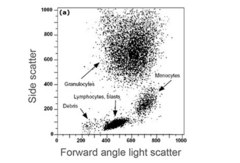
Forward Light Scatter (FSC)
light that scatters in the forward direction
reflects the SIZE of the cells
monocytes & neutrophils
Which cells make the most FSC?
**hint: larger cells
Side Light Scatter (SSC)
light that scatters at a 90° angle
reflects GRANULARITY, or internal complexity, of the cell
granulocytes
Which cells have the highest SSC?
lymphocytes
Which cells have the lowest SSC?
populations
FSC and SSC data are used together to create
Principle of fluorescence
Laser light excites fluorescent molecules used as labels (fluorochromes) and the exciting causes fluorochromes to emit light at specific wavelength
Fluorochromes
Labels that are attached to cell antigens using fluorescently tagged antibodies
propidium iodine
used for visibility staining
photomultipler
light emitted by fluorchormes are detected by
true
T or F:
The number of lasers determine the number of fluorochromes that can be used
Compensation
the correction between the excitation wavelength and the detection wavelength
can be done manually by adjusting settings or using a mathematical correction
Histogram
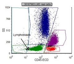
a graph used to differentiate populations of cells
can also use dot plts (provide 2D way to view data
some fluorochromes have the same excitation wavelength but a unique detection wavelength
Why must compensation occur?
Gating
ensures that populations are as pure as possible
- allows for immunophenotyping of cells
immunophenotyping
the detection of Ag that are being expressed on the cell surfaces using detection antibodies
CD markers
cluster of differentiation markers
ags on cell surfaces that can be tagged with abs
CD45
ab that is present on ALL leukocytes
used against SSC to separate populations further
Lymphocyte antigens
B-lymphocyte markers
NK markers
T-large granular
lymphocyte markers
Monocyte/Granulocyte antigens
evaluated for normal cell markers
Blasts antigens
evaluated for all cell markers to determine what type of blasts they are
True, dx Non-Hodgkin's Lymphoma
T or F:
Flow Cytometry CANNOT diagnose Hodgkin's Lymphoma
lymphs look normal
Why can't flow cytometry diagnose Hodgkin's Lymphoma =
CD45
all leukocytes
CD3
T-lymphocytes
CD4
Helper T-lymphocytes (CD3+)
CD8
Cytotoxic T-lymphocytes (CD3+)
CD56
Natural Killer Cells (CD3-)
CD19 & CD20
B-lymphocytes
CD14
Monocytes
CD34
Blasts. Stem cells
CD117
Myeloblasts
CD55
Decay Accelerating Factor (DAF)
CD59
Membrane Inhibitor of Reactive Lysis (MIRL)
Surface Kappa:Lambda
other markers expressed as ratio on B-lymphocytes
looked at against CD19 to confirm they are B cells
0.8 - 2.0
normal Kappa:Lambda ratio
B-lymphs with no K:L chains ("baldies")
Kappa:Lambda ratio
neoplastic process (NHL, CLL/SLL)
Kappa:Lambda ratio >2.0
CD19, CD20, surface Kappa, surface Lambda
What 4 antibodies are expressed on normal, mature B-lymphs?
hematogones
normal immature B-lymphs
2:1
What is a normal CD4:CD8 ratio?
AIDS crisis patient (can be as low as 0.1)
What does a low CD4:CD8 ratio indicate?
CD34+
used to determine the success of a stem cell harvest
CD55, CD59, CD14, CD24
PNH markers
neg for CD3 & CD4
Read the quadrants

CD3 pos
CD4 neg
Read the quadrants

pos for CD3 & CD4
Read the quadrants

CD3 neg
CD4 pos
Read the quadrants
