Module 1: NS/Lungs/Heart
 Call Kai
 Learn
Learn Practice Test
 Spaced Repetition
 Match
Match Flashcards
 Knowt Play
1/40

Earn XP
Description and Tags
OT881
Name | Mastery | Learn | Test | Matching | Spaced | Call with Kai | Chat |
|---|
No analytics yet
Send a link to your students to track their progress
41 Terms
The 3 planes
1. Sagittal (or median) plane
2. frontal (coronal) plane
3. transverse (axial or horizontal) plane
Sagittal plane
Right and Left halves

Frontal plane
Front and back

Transverse plane
Top and Bottom halves

Anatomical terms
superior and inferior
→ up and down
anterior and posterior
→ forward and backward
proximal and distal
→ close to midline and further from midline
medial and lateral
→ right and left

Function of the nervous system
enables the body to react to continuous change in its internal and external environments
Controls and integrates bodily activity
Anatomical Nervous system structure
CNS
brain
spinal cord
PNS
everything else
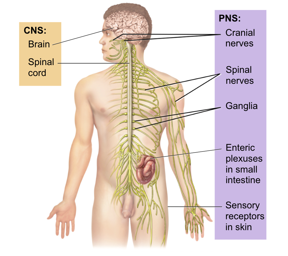
Spinal Cord
CNS
all the information inside the spinal cord are CNS
BUT
As soon as that information leaves the spinal cord (out through spinal nerve to get to muscles) it becomes PNS
Sensory information will come in and synapse in the ganglia
CNS vs PNS
Somatic: Voluntary (you have to think)
Autonomic: Involuntary (smooth + cardiac muscle → heart and digestion)
Sympathetic (fight/flight)
Parasympathetic (rest + digest)
Sympathetic and parasympathetic work opposite to each other, meaning if one is regulated, the other is not

Sensory neurons
Afferent (arrives)
Motor neurons
efferent (exists)
Spinal nerves
31 spinal nerves
union of posterior and anterior roots of the spinal cord
includes sensory, motor + autonomic info
Spinal nerve sections
C1-C7
nerves emerge above vertebrae
T1-T12
Nerves emerge inferior
L1-L5
cauda equina
S1-S5
Breakfast @ 7
Lunch @ 12
Dinner @ 5

Motor and sensory pathway
Sensory information
→ coming into the back (dorsal or posterior)
Motor information
→ out through the front (ventral or anterior)
THINK:
root→ nerve→ rami
In the DOR, out the VENT

Motor neurons are:
anterior
ventral
efferent
out the VENT(ral)
Motor pathway
1. starts in Ventral horn
2. out through the ventral root
3. through the spinal nerve
4. and then SPLIT into dorsal and ventral rami

Sensory neurons are:
posterior
dorsal
afferent
In the DOR(sal)
Sensory pathway
ALL information entering the spinal cord will enter through the:
- Dorsal Rami
- Ventral Rami
2. travel through the spinal nerve
3. then go through the dorsal root to the Dorsal horn
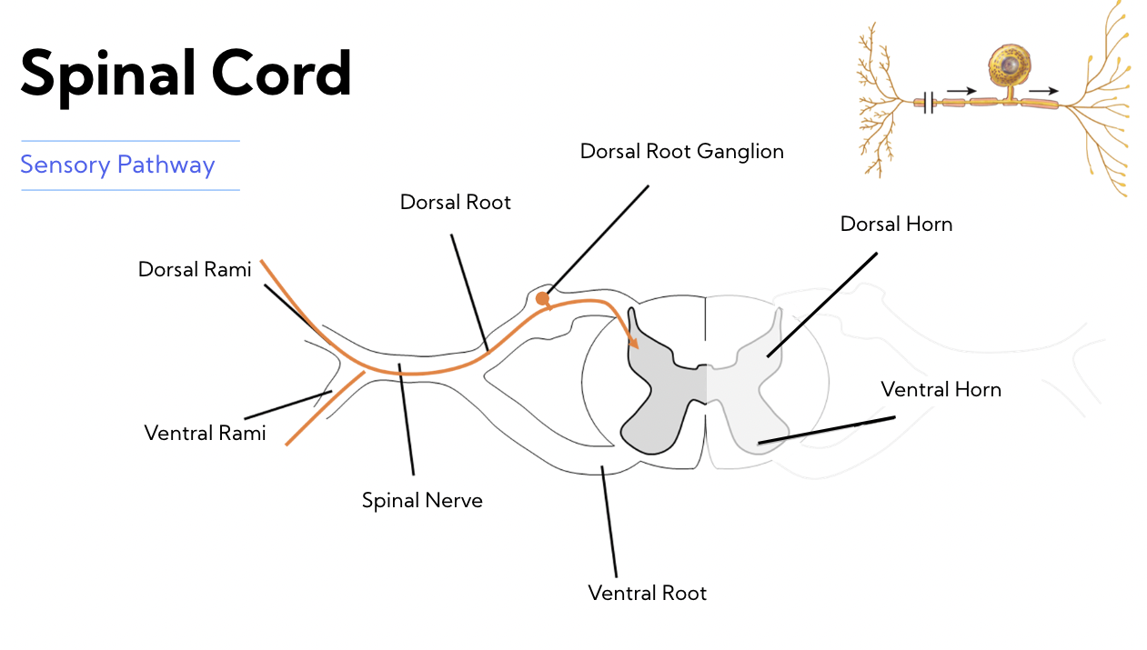
Neuron overview

Thoracic pleural cavities
pleural fluid allows lungs to expand

pleura
A serous membrane forming a closed sac on the outer surface of the lung and lining the thoracic cavity
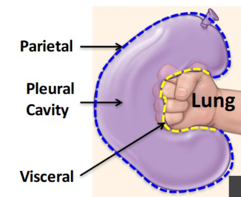
Parietal vs visceral pleura
Pleura are 2-layered sacs surrounding the lungs
- visceral = next to lung
- Parietal = next to thoracic wall
think of a pleural cavity as punching a balloon that isn’t very blown up
*fist = lung
hilum = wrist
pulmonary ligament = below wrist
Pulmonary ligament
extension of the right hilum in the inferior aspect
- double layer of pleura transitioning from visceral to parietal
Hilum
a place of transition…
* where tubes and vessels transition from the mediastinum to the lungs
contains:
pulmonary viens
pulmonary arteries
primary bronchi

The left lung has lobes _ and right lung has _
Left: 2
Right: 3
Each lung has 3 surfaces:
costal surface (against the ribs)
Diaphragmatic surface (against the diaphragm)
Mediastinal surface (against the mediastinum)
top of lung is the: Apex
Right lung
Landmarks:
oblique fissure
horizontal fissure
Lobes:
Superior lobe
Middle lobe
inferior lobe

Left Lung
Landmarks
1 fissure: Oblique fissure
the Lingula
- wraps around the heart (looks like a tongue👅)cardiac notch: allows for heart
Lobes:
Superior lobe
Inferior lobe

Trachea
Made of cartilage to hold open trachea
‘C- shaped’ rings so the posterior side can allow for the esophagus
Basic air flow from nose to trachea
Air
nasal cavity
pharynx
larynx
trachea
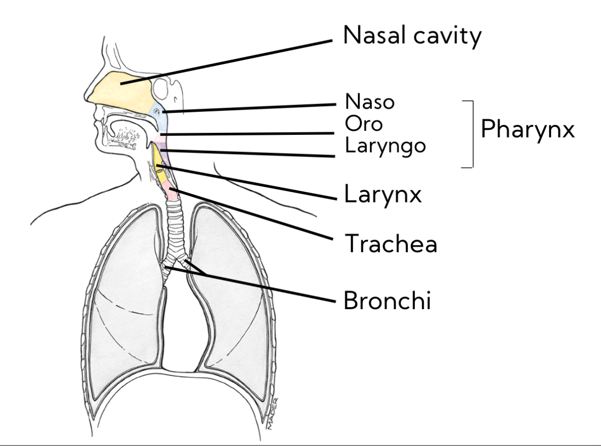
Tracheobronchial tree
Trachea divides into primary bronchi at the carina (black star on image)
Left = longer, more horizontal
Right = shorter, more vertical, wider
Secondary (Lobar)
Left = 2
Right = 3
Tertiary (segmental)
Bronchopulmonary segments
no longer has cartilage - smooth muslce only to allow for airflow
(segmental bronchus + vessels)

Respiratory/ terminal bronchiole
smooth muscle
no cartilage
1st structures to do gas exchange + oxygenate blood

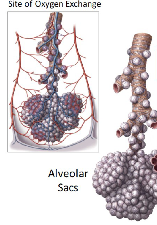
Alveolar sacs
site of gas exchange
GIANT surface area for blood to be deoxygenated
Heart halves
right = thinner walls - only pumping blood to lungs
Left = thicker walls - pumping blood all around body with greater force
Heart chambers + valves
4 chambers
* 2 atria (L+R) - Superior to ventricles
* 2 ventricles (L+R)
4 valves
* 2 atrioventricular valves (atrium → ventricles)
* 2 semilunar valves (ventricles → out to body)
communicates with:
* the lungs (pulmonary)
* the body (systemic)
* itself (coronary)

2 atrioventricular (AV) valves
Atrium → ventricle
right = tricuspid
left = bicuspid/mitral
* chordae tendineae prevent backflow
LAB RAT:
R = Right = T (tricuspid)
L = Left = B (bicuspid)

Vessels of the heart
blood travelling away from the heart travels via arteries
blood travelling to the heart comes in veins (and coronary sinus)

Blood flow
1. blood flows into right atrium
- via Sup. VC + Inf. VC
2. moves through tricuspid valve (R atrioventricular)into right ventricle
3. heads out through pulmonary semilunar valve
4. reaches the pulmonary trunk
5. which divides to become L + R pulmonary arteries (only artery to carry deox blood)
6. blood travels through pulmonary capillaries
7. back to heart through pulmonary veins
8. enters Left atrium
9. travels through the bicuspid valve (L atrioventricular)
10. to reach the left ventricle
11. exits the heart through the aortic semilunar valve
12. through the aorta to reach systemic circulation
15. arrive back at heart and enter through sup. and inf. vena cava

coronary circulation

Heart attack
R coronary artery or L coronary artery gets blocked
Where does the coronary sinus drain into?
the right atrium
(so do the anterior cardiac veins, bypassing the coronary sinus)