A&P Ch 10. The muscular system
 Call Kai
 Learn
 Practice Test
 Spaced Repetition
 Match
 Flashcards
 Knowt Play
1/71

There's no tags or description
Looks like no tags are added yet.
72 Terms
Prime mover (agonist)
Major responsibility for producing specific movement
Antagonist
Opposes or reverses particular movement
– Prime mover and antagonist are located on opposite sides of joint across which they act
Synergist
helps prime movers
– Adds extra force to the same movement
– Reduces undesirable or unnecessary movement
Fixators
type of synergist that immobilizes bone or
muscle’s origin rather than enhancing movement of
prime movers
▪ Gives prime mover stable base on which to act
example of muscle actions and interactions
For elbow flexion:
• Prime mover—brachialis
• Synergist—biceps brachii
• Antagonist—triceps brachii
• Fixator—muscle that holds
scapula firmly in place
– Rhomboids

Pennate
Three forms
▪ Unipennate: fascicles attach only to one side of
tendon (example: extensor digitorum longus)
▪ Bipennate: fascicles insert from opposite sides of
tendon (example: rectus femoris)
▪ Multipennate: appears as feathers inserting into
one tendon (example: deltoid), strongest

Patterns of Fascicle Arrangement in Muscles

Lever Systems
– Lever: rigid bar (bone) that moves on a fixed point
called fulcrum (joint)
– Effort: force (supplied by muscle contraction) applied
to lever to move resistance (load)
– Load: resistance (bone + tissues + any added weight)
moved by the effort
Basic principle of levers
– Effort farther than load from fulcrum = lever operates
at mechanical advantage
– Effort nearer than load to fulcrum = lever operates at
mechanical disadvantage
Mechanical advantage (power lever)
load is close to fulcrum, with effort far from fulcrum
– Small effort can move large load
slower, but more stable
▪ Used where strength is a priority

Mechanical disadvantage (speed lever)
load is far from fulcrum, with effort close to fulcrum
– Load moved rapidly over large distance; offers wider
range of motion
force is lost, but speed and range of movement are gained

First-class lever
– Fulcrum is between load and
effort
– Example: seesaw, scissors

second-class lever
– Load is between fulcrum
and effort
– Example: wheelbarrow,
standing on toes

Third-class lever
– Effort is applied between fulcrum and load
– Example: tweezers, forceps, most skeletal muscles

Muscles of Facial Expression
frontalis- elevator and depression of eyebrows
Orbicularis oculi
Zygomaticus minor
Zygomaticus major
Platysma
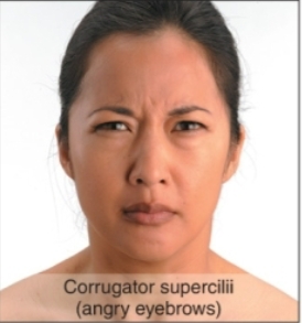
Corrugator supercilii- furrowed brow
buccinator
Orbicularis oris

Zygomaticus major

orbicularis oris

mentalis

platysma

corrugator supercilii
orbicularis oculi

frontal belly of epicranius

Muscles Promoting Mastication
Prime movers of jaw closure: temporalis (elevation and retraction) and masseter (elevation and retraction)
▪ Chewing role: buccinator (elevation of mandible)
push food back toward pharynx, where muscles in posterior mouth and pharynx
complete swallowing process

Muscles promoting tongue movements
▪ Genioglossus (goes from tongue to chin)-
Tongue Protrusion (sticking tongue out), Tongue Depression
hyoglossus- pulls tongue back and down
Depression: Pulls the side and lower third of the tongue downward, flattening the tongue.
Retraction (Retrusion): Pulls the tongue backward into the oral cavity.
styloglossus- pulls tongue back and up
Retraction (Retrusion): Draws the tongue backward into the oral cavity and oropharynx.
Elevation: Pulls the tongue upward and poster superiorly.
all help with swallowing

Suprahyoid muscles
– Four deep muscles involved in swallowing (move
hyoid bone and larynx)-above
▪ Form floor of oral cavity
▪ Anchor tongue
▪ Elevate hyoid bone
▪ Move larynx during swallowing
Infrahyoid muscles
– Four straplike muscles
– Depress hyoid bone and larynx during swallowing and
speaking, below
think thyroid cartilage
Muscles of the Anterior Neck and Throat Used in Swallowing
Suprahyoid
digastric (mandible to hyoid) - elevates/retracts hyoid, depresses mandible (right lateral side of neck)
stylohyoid (connects to styloid process)- hyoid elevation
Infrahyoid
thyrohyoid (thyroid cartilage to hyoid)- depresses hyoid or elevates larynx if hyoid is fixed
sternohyoid (sternum to hyoid)- depresses thyroid cartilage and larynx

Muscles of the Anterior Neck and
Throat Used in Swallowing
three stacked, fan-shaped skeletal muscles that form the circular muscular wall of the pharynx
suprahyoid squeezes food and pushes it further down
superior pharyngeal constrictor- pushes food into mpc
middle pharyngeal constrictor pushes food into ipc
inferior pharyngeal constrictor- pushes food into esophagus

Sternocleido-mastoid (2)
on lateral sides of neck-neck movement
activates and head is turned to opposite side

posterior side of neck
splenius capitus (goes to skull)
splenius cervicus (goes to cervical spine)
both turn head and help it flex

back muscles
erector spinae (split into 3)- keep spine straight and erect
iliocostalis: ilium to ribs
longissimus: runs all the way up to skull
spinalis: entire length of spine

diaphragm
breathing prime mover
exhale- pulls up
inhale- pulls down

external intercostal and internal intercostal muscles
control rib position during breathing

Muscles of the Abdominal Wall
rectus abdominus- runs straight down abs and engages in flexion
internal oblique and external oblique- run across abdominal cavity, rotation and lateral flexion
Transversus abdominius(deepest abdomen muscle)- wrapping horizontally around the torso from the ribs and spine to the pelvis like a natural corset

Muscles of the Pelvic Floor and Perineum
pelvic diaphragm
levator ani- elevates anus
coccygeus (coccyx to ischium)- form supportive part of pelvic diaphragm and stabilize pelvic floor
perineum
diamond-shaped region at the bottom of the pelvis, located between the thighs

scapula and arm (anterior)
pectoralis minor (ribs to coracoid process of scapula)- protraction, depression, downward rotation
serratus anterior (connects to 9 ribs)- attached to scapula on back (protraction and rotation upward), stabilizes scapula

scapula and arm (posterior)
trapezius- elevation, depression, retraction of scapula
rhomboid minor and rhomboid major- retraction of scapula
latissimus dorsi (goes to arm and across back except midline)- adduction, extension, internal rotation, sometimes hyperextension
levator scapulae- elevates scapula

Muscles Crossing the Shoulder Joint (anterior)
pectoralis major- adduction and flexion
Deltoid-
front: flexion
lateral: abduction
posterior: extension
coracobrachialis- flexion and adduction of upper arm, humeral head stabilization
triceps brachii- extension of forearm at elbow joint
long head of triceps-extension of shoulder
biceps brachii- flexion of elbow, supination
brachialis- pronation
brachioradialis- flexes elbow joint, supination and pronation

Muscles Crossing the Shoulder Joint (deep)
infraspinatus
teres minor
(both laterally rotate shoulder joint)
teres major- internal rotation, adduction, extension
triceps brachii- extension of forearm at elbow joint
long head of triceps-extension of shoulder
latissimus dorsi (goes to arm and across back except midline)- adduction, extension, internal rotation, sometimes hyperextension

Biceps Brachii

triceps brachii

Brachioradialis

Brachialis

rotator cuff (SITS)
originate on scapula; reinforce shoulder capsule; prevent dislocation, stabilizes humerus in glenoid facet of scapula
– Supraspinatus- arm abduction (lifting the arm out to the side)
– Infraspinatus- externally rotates shoulder joint
– Teres minor- externally rotates shoulder joint
– Subscapularis- internal rotation and adduction

flexor digitorum superficialis
flexes middle joint of second through fifth fingers (index, middle, ring, and little fingers) and wrist flexion

Flexor Pollicus Longus
deep muscle in the anterior forearm that flexes the thumb (flexion)

pronator teres and pronator quadratus
pronators

Wrist and Fingers
flexor pollicis longus- deep muscle in posterior forearm that flexes the thumb (flexion)
flexor digitorum profundus: flexing the fingers and gripping power
supinator- sits deep under the superficial skin and muscles of upper forearm, near the elbow joint, supination
pronator quadratus- Forearm Pronation: Rotates radius over ulna to turn the palm downward

supinator
deep under the superficial skin and muscles of the upper forearm, supination
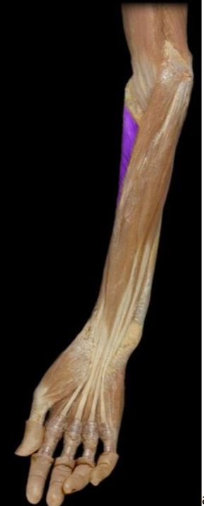
Flexor Digitorum Profundus
flexing the fingers and primary gripping power

Palmaris Longus
flexes the wrist and tenses the palmar fascia, for grip

Flexor retinaculum
bracelet-like fibrous sheet on wrist where most tendons of the extrinsic muscles pass under

carpal tunnel syndrome
prolonged, repetitive motions of wrist and fingers cause tissues in the
carpal tunnel to become inflamed, swollen
–Puts pressure on median nerve of wrist that passes through the carpal tunnel along with flexor tendons
–Tingling and muscular weakness in the palm and medial side of the hand
–Pain may radiate to arm and shoulder
–Treatment: anti-inflammatory drugs, immobilization of the wrist, and sometimes surgery to remove part or all of flexor retinaculum
posterior wrist and fingers muscles
extensor digitorum: Extends the metacarpophalangeal (MCP) and interphalangeal (PIP/DIP) joints of fingers 2–5
extensor pollicis longus- Extends all joints of the thumb

Extensor Digitorum
extending the wrist. When it contracts, it pulls the tendons on the back of the hand to lift the index, middle, ring, and little fingers

Posterior Compartment - Deep Extensor Pollicis Longus
a deep forearm muscle, extends the distal phalanx of the thumb

movements of hand

muscles of hand
second lumbrical (central compartment of hand): flexion and extension simultaneously, allows to bend the knuckle while keeping the finger straight
first lumbrical- worm-like intrinsic muscle of the hand, fine motor control and precision grip (flexion and extension)
dorsal interossei (4 muscles): located in the spaces between the metacarpal bones of the hand- (Dorsal ABduct)
opponens pollicis (index finger)- Flexes, medially rotates, and opposes the 1st metacarpal toward the palm
opponens digiti mini (pinky)- Flexes and laterally rotates the 5th metacarpal to bring the pinky toward the thumb (deepens the palm)

dorsal interossei (4 muscles-DAB)
located in the spaces between the metacarpal bones of the hand
the index, middle, and ring fingers away from the middle finger's central axis, aided by the mnemonic DAB (Dorsal ABduct)

palmar interossei (PAD)
spaces between the metacarpal bones of the hand (palmar ADuct)

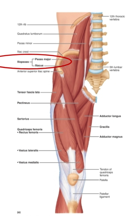
iliopsoas (iliacus and psoas major)
hip flexion and lateral rotation
hand muscle groups
Thenar group—form thick, fleshy mass at base of thumb
– Flexor pollicis brevis
– Opponens pollicis
• Hypothenar group—fleshy base of the little finger
– Abductor digiti minimi
– Flexor digiti minimi brevis
– Opponens digiti minimi
• Midpalmar group—hollow of palm
– Dorsal interosseous muscles (4)
– Palmar interosseous muscles (3)
– Lumbricals (4 muscles
Thigh muscles
adductor brevis: Adducts, lateral rotation and flexion of the hip
adductor longus: Adducts, flexes, and medially rotates the thigh.
adductor magnus: Powerful thigh adductor, hip extensor, and stabilizer

leg muscles (anterior)
vastus lateralis (largest and strongest): extends knee and helps stabilize patellar tracking
vastus intermedialis: Extends leg at knee joint
vastus medialis: Extends knee
all extensors
sartorius: flexor, abductor, and lateral rotator of the hip (crossing your legs)
quadriceps femoris (rectus femoris): Extends leg at knee and flexes thigh hip joint

glutes
Gluteus maximus: hip extension, lateral rotation of thigh and knee
– Gluteus medius and Gluteus minimus: abduction and medial and lateral rotation of thigh

Piriformis (also laterally rotates thigh, posterior)

biceps femoris
a two-headed muscle in the posterior compartment of the thigh, primary component of the hamstrings
flexes knee, extends hip (long head only), and laterally rotates lower leg or thigh

semitendinosus, semimembranosus
extension at thigh by hip, flex leg at the knee, and internally rotate the lower leg when the knee is bent

tibialis anterior (dorsiflexion)

Fibularis longus (on lateral side of leg)
Plantar flexion and eversion of the foot; stabilize lateral
ankle and lateral longitudinal arch of foot

posterior compartment (lower leg), goes deeper and deeper
gastrocnemius (forms visible shape of calf): plantar flexion, knee flexion, propulsion (walking, running)
soleus (deep to the gastrocnemius): plantar flexion, posture and balance
flexor hallucius longus: flexes the great toe (hallux) and stabilizes the foot during movement

flexor digitorum brevis
flexion of 4 toes
