Rheumatic Disorder (copy)
 Call Kai
 Learn
 Practice Test
 Spaced Repetition
 Match
 Flashcards
 Knowt Play
1/37

Earn XP
Description and Tags
38 Terms
Types of articular rheumatic diseases
Gout, CPPD, Infectious arthritis, OA, soft tissue rheumatism
Types of systemic connective tissue disease
SLE, RA, SSc, SNSA, SS, PM and DM myopathies, vasculitis
Symptoms of ARTICULAR disorders
Joint pain and edema, stiffness, limited ROM, crackling sound, joint deformity, pain/limit ROM in active AND passive movement
Articular structures
Synovium, synovial fluid, articular cartilage, intraarticular ligaments, joint capsule and juxtaarticular bones
Non articular/periarticular structures
Supportive extraarticular ligaments, tendon, bursae, muscle, fascia, bone, nerve and overlying skin
NON articular disorder symptoms
Point/focal tenderness near articular structure elicit by SPECIFIC movement/position, pain/limit ROM ONLY IN ACTIVE MOVEMENT, lack of swelling/crepitation/instability, deformation
Inflammatory disorder characteristic
Cardinal sign of inflammation, systemic symptom, lab evidence of inflammation (ESR, CRP etc), morning stiffness that improve after activity
Cut off for acute and chronic rheumatic disease
Less than 6 wk = acute
More than 6 wk = chronic
Causes of acute monoarthritis
infectious or crystal induced (Gout, CPPD) → NEED TO DO SYNOVIAL FLUID ANALYSIS
Acute polyarthritis causes
Acute viral infection or early presentation of SLE or RA
Ddx of chronic mono oligoarthritis
Indolent infectious arthritis, reactive arthritis, psoriatic arthritis, OA
RA involved joints
SYMMETRIC knee and ankle
MCP and PIP + feet MTP
Cervical spine
OA involved joints
Hip, knees ankle
DIP, PIP and 1st CMP + 1st MTP in feet
Cervical and LS spine
Psoriatic arthritis involved joints
ASYMMETRIC knee and ankle
DIP + nail
LS spine and SI joint
Gout/CPPD involved joints
Knees and ankles + wrist in CPPD
Small joint of feet + MCP in CPPD
NO SPINAL INVOLVEMENT

Describe these lesions and the associated condition
Malar rash and discoid rash (annular polycyclic type) → SLE signs

Describe the lesion and the associated condition
Discoid rash of scalp + scarring alopecia + central hypopigmentation ; lupus profundus (Panniculitis + atrophy) → SLE

What is this and what disorder is associated with it
Rheumatoid nodule → firm painless over area of pressure and trauma → sign of RA

Describe the lesion and associated disorder
Guttage psoriasis (scale papules with erythema) and plaque psoriasis → psoriatic arthritis sign

Describe the finding and associated disorder
A = nail pitting
B = onycholysis
C = destructive change with nail loss and pustule formation
Signs of psoriatic arthritis

What is this
Circinate balanitis → painless erythematous papules → reactive arthritis

What is this
Keratoderma blennorrhagicum (early) → Reactive arthritis

What is this
Keratoderma blennorrhagicum (late stage see hyperkeratotic plaque) → Reactive arthritis

Describe the lesion and associated disorder
Gottron papule over IP and MCP; heliotrope with edema → Dermatomyositis sign

Describe the lesion and associated disorder
Leukocytoclastic vasculitis and palpable purpura → primary vasculitis

What is this
Saddle nose deformity → granulomatosis with polyangitis
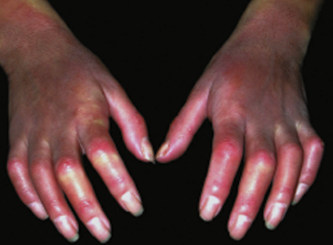
What is this
Flexion contracture sclerodactyly → Systemic sclerosis
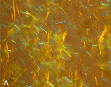
Synovial fluid shows urate crystals like this what disorder do you suspect
Needle shaped crystal → GOUT

Synovial fluid shows this what should you suspect
Rhomboid shaped → CPPD
Auto Ab SLE
Anti dsDNA and Sm
Auto Ab SSc
Anti topoisomerase 1 in dscSSc and anti centromere in lcSSc
Auto Ab PM
anti Jo-1
Auto Ab DM
Anti Mi-2
Auto Ab SS
Anti Ro anti La (SS-A and SS-B)
Auto Ab RA
ACPA and RF
When should you send ANA
Only if the there is CLEAR INDICATION → not for screening bc normal people can have ANA postiive

Describe the finding
Lt = bone erosion and cartilage loss
Rt = erosion and ankylosis
RA signs

Compare the left to right image
Normal on Lt vs OA on right