Lower Extremity Muscles
 Call Kai
 Learn
 Practice Test
 Spaced Repetition
 Match
 Flashcards
 Knowt Play
1/54

There's no tags or description
Looks like no tags are added yet.
55 Terms
Psoas major
O: transverse processes of lumbar vertebrae (bodies, intervertebral disks)
In: lesser trochanter of femur
A: flexes thigh + trunk
I: L2 and L3 branches of lumbar plexus
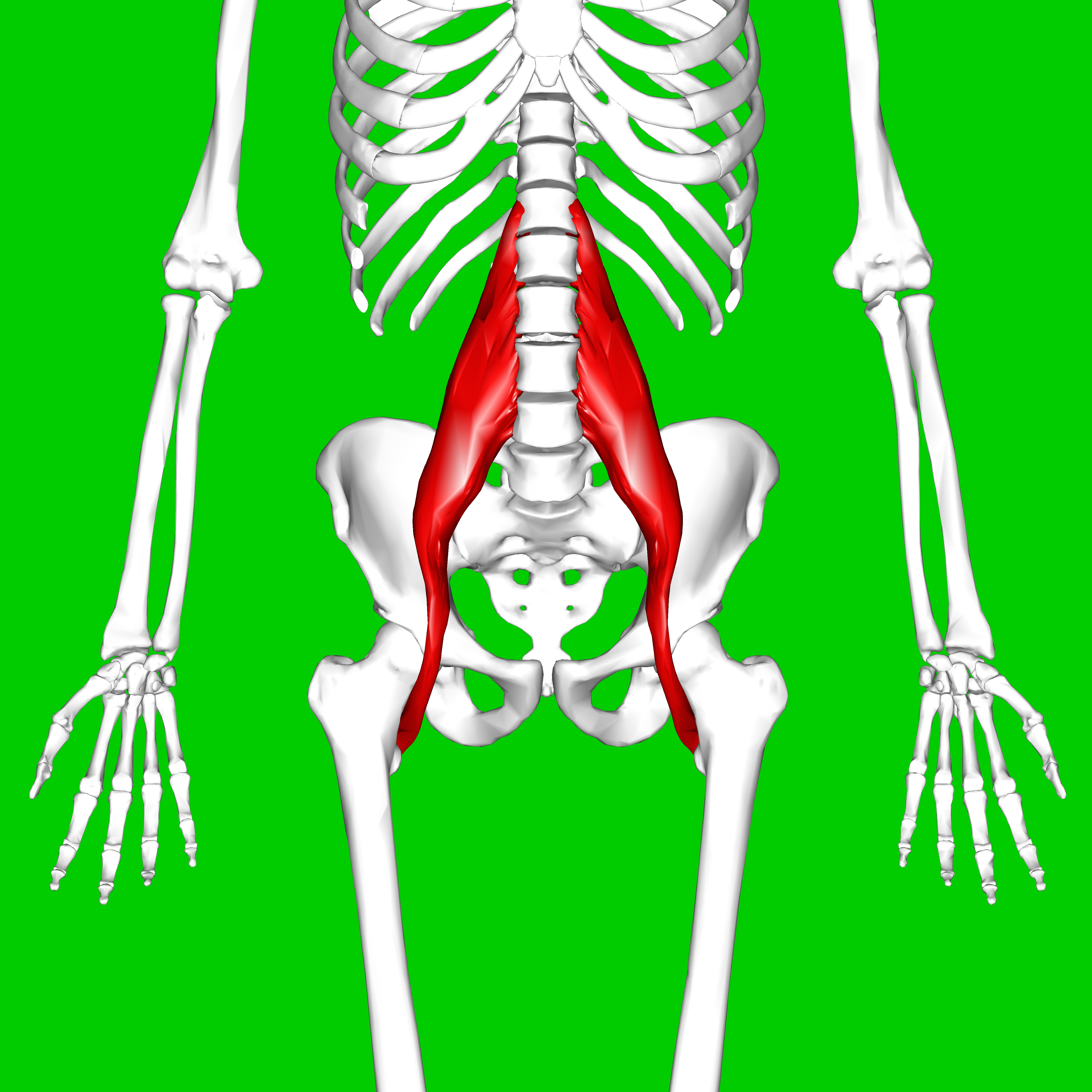
Iliacus
O: upper 2/3 of iliac fossa
In: lesser trochanter with tendon of psoas major
A: flexes thigh
I: femoral nerve

Gluteus Maximus
O: outer surface of ilium, adjacent posterior surface of sacrum + coccyx
In: iliotibial tract; gluteal tuberosity
A: abducts, extends, and laterally rotates thigh
I: inferior gluteal nerve

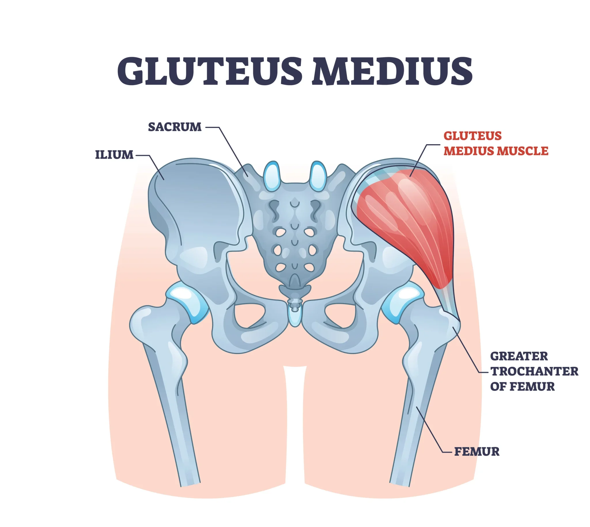
Gluteus Medius
O: outer surface of ilium inferior to iliac crest
In: lateral surface of greater trochanter
A: abducts and medially rotates thigh
I: superior gluteal nerve
Gluteus minimus
O: outer surface of ilium between anterior and inferior gluteal line
In: anterior surface of greater trochanter
A: abducts and medially rotates thigh
I: superior gluteal nerve

Tensor Fascia Latae
O: outer lip of iliac crest between ASIS and iliac tubercle
In: upper part of iliotibial tract
A: abducts + flexes thigh
I: superior gluteal nerve

Piriformis
O: internal surface of sacrum
In: upper border of greater trochanter
A: laterally rotates + abducts thigh
I: 1st and 2nd sacral nerve

Obturator Internus
O: internal pelvic surface of obturator membrane
In: common tendon w/ superior + inferior gemelli to medial surface of greater trochanter
A: laterally rotates thigh + stabilize femoral head
I: nerve to obturator internus

Gemellus Superior
O: spine of ischium
In: with the tendon of obturator internus into upper border of greater trochanter
A: laterally rotates thigh + stabilizes hip
I: branch of nerve to obturator internus from sacral plexus

Gemellus Inferior
O: upper margin of ischial tuberosity
In: w/ tendon of obturator internus into upper border of greater trochanter
A: laterally rotates thigh + stabilize femoral head
I: branch of nerve to quadratus femoris from sacral plexus

Quadratus Femoris
O: lateral border of ischial tuberosity
In: below intertrochanteric crest
A: laterally rotates thigh
I: nerve to quadratus femoris

Obturator Externus
O: outer surface of inferior + superior rami of pubis, ramus of ischium
In: trochanteric fossa of femur
A: lateral thigh
I: obturator nerve

Sartorius
(crosses hip to knee)
(longest muscle in body, called “tailor’s muscle”)
O: ASIS (anterior superior iliac spine)
In: upper part of medial surface of shaft of tibia
A: flexes, abducts, external rotation of femur; slight internal rotation of leg at knee joint after flexion
I: femoral nerve

Rectus Femoris
(crosses hip and knee)
O: AIIS (anterior interior iliac spine)
In: tibial tuberosity via patellar tendon
A: flexes thigh, extends leg
I: femoral nerve

Vastus Lateralis
O: Intertrochanteric line
In: tibial tuberosity via patellar tendon
A: extends leg
I: femoral nerve

Vastus Medialis
O: intertrochanteric line
In: tibial tuberosity via patellar tendon
A: extends leg
I: femoral nerve

Vastus Intermedius
O: anterior and lateral 2/3 of femur
In: tibial tuberosity via patellar tendon
A: extends leg
I: femoral nerve

Gracilis
O: pubic ramus
In: upper, medial shaft of tibia
A: adducts thigh, also flexes knee + medially rotates leg when knee is flexed
I: obturator nerve

Pectineus
O: superior ramus of pubis
In: lesser trochanter
A: flexes thigh, assists in adduction when hip flexed
I: femoral nerve (some people receive contribution from obturator nerve)

Adductor Longus
O: anterior pubis
In: linea aspera
A: adducts thigh, assists w/ hip flexion + medial rotation
I: obturator nerve

Adductor Brevis
(deep to adductor longus)
O: inferior ramus of pubis
In: linea aspera
A: adducts thigh
I: obturator nerve

Adductor Magnus
O: inferior ramus of pubis
In: linea aspera, adductor tubercle of femur
A: adducts + extends thigh
I: obturator nerve

Biceps Femoris
O: Long Head: ischial tuberosity - Short Head: linea aspera
In: head of fibula
A: flexes knee; Long head: extends thigh
I: Long Head: tibial nerve; Short: common peroneal nerve

Semitendinosus
O: ischial tuberosity
In: medial, superior tibia
A: flexes knee, extends thigh
I: tibial nerve

Semimembranosus
O: ischial tuberosity
In: posterior, superior thigh
A: flexes knee, extends thigh
I: tibial nerve

anterior thigh muscles
extends knee
medial thigh muscles
adducts thigh
posterior thigh muscles
flexes knee, extends hip
Lower Leg - Anterior Compartment
all assist dorsiflexion
innervated by deep peroneal/fibular nerve
Tibialis Anterior (anterior compartment)
tendon passes over ankle
O: lateral condyle of tibia, upper ½ of lateral surface of tibia, interosseous membrane
In: medial and plantar surface of medial cuneiform, base of 1st metatarsal
A: dorsiflexion, inversion of the foot
I: deep peroneal

Extensor Hallucis Longus (anterior compartment)
thinner + lies deeper than tibialis anterior
crosses to great toe
O: middle half of anterior fibula, interosseous membrane
In: base of distal phalanx of great toe
A: extends + hyperextends great toe, dorsiflexion + inverts foot
I: deep peroneal

Extensor Digitorum Longus (anterior compartment)
O: upper 2/3 of anterior fibula, interosseous membrane, lateral condyle of TIBIA
In: dorsal surface of lateral 4 toes, base of middle and distal phalanges
A: extends toes 2-5, dorsiflexion an eversion of foot
I: deep peroneal

Peroneus (fibularis) tertius [anterior compartment]
O: lower 1/3 of fibula; interosseous membrane
In: dorsal surface of the base of the 5th metatarsal
A: dorsiflexion and eversion of foot
I: deep peroneal

Lower Leg - Superficial Posterior Compartment
all contribute to plantarflexion
innervated by tibial nerve
Gastrocnemius (superficial posterior compartment)
O: Lateral Head: lateral condyle and posterior surface of femur; Medial Head: popliteal surface of femur above medial condyle
In: posterior surface of calcaneus through the Achilles tendon
A: plantarflexion of foot, flexes leg at knee
I: tibial nerve

Soleus (superficial posterior compartment)
lies deep to gastrocnemius
does NOT cross knee\
primary for plantarflexion when knee is flexed (seated)
O: posterior surface of tibia, upper 2/3 of posterior surface of the fibula, fibrous arch between tibia + fibula
In: posterior surface of calcaneus via the Achilles tendon
A: plantarflexion of foot
I: tibial nerve

Plantaris (superficial posterior compartment)
O: lateral supracondylar ridge of femur, oblique popliteal ligament
In: posterior surface of calcaneus
A: plantarflexion of foot; flexes leg at knee (weakly assists both)
I: tibial nerve

Lower Leg - (deep posterior compartment)
all receive the tibial nerve
Popliteus (deep posterior compartment)
O: lateral surface of the lateral condyle of the femur
In: upper part of the posterior surface of the tibia
A: “unlocks,” medial rotation of leg, flexion of the leg, also helps stabilize knee
I: tibial nerve

Flexor Hallucis Longus (deep posterior compartment)
O: lower 2/3 of posterior surface of the shaft of the fibula, posterior intermuscular septum, interosseous membrane
In: base of distal phalanx of the great toe
A: flexion of great toe (distal phalanx), assist PF and inversion of foot
I: tibial nerve

Flexor Digitorum Longus (deep posterior compartment)
O: medial part of the posterior surface of the tibia
In: base of 2-5 distal phalanges
A: flexion of 2-5 distal phalanges, assist PF and inversion of foot
I: tibial nerve

Tibialis Posterior (deep posterior compartment)
O: lateral part of the posterior surface of the tibia, proximal ½ of the posterior surface of the fibula, interosseous membrane
In: navicular tuberosity, cuboid, cuneiforms, 2-4 metatarsals, sustentaculum tali
A: plantarflexion + inversion of foot; supports the medial longitudinal arch
I: tibial nerve

Lower Leg - Lateral Compartment
plantarflexion + eversion
superficial peroneal nerve
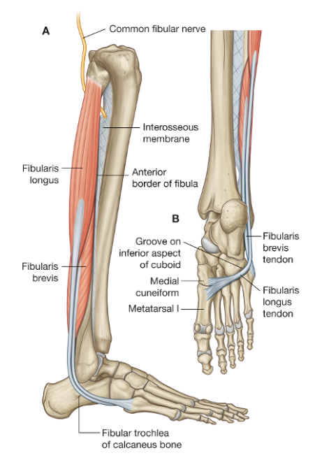
Peroneus (fibularis) longus [lateral compartment]
O: upper 2/3 of lateral surface of the fibula
In: lateral surface medial cuneiform, base of 1st metatarsal
A: plantarflexion + eversion of foot
I: superficial peroneal nerve
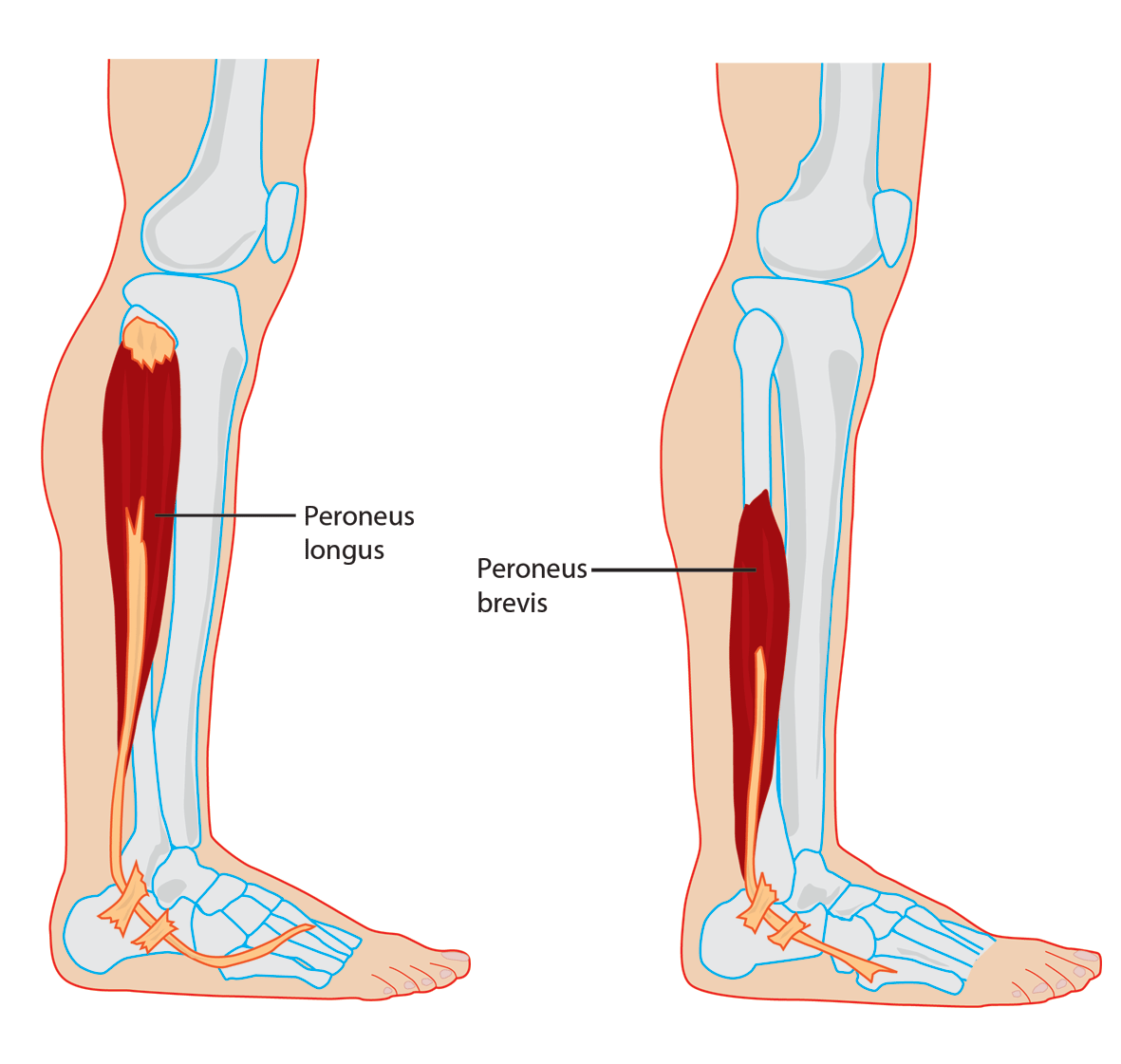
Peroneus (fibularis) Brevis [lateral compartment]
stays lateral
O: lower 2/3 of lateral surface of the fibula
In: lateral side of the base of the 5th metatarsal
A: plantarflexion + eversion of foot
I: superficial peroneal nerve
Extensor Digitorum Brevis
O: anterior + lateral surface of calcaneus; lateral talocalcaneal ligament, inferior extensor retinaculum
In: extensor digitorum longus tendons + extensor expansions of toes 2-4 (some muscles attach to other muscle’s tendons
A: extension of 2-4 toes
I: deep peroneal nerve

Abductor Hallucis
O: tuberosity of the calcaneus, flexor retinaculum and plantar aponeurosis
In: medial side of base of 1st proximal phalanx
A: abducts and stabilizes the great toe, flexion at MT joint
I: medial plantar nerve

Flexor Digitorum Brevis
O: tuberosity of the calcaneus; plantar aponeurosis
In: sides of middle phalanges of toes 2-5
A: flexes proximal interphalangeal (PIP) joints of toes 2-5; assists metatarsophalangeal (MTP) joint flexion
I: medial plantar nerve

Abductor Digiti Minimi
O: tuberosity of the calcaneus; plantar aponeurosis
In: lateral side of 5th proximal phalanx
A: abducts 5th toe, assists flexion a MTP joint
I: lateral plantar nerve

Lumbricales
O: tendons of flexor digitorum longus
In: medial sides of extensor expansions of toes 2-5
A: flexes MTP joints and extends PIP joints of toes 2-5 (think of hand lumbricales; bend at knuckle, keep fingers straight)
I: 1st lumbrical (most medial) - medial plantar nerve; 2-4 (lateral 3) - lateral plantar nerve

Flexor Hallucis Brevis
O: cuboid, lateral cuneiform
In: medial + lateral sides of the base of 1st proximal phalanx
A: flex proximal phalanx of great toe (MTP joint)
I: medial plantar nerve

Adductor Hallucis
O: Oblique head: 2-4 metatarsal bones, sheath of peroneus longus tendon; Transverse head: plantar metatarsophalangeal ligaments of 3, 4, 5. transverse metatarsal ligament
In: lateral side of base of 1st proximal phalanx
A: adducts and stabilizes the great toe
I: lateral plantar nerve

Flexor Digiti Minimi Brevis
O: Base of 5th metatarsal; sheath of peroneus longus tendon
In: lateral sides of the base of the 5th proximal phalanx
A: flex proximal phalanx of 5th toe
I: lateral plantar nerve

Dorsal Interossei
DAB - dorsal
ABduct
O: adjacent sides of metatarsal bones
In: 1st - medial side of proximal phalanx of 2nd toe; 2-4: lateral sides of proximal phalanges of 2-4 toes
A: abduct toes, flex proximal phalanges
I: lateral plantar nerve

Plantar Interossei
PAD - plantar ADduct
O: medial aspect of metatarsals 3-5
In: proximal phalanges 3-5
A: adduct toes, flex proximal phalanges
I: lateral planter nerve
