CB. 16 The cell cycle and division

0.0(0)
Studied by 0 people
call kaiCall Kai
learnLearn
examPractice Test
spaced repetitionSpaced Repetition
heart puzzleMatch
flashcardsFlashcards
GameKnowt Play
Card Sorting

1/29

flashcard set

Earn XP

Description and Tags

To identify the phases of the cell cycle, and their purpose To describe the process of cell division (mitosis) To outline the role of cytoskeletal molecules in cell division To outline the purpose of G and S phases of the cell cycle To describe the regulation of the cell cycle by cyclin dependent kinases (CDKs) To explain the cell cycle control system and the role of checkpoints

Last updated 9:25 AM on 10/14/26
Name
Mastery
Learn
Test
Matching
Spaced
Call with Kai
Chat

No analytics yet

Send a link to your students to track their progress

30 Terms

1
New cards

What is the cell cycle? 

  • The cell cycle is the coordinated process through which a cell grows, duplicates its contents and divides to produce two daughter cells. 

  • Essential for growth/development/repair/tissue maintenance and cell renewal e.g. oral epithelium as it loses cells.  


<ul><li><p class="Paragraph SCXW93574407 BCX0" style="text-align: left;"><span style="line-height: 16.2167px;">The cell cycle is the coordinated process through which a cell grows, <strong>duplicates </strong>its contents and divides to <strong>produce two daughter cells.</strong>&nbsp;</span></p></li></ul><ul><li><p class="Paragraph SCXW93574407 BCX0" style="text-align: left;"><span style="line-height: 16.2167px;">Essential for growth/development/repair/tissue maintenance and cell renewal e.g. oral epithelium as it loses cells.&nbsp;&nbsp;</span></p></li></ul><p></p>
2
New cards

How is the cell cycle divided?  

  • Two main stages Interphase + Mitotic (M) phase  

  • Within interphase: G1, S, and G2.  

  • Then mitotic (M) phase is followed by cytokinesis.  


<ul><li><p class="Paragraph SCXW185508431 BCX0" style="text-align: left;"><span style="line-height: 16.2167px;">Two main stages <strong>Interphase</strong> + <strong>Mitotic (M) phase</strong>&nbsp;&nbsp;</span></p></li></ul><ul><li><p class="Paragraph SCXW185508431 BCX0" style="text-align: left;"><span style="line-height: 16.2167px;">Within interphase: G1, S, and G2.&nbsp;&nbsp;</span></p></li></ul><ul><li><p class="Paragraph SCXW185508431 BCX0" style="text-align: left;"><span style="line-height: 16.2167px;">Then mitotic (M) phase is followed by cytokinesis.&nbsp;&nbsp;</span></p></li></ul><p></p>
3
New cards

What are the 3 stages of Interphase: 

  1. Interphase G1 – Cell GROWs and preparation = synthesises proteins and organelles needed for DNA replication.  

  1. Interphase S – Cells DUPLICATE its genome: DNA synthesis and replication.  

  1. Interphase G2 - cells GROW and prep for cell division  
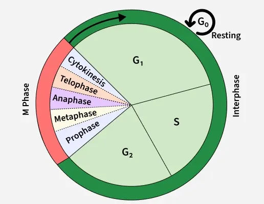

<ol><li><p class="Paragraph SCXW124022708 BCX0" style="text-align: left;"><span style="line-height: 16.2167px;"><strong>Interphase G1</strong> – Cell GROWs and preparation = synthesises proteins and organelles needed for DNA replication.&nbsp;&nbsp;</span></p></li></ol><ol start="2"><li><p class="Paragraph SCXW124022708 BCX0" style="text-align: left;"><span style="line-height: 16.2167px;"><strong>Interphase S</strong> – Cells DUPLICATE its genome: DNA synthesis and replication.&nbsp;&nbsp;</span></p></li></ol><ol start="3"><li><p class="Paragraph SCXW124022708 BCX0" style="text-align: left;"><span style="line-height: 16.2167px;"><strong>Interphase G2</strong> - cells GROW and prep for cell division&nbsp;&nbsp;</span></p></li></ol><p></p>
4
New cards

What processes are occurring in G1?  

  • G1 is the first growth phase in interphase.  

  • The cell prepares for DNA synthesis occurring in S phase.  

  • In G1: cell increases in size, synthesises more proteins/cellular components, respond to signals from environment. Assess whether conditions are suitable for DNA replication.  


<ul><li><p class="Paragraph SCXW235206008 BCX0" style="text-align: left;"><span style="line-height: 16.2167px;">G1 is the first growth phase in interphase.&nbsp;&nbsp;</span></p></li></ul><ul><li><p class="Paragraph SCXW235206008 BCX0" style="text-align: left;"><span style="line-height: 16.2167px;">The cell prepares for DNA synthesis occurring in S phase.&nbsp;&nbsp;</span></p></li></ul><ul><li><p class="Paragraph SCXW235206008 BCX0" style="text-align: left;"><span style="line-height: 16.2167px;">In G1: cell increases in size, synthesises more proteins/cellular components, respond to signals from environment. Assess whether conditions are suitable for DNA replication.&nbsp;&nbsp;</span></p></li></ul><p></p>
5
New cards

What processes are occurring in S? 

  • Second stage of interphase (G1, S, G2)  

  • The cell duplicates its DNA. Each chromosome now consists of two identical copies called sister chromatids, which remain joined together, particularly at the centromere region. 


6
New cards

What processes are occurring in G2?  

  • Final stage of interphase (G1, S, G2) 

  • Post DNA synthesis.

  • Cell prepares to enter mitosis. Cell grows, produces more proteins, checks DNA replication was accurate.


7
New cards

What is G0? E.g. neurons vs immune cells.  

  • G0 is not simply another stage that every dividing cell must pass through. It is an alternative state outside the main G1–S–G2–M sequence. 

  • Some cells enter G0 temporarily/remain there for a long time --> Then re-enter the cell cycle when they receive appropriate signals.  

  • E.g. Neurons remain outside the active cell cycle. E.g. immune cells can be activated and proliferate when required.  


<ul><li><p class="Paragraph SCXW206544160 BCX0" style="text-align: left;"><span style="line-height: 16.2167px;">G0 is not simply another stage that every dividing cell must pass through. It is an alternative state outside the main G1–S–G2–M sequence.&nbsp;</span></p></li></ul><ul><li><p class="Paragraph SCXW206544160 BCX0" style="text-align: left;"><span style="line-height: 16.2167px;">Some cells enter G0 temporarily/remain there for a long time --&gt; Then re-enter the cell cycle when they receive appropriate signals.&nbsp;&nbsp;</span></p></li></ul><ul><li><p class="Paragraph SCXW206544160 BCX0" style="text-align: left;"><span style="line-height: 16.2167px;">E.g. Neurons remain outside the active cell cycle. E.g. immune cells can be activated and proliferate when required.&nbsp;&nbsp;</span></p></li></ul><p></p>
8
New cards

What is mitosis and what are the 5 stages? 

  • Mitosis is the division of one cell into 2x identical daughter cells.  

  • Five stages (prophase, prometaphase, metaphase, anaphase, telophase) followed by cytokinesis.  

  • PPMAT 


<ul><li><p class="Paragraph SCXW64391147 BCX0" style="text-align: left;"><span style="line-height: 16.2167px;">Mitosis is the division of one cell into <strong>2x </strong>identical <strong>daughter cells</strong>.&nbsp;&nbsp;</span></p></li></ul><ul><li><p class="Paragraph SCXW64391147 BCX0" style="text-align: left;"><span style="line-height: 16.2167px;">Five stages (<strong>prophase, prometaphase, metaphase, anaphase, telophase</strong>) followed by <strong>cytokinesis.&nbsp;</strong>&nbsp;</span></p></li></ul><ul><li><p class="Paragraph SCXW64391147 BCX0" style="text-align: left;"><span style="line-height: 16.2167px;"><strong>PPMAT</strong>&nbsp;</span></p></li></ul><p></p>
9
New cards

In Prophase how does the cell begin and end? What processes occur to achieve this?  

  • Beginning: Each chromosome = consists of 2x identical chromatids joined by a centromere.  

Prophase process:  

  • DNA is condensed – chromatin condenses the chromosomes.  

  • Mitotic spindles (made from microtubules) form and the centrioles migrate to opposite poles of the cell.  

  • Nucleolus gradually disintegrates – end of prophase.


<ul><li><p class="Paragraph SCXW87629425 BCX0" style="text-align: left;"><span style="line-height: 16.2167px;">Beginning: Each chromosome = consists of 2x identical chromatids joined by a centromere.&nbsp;&nbsp;</span></p></li></ul><p class="Paragraph SCXW87629425 BCX0" style="text-align: left;"><span style="line-height: 16.2167px;">Prophase process:&nbsp;&nbsp;</span></p><ul><li><p class="Paragraph SCXW87629425 BCX0" style="text-align: left;"><span style="line-height: 16.2167px;">DNA is <strong>condensed</strong> – chromatin condenses the chromosomes.&nbsp;&nbsp;</span></p></li></ul><ul><li><p class="Paragraph SCXW87629425 BCX0" style="text-align: left;"><span style="line-height: 16.2167px;"><strong>Mitotic spindles</strong> (made from <strong>microtubules</strong>) form and the <strong>centrioles</strong> migrate to opposite poles of the cell.&nbsp;&nbsp;</span></p></li></ul><ul><li><p class="Paragraph SCXW87629425 BCX0" style="text-align: left;"><span style="line-height: 16.2167px;">Nucleolus gradually disintegrates – end of prophase.</span></p></li></ul><p></p>
10
New cards

In Prometaphase how does the cell begin and end? What processes occur to achieve this? 

  • Start – break down of nuclear envelope.  

  • Mitotic spindles attach to the kinetochores (region on centromeres).


<ul><li><p class="Paragraph SCXW236626312 BCX0" style="text-align: left;"><span style="line-height: 16.2167px;">Start – break down of nuclear envelope.&nbsp;&nbsp;</span></p></li></ul><ul><li><p class="Paragraph SCXW236626312 BCX0" style="text-align: left;"><span style="line-height: 16.2167px;">Mitotic spindles attach to the kinetochores (region on centromeres).</span></p></li></ul><p></p>
11
New cards

In Metaphase how does the cell begin and end? What processes occur to achieve this? 

  • Centromeres of sister chromatids line up at the equator of the cell.  (Metaophase-middle).

  • The cells check the chromosomes are properly attached to the spindle before allowing separation.  


<ul><li><p class="Paragraph SCXW138806945 BCX0" style="text-align: left;"><span style="line-height: 16.2167px;">Centromeres of sister chromatids line up at the equator of the cell<strong>. </strong>&nbsp;<em>(Metaophase-middle). </em></span></p></li></ul><ul><li><p class="Paragraph SCXW138806945 BCX0" style="text-align: left;"><span style="line-height: 16.2167px;">The cells check the chromosomes are properly attached to the spindle before allowing separation.&nbsp;&nbsp;</span></p></li></ul><p></p>
12
New cards

In Anaphase how does the cell begin and end? What processes occur to achieve this? 

  • Two sister chromatids split towards the pole cause by shortening of the microtubules.  

  • Once separated each chromatid is considered its own individual chromosome.  


<ul><li><p class="Paragraph SCXW30701605 BCX0" style="text-align: left;"><span style="line-height: 16.2167px;">Two sister chromatids split towards the pole cause by <strong>shortening of the microtubules</strong>. &nbsp;</span></p></li></ul><ul><li><p class="Paragraph SCXW30701605 BCX0" style="text-align: left;"><span style="line-height: 16.2167px;"><strong>Once separated each chromatid is considered its own individual chromosome.&nbsp;</strong>&nbsp;</span></p></li></ul><p></p>
13
New cards

In Telophase how does the cell begin and end? What processes occur to achieve this? 

  • Chromosomes reach opposite poles and the Nuclear envelope reforms. Chromosomes de condense (reverse of prophase). 

  • End of telophase marked by daughter chromosomes being at the poles.


<ul><li><p class="Paragraph SCXW87294471 BCX0" style="text-align: left;"><span style="line-height: 16.2167px;">Chromosomes reach opposite poles and the Nuclear envelope reforms. Chromosomes de condense (reverse of prophase).&nbsp;</span></p></li></ul><ul><li><p class="Paragraph SCXW87294471 BCX0" style="text-align: left;"><span style="line-height: 16.2167px;">End of telophase marked by<strong> daughter chromosomes being at the poles.</strong></span></p></li></ul><p></p>
14
New cards

What happens to sister chromatids during anaphase, and how many chromosomes does each daughter nucleus receive in a typical human somatic cell? 

Sister chromatids separate and move to opposite poles. Each daughter nucleus receives 46 chromosomes, ensuring both daughter cells have a complete genome. 

15
New cards

What occurs in Cytokinesis? 

Cytokinesis follows mitosis. Cell divides through a contractile ring made primarily of actin and myosin --> producing two daughter cells. 

16
New cards

How does the contractile ring produce two daughter cells? 

Actin filaments assemble into a ring beneath the plasma membrane. Myosin motor proteins interact with the actin filaments and generate contractile force. The ring tightens, producing an indentation called the cleavage furrow. The furrow deepens until the cell separates into two daughter cells. 

<p><span style="line-height: 16.2167px;">Actin filaments assemble into a ring beneath the plasma membrane. Myosin motor proteins interact with the actin filaments and generate contractile force. The ring tightens, producing an indentation called the cleavage furrow. The furrow deepens until the cell separates into two daughter cells.&nbsp;</span></p>
17
New cards

How are microtubules used in cell division?  

  • Microtubules form the mitotic spindle, which is organised around the spindle poles associated with centrosomes. Spindle microtubules attach to kinetochores on chromosomes. 

  • The spindle ensures that chromosomes move towards opposite poles. 


<ul><li><p class="Paragraph SCXW110060463 BCX0" style="text-align: left;"><span style="line-height: 16.2167px;">Microtubules form the mitotic spindle, which is organised around the spindle poles associated with centrosomes. Spindle microtubules attach to kinetochores on chromosomes.&nbsp;</span></p></li></ul><ul><li><p class="Paragraph SCXW110060463 BCX0" style="text-align: left;"><span style="line-height: 16.2167px;">The spindle ensures that chromosomes move towards opposite poles.&nbsp;</span></p></li></ul><p></p>
18
New cards

How is the actin-myosin contractile ring used in cell division? 

Actin forms filaments. Myosin generates force through its interaction with actin.The ring constricts the cell to complete cytokinesis. 

19
New cards

How is the cell cycle regulated/controlled? 

Cell cycle is controlled by cyclins and cyclin-dependent kinases (CDKs). 

20
New cards

What are cyclins and cyclin-dependent kinases (CDKs)? 

  • Cyclins = regulatory proteins. The cyclin concentrations rise and fall during the cell cycle. Different cyclins are associated with different stages and help determine when particular processes should occur. 

  • CDKs = enzymes. Cyclin-dependent kinases are protein kinases. When activated, they phosphorylate target proteins involved in cell-cycle progression. 

  • Cyclin–CDK complex = active regulator Cyclin binding activates the corresponding CDK, subject to additional regulatory mechanisms. The activated complex modifies target proteins to help initiate or coordinate specific cell-cycle events. 


<ul><li><p class="Paragraph SCXW54021940 BCX0" style="text-align: left;"><span style="line-height: 16.2167px;">Cyclins = regulatory proteins. The cyclin concentrations rise and fall during the cell cycle. Different cyclins are associated with different stages and help determine when particular processes should occur.&nbsp;</span></p></li></ul><ul><li><p class="Paragraph SCXW54021940 BCX0" style="text-align: left;"><span style="line-height: 16.2167px;">CDKs = enzymes. Cyclin-dependent kinases are protein kinases. When activated, they phosphorylate target proteins involved in cell-cycle progression.&nbsp;</span></p></li></ul><ul><li><p class="Paragraph SCXW54021940 BCX0" style="text-align: left;"><span style="line-height: 16.2167px;">Cyclin–CDK complex = active regulator Cyclin binding activates the corresponding CDK, subject to additional regulatory mechanisms. The activated complex modifies target proteins to help initiate or coordinate specific cell-cycle events.&nbsp;</span></p></li></ul><p></p>
21
New cards

How do cyclin-dependent kinases utilise phosphorylation?  

  • Phosphorylation = addition of a phosphate group to a molecule. 

  • CDKs transfer phosphate groups from ATP to target proteins. This can change a protein's activity (activate it or deactivate it), interactions or location. 

  • For example, phosphorylation of particular target proteins can help initiate DNA replication or promote the events of mitosis. 


<ul><li><p class="Paragraph SCXW89624521 BCX0" style="text-align: left;"><span style="line-height: 16.2167px;">Phosphorylation = addition of a phosphate group to a molecule.&nbsp;</span></p></li></ul><ul><li><p class="Paragraph SCXW89624521 BCX0" style="text-align: left;"><span style="line-height: 16.2167px;">CDKs transfer phosphate groups from ATP to target proteins. This can change a<strong> protein's activity (activate it or deactivate i</strong>t), <strong>interactions </strong>or <strong>location</strong>.&nbsp;</span></p></li></ul><ul><li><p class="Paragraph SCXW89624521 BCX0" style="text-align: left;"><span style="line-height: 16.2167px;">For example, phosphorylation of particular target proteins can help initiate DNA replication or promote the events of mitosis.&nbsp;</span></p></li></ul><p></p>
22
New cards

How do cyclins and CDKs work together? 

  • Cyclin concentration rises...Then Cyclin binds to its CDK. The cyclin–CDK complex becomes active....The Target proteins are then phosphorylated....So Specific cell-cycle events can be promoted. 

  • Think of the cyclin as a timing signal and the CDK as the enzyme that carries out the regulatory action. 


<ul><li><p class="Paragraph SCXW181743965 BCX0" style="text-align: left;"><span style="line-height: 16.2167px;">Cyclin concentration rises...Then Cyclin binds to its CDK. The cyclin–CDK complex becomes active....The Target proteins are then phosphorylated....So Specific cell-cycle events can be promoted.&nbsp;</span></p></li></ul><ul><li><p class="Paragraph SCXW181743965 BCX0" style="text-align: left;"><span style="line-height: 16.2167px;"><strong>Think of the cyclin as a timing signal and the CDK as the enzyme that carries out the regulatory action.</strong>&nbsp;</span></p></li></ul><p></p>
23
New cards

Why must cyclins be degraded? 

  • Anaphase-promoting complex (APC) help regulate the cell cycle by promoting the degradation of particular proteins. APCs attach ubiquitin to specific target proteins, marking them for degradation by the proteasome. This is vital so certain processes can be stopped helping the cell exit mitosis. 

  • Cyclin degradation is not a random removal of proteins. It is a regulated way of switching off specific cell-cycle activities at the appropriate time. 


24
New cards

What other role do anaphase-promoting complexes serve in anaphase specifically?  

  • The APC/C also promotes the destruction of securin, a protein that restrains separase.  

  • When securin is degraded, separase can cleave cohesin, the protein complex holding sister chromatids together. This allows the chromatids to separate at anaphase. 


<ul><li><p class="Paragraph SCXW12458704 BCX0" style="text-align: left;"><span style="line-height: 16.2167px;">The APC/C also promotes the destruction of securin, a protein that restrains separase. &nbsp;</span></p></li></ul><ul><li><p class="Paragraph SCXW12458704 BCX0" style="text-align: left;"><span style="line-height: 16.2167px;">When securin is degraded, separase can cleave cohesin, the protein complex holding sister chromatids together.<strong> This allows the chromatids to separate at anaphase.</strong>&nbsp;</span></p></li></ul><p></p>
25
New cards

What are cell-cycle checkpoints? 

  • The cell cycle needs more than signals to proceed. It also needs mechanisms to prevent progression when conditions are unsuitable. 

  • A checkpoint is a control point at which cell-cycle progression can be delayed or stopped until a particular set of requirements have been met. 

  • Uses negative regulation: in the form of an inhibitory protein. e.g. p27.  


<ul><li><p class="Paragraph SCXW174679302 BCX0" style="text-align: left;"><span style="line-height: 16.2167px;">The cell cycle <strong>needs more than signals to proceed.</strong> It also needs me<strong>chanisms to prevent progression when conditions are unsuitable.</strong>&nbsp;</span></p></li></ul><ul><li><p class="Paragraph SCXW174679302 BCX0" style="text-align: left;"><span style="line-height: 16.2167px;">A checkpoint is a control point at which cell-cycle progression <strong>can be delayed or stopped until a particular set of requirements have been met.</strong>&nbsp;</span></p></li></ul><ul><li><p class="Paragraph SCXW174679302 BCX0" style="text-align: left;"><span style="line-height: 16.2167px;"><strong>Uses negative regulation: </strong>in the form of an <strong>inhibitory protein. </strong>e.g. p27. &nbsp;</span></p></li></ul><p></p>
26
New cards

What role do the checkpoints in each of these stages serve – G1, G2/M, spindle assembly?  

  • G1 checkpoint- CHECKS Cell size, nutrients, growth signals and DNA integrity. STOPS unsuitable cells from committing to DNA replication. 

  • G2/M checkpoint- CHECKS Whether DNA replication is complete and whether DNA is damaged. STOPS entry into mitosis with incomplete or damaged DNA. 

  • Spindle assembly checkpoint – CHECKS whether chromosomes are correctly attached to spindle microtubules. STOPS unequal chromosome distribution. 
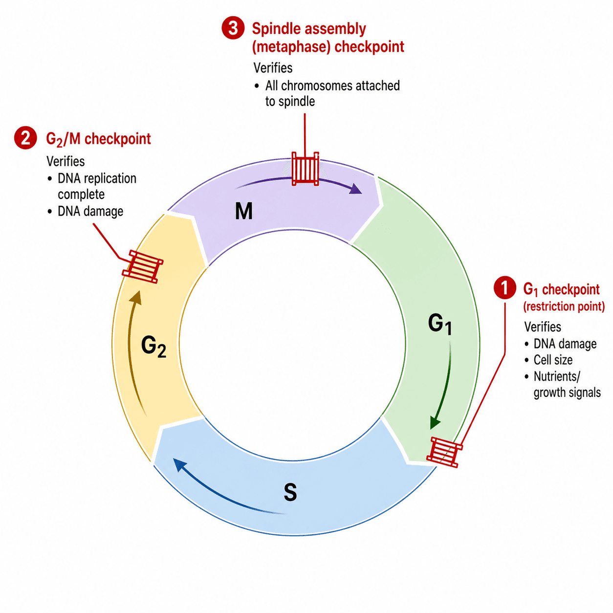

<ul><li><p class="Paragraph SCXW254372297 BCX0" style="text-align: left;"><span style="line-height: 16.2167px;">G1 checkpoint- CHECKS Cell size, nutrients, growth signals and DNA integrity. <strong>STOPS unsuitable cells from committing to DNA replication.</strong>&nbsp;</span></p></li></ul><ul><li><p class="Paragraph SCXW254372297 BCX0" style="text-align: left;"><span style="line-height: 16.2167px;">G2/M checkpoint- CHECKS Whether DNA replication is complete and whether DNA is damaged. <strong>STOPS entry into mitosis with incomplete or damaged DNA.</strong>&nbsp;</span></p></li></ul><ul><li><p class="Paragraph SCXW254372297 BCX0" style="text-align: left;"><span style="line-height: 16.2167px;">Spindle assembly checkpoint – CHECKS whether chromosomes are correctly attached to spindle microtubules. <strong>STOPS unequal chromosome distribution.</strong>&nbsp;</span></p></li></ul><p></p>
27
New cards

How does p27 regulate at a checkpoint? E.g. G1 checkpoint. 

P27 is an inhibitory protein. P27 binds to CDK and inhibits the complex – it stops CDK phosphorylating their target proteins.  

<p><span style="line-height: 16.2167px;">P27 is an inhibitory protein. P27 binds to CDK and inhibits the complex – it stops CDK phosphorylating their target proteins. &nbsp;</span></p>
28
New cards

What is the role of mitogens and growth factors in the cell cycle?  

  • Essentially the opposite of p27.  

  • Mitogens/growth factors stimulate cells to progress through the cell cycle: (1) The cell receives appropriate external signals. (2) Intracellular signalling pathways respond. (3)  Cyclin production or activity changes. (4) Cyclin–CDK complexes promote progression through G1 towards S phase. 


<ul><li><p class="Paragraph SCXW88665758 BCX0" style="text-align: left;"><span style="line-height: 16.2167px;">Essentially the opposite of p27. &nbsp;</span></p></li></ul><ul><li><p class="Paragraph SCXW88665758 BCX0" style="text-align: left;"><span style="line-height: 16.2167px;">Mitogens/growth factors stimulate cells to progress through the cell cycle: (1) The cell receives appropriate external signals. (2) Intracellular signalling pathways respond. (3) &nbsp;Cyclin production or activity changes. (4) Cyclin–CDK complexes promote progression through G1 towards S phase.&nbsp;</span></p></li></ul><p></p>
29
New cards

Why does cell-cycle regulation matter in cancer? How could this be disturbed?

Cell-cycle checkpoints directly correlate with the pathogenesis of cancer: alterations in the genes and proteins controlling cell cycle processes allow abnormal cells to survive and proliferate. E.g.: 

  • Overactive growth signalling: cells may receive excessive signals to divide. 

  • Overactive cyclin–CDK activity: cells may progress through checkpoints too readily. 

  • Loss of inhibitory proteins: cell-cycle brakes may no longer function properly. 

  • Defective DNA-damage responses: cells with damaged DNA may continue dividing. 

  • Defective chromosome segregation: daughter cells may acquire abnormal chromosome numbers. 


30
New cards

Explain the pathophysiology of p53? 

  • P53 is a tumour suppressor protein = NORMALLY = damaged DNA is detected p53 promotes expression p21 a CDK inhibitor. This supresses the cyclin-CDK activity and cell cycle is HALTED, if the DNA damage can't be fixed the causes CELL DEATH.  

  • BUT in cancer = p53 disrupted. Damaged cells continue to proliferate.



