Skeletal Muscle Biology Exam 1
 Call Kai
 Learn
 Practice Test
 Spaced Repetition
 Match
 Flashcards
 Knowt Play
1/71

There's no tags or description
Looks like no tags are added yet.
72 Terms
Importance of skeletal muscle (Basic level)
Body movements/locomotion
Daily living activities
Exercise
Posture/stability
Protection/support
Metabolism and heat production (thermogenesis)
Importance of skeletal muscle (Complex level)
Energy storage and supply (stores glycogen and amino acids)
Metabolic regulation (major site of glucose uptake in response to insulin)
Amino acid reservoir (in catabolism, skeletal muscle provides AA for gluconeogenesis and protein synthesis)
Major site of lipid metabolism (fatty acid uptake and oxidation)
Venous return (muscle pump acting during contraction helping return blood to heart- especially calf muscles)
Economic burden of low muscle mass
Increased strain on healthcare system
Increased financial strains for patients
Polypharmacy
Future challenges involving muscle research
Aging societies: Sarcopenia
Chronic diseases and metabolism: Type 2 diabetes, obesity, cancer cachexia, etc.
Neurodegeneration and brain-muscle crosstalk: Therapies for Alzheimer’s and Parkinson’s
Space: Muscle atrophy induced by microgravity
Greenhouse gas x global food insecurity: Burden on meat production
Artificial meat
Myosin and its properties
Superfamily of motor proteins
Actin binding
ATPase
Force transduction
Evolutionary precursors to true muscle cells (sponges, cnidarians, and bilaterians)

Evolution of contractile apparatus until modern muscle
Muscle-like contractile cells evolved in simple animals - sponges (lack nervous system
Nervous systems evolved later to coordinate muscle activity - cnidarians (jellyfish)
Genetic evidence shows that genes that control muscle contraction (actin/myosin) are ancient
Muscle-like tissues have evolved multiple times in early animals, possibly in cnidarians and bilaterians separately
General features of a mature muscle cell (myofiber)
Multinucleated: A single myofiber contains hundreds of myonuclei- comes from fusion of myoblasts from embryonic development
Synctial cells: A large cell-like structure formed by the joining of two or more cells. One skeletal muscle fiber is a single multinucleated synctium. Myonuclei are located in periphery just beneath sarcolemma.
Striated: Myofibrils (and sarcomeres), organization of myosin and actin filaments make striations visible under light microscopy.
Sarcolemma
The cell membrane
Has invaginations called transversal tubes, or T-tubules. Important to conduct action potentials.
Covered by the basement membrane (basal lamina)
Basal lamina
Outside sarcolemma
Provides protection to each muscle fiber and anchors muscle fibers to the connective tissue (ECM)
Myonucleus
Post-mitotic and terminally differentiated (incapable of proliferation)
Right beneath sarcolemma, residing in periphery of muscle cells (except when myofibers are regenerating, when they can be found in the center of myofiber)
Myonuclear domain theory
Each myonucleus in a skeletal muscle fiber governs a fixed volume of cytoplasm
Satellite cell
Myoblast that resides between basal lamina and sarcolemma (outside actual myofiber)
In mature organisms, SCs are in resting and will wake up during injury.
Once activated, SC can divide and one fuses with damaged myofiber while other replenishes SC pool.
Identified by Pax7+ staining (txn factor)
Sarcoplasm
Primarily occupied by myofibrils
Contains sarcoplasmic reticulum, mitochondria, glycogen and lipid droplets, and myoglobins
Myofibrils
Long rod-like bundles of contractile proteins
Tandem repeats of sarcomeres
Gives striated appearance
Connective tissues
Epimysium: “above”
Perimysium: “around”
Endomysium: “within”
Sarcomeres
Myofibrils (sarcomeres) are composed of actin and myosin filaments
Each myofiber contains 100-1000 myofibrils
Sarcomere anatomy

Muscle fiber types
Type I: slow fibers, “red”
Type II: fast fibers, “white”
Type IIA
Type IIB (only rodents)
Type IIX
Type I Fibers
Slow fibers, red muscle
Rely more on oxidative metabolism
Smaller CSA
More extensive blood vessel system and more capillaries to supply extra amount of oxygen
More mitochondria, high levels of oxidative metabolism
Large amounts of myoglobin
Type II Fibers
Fast fibers, white muscle
Rely more on glycolysis
Larger CSA
Extensive sarcoplasmic reticulum for rapid release of Ca ions to initiate contraction
Larger amounts of glycolytic enzymes and fewer mitochondria than slow fibers
Less extensive blood supply
Less myoglobin
Hematoxylin and Eosin staining
Hematoxylin: stains cell nuclei blue/purple. Basic dye (+) binds to acidic (-) chromatin
Eosin: stains cytoplasm pink. Acidic dye binds to positively charged proteins
Myofibril protein composition
70% of myofiber volume is occupied by myofibrils
80% of its dry weight is protein
50% of that is myofibrillar protein
Sarcoplasmic reticulum
Stores, releases, and reabsorbs Ca2+ ions, which enables control of muscle contraction and relaxation
Oxidative vs glycolytic metabolism
Glycolytic: rapid, oxygen-independent (anaerobic) breakdown of glycose in cytosol to produce small amount ATP. (quick ATP for short, intense efforts like sprinting and heavy lifting)
Oxidative: aerobic, mitochondria based process that yields far more ATP (sustains prolonged, moderate-intensity activity like endurance training)
Motor unit
Consists of a motor neuron and the group of skeletal muscle fibers it innervates. A single motor axon may branch to innervate several muscle fibers that function together as a group. Although each muscle fiber is innervated by a single motor neuron, an entire muscle may receive input from hundreds of different motor neurons.
Neuromuscular junction (NMJ)
Nerve ending + myofiber
Myonuclei neighboring the NMJ are specialized nuclei, called fundamental myonuclei (express AChE - acetylcholinesterase)
Transverse tubules + Sarcoplasmic reticulum role in muscle contraction
Transmission of action potentials flows along t tubules, which causes release of Ca+ inside myofiber close to the myofibrils, causing contraction.
SR contains excess Ca+ which are released when an AP occurs in adjacent T tubule
General mechanism of muscle contraction
AP travels along motor nerve to its endings on muscle fibers
At each ending, nerve secretes neurotransmitter acetylcholine
Acetylcholine acts on a local area of the muscle fiber membrane to open acetylcholine-gated cation channels in membrane.
Opening of channels allows large quantities of sodium ions to diffuse into interior of muscle fiber membrane, causing local depolarization. This leads to opening of voltage-gated sodium channels, which initiates AP at the membrane
AP travels along muscle fiber membrane in the same way AP travels along nerve fiber membrane
AP depolarizes the muscle membrane, and the AP electricity flows thru center of the muscle fiber. This causes sarcoplasmic reticulum to release large quantities of calcium ions
Calcium ions initiate attractive forces between the actin and myosin filaments, causing them to slide alongside each other, which is a contractile process
Ca ions are pumped back into the sarcoplasmic reticulum by a Ca2+ membrane pump and remain stored in the reticulum until a new muscle AP comes along
Sarcomeric proteins
Actinin: binds actin to Z-lines
Titin: largest protein connects the Z-lines to the M-lines and provides scaffolding to the sarcomere
Desmin: adds structure to the Z-lines in part by binding Z-lines to the plasma membrane
Action potentials
Resting membrane potential of skeletal muscle cells in roughly -90 mV
Action potential (AP) lasts 2-4 ms and is conducted along the muscle fiber at about 5 m/s
Absolute refractory period is 1-3 ms
Similar to nerve cell membrane, membrane depolarization is chiefly driven by Na+ influx and repolarization by K+ efflux
Steps in skeletal muscle contraction and relaxation
Ca2+ is unbound from troponin on the thin filament, tropomyosin is in the blocked state, blocking myosin-binding sites on F-actin. Myosin in resting conformation, folded onto thick filament backbone
Upon Ca2+ binding to troponin, tropomyosin is displaced. Closed state partially exposes myosin binding sites on F-actin, permitting weak actomyosin interaction. Myosin is loaded with its energy-bearing hydrolysis products, ADP and Pi (inorganic phosphate)
Tropomyosin is further displaced to an open state that permits strong myosin binding. Powerstroke occurs when myosin undergoes conformational change while maintaining its strong interaction with actin, which results in force production. ADP released post powerstroke, producing rigor state until ATP binds to nucleotide-binding pocket of myosin
With ATP bound, affinity of myosin and actin is reduced, and myosin dissociates from actin. ATP is hydrolyzed again, priming myosin for another interaction with actin.
General mechanism of muscle contraction (less specific)

Skeletal muscle environment
Skeletal muscle is a complex tissue composed of a myriad of different cells. Many of these cells interact with the muscle fibers, myonucleus, SC, and between themselves
Intramuscular crosstalk
Neurons: form neuromuscular junctions with myofibers, allowing voluntary contraction
Fibroblasts: synthesize extracellular matrix proteins (such as collagen) that provide structural support around myofibers
Satellite cells: lie between the basal lamina and sarcolemma of myofibers and contribute to muscle repair
Endothelial cells: form the capillaries that deliver oxygen and nutrients to myofibers
Macrophages: important for immune surveillance and tissue repair
Muscle as an endocrine organ
Exercise stimulates the release of proteins with autocrine, paracrine, and endocrine functions (they are known as myokines)
Endocrine functions attributed to myokines are involved in body weight reg, inflammation, insulin sensitivity, suppression of tumor growth, and improvement of cognitive function!
Irisin and PCG1 alpha
PCG1 alpha: Exercise increases PCG1-a in skeletal muscle. It transcriptionally upregulates FNDC5 and modulates its clevage
Irisin: Produced from the clevage of FNDC5, induces “browning” of white adipose tissue to increase energy expenditure
FAPs: Fibro-Adipogenic Progenitors
Central drivers of muscle pathology and fibro-fatty infiltration. In homeostasis, a high proportion of FAPs remain quiescent. Injury activates them and induces their differentiation into fibroblasts, adipocytes, and bone-like cells. Muscle degradation associates with increased amount of FAPs
Endurance exercise
Relies on aerobic energy system
Low to moderate intensity exercise
Prolonged duration
Repetitive, rhythmic contractions of large muscle groups
Depends heavily on oxidative metabolism
Ex. —> Running, cycling, swimming, rowing
Size principle of recruitment (Henneman’s size principle)
Small, low threshold motor units (Type I) are recruited first
As intensity increases —> Type IIa recruited
Type IIx only used during near max or all out efforts
Lactate threshold
(Or anaerobic threshold) defined as the point at which oxidative phosphorylation begins to lag behind anaerobic (lactate-generating) forms of energy production
Exercising at levels slightly above the threshold overloads the aerobic system, upregulating oxidative enzyme synthesis (leading to muscle adaption)
Substrate metabolism and fuel sources according to exercise intensity and duration
Short and maximal power activity is fueled by anaerobic metabolism phosphocreatine stores, which are quickly depleted, and subsequent glucose and glycogen stores, whereas sustained activity at lower intensities is primarily fueled by fatty acid oxidation
Exercise stimulates lipolysis and liberates free fatty acids FFAs that become fuel sources for working skeletal muscle
Cellular and molecular adaptions to different modalities (endurance vs resistance)
Each individual bout of exercise is necessary as stimulus for adaption, but alone is insufficient to alter phenotype
Phenotypic adaption is the consequence of repetition of stimulus
Muscle angiogenesis
More capillaries observed following chronic endurance training
Capillary sprouting is the main way, but intussusceptive angiogenesis (splitting of existing vessels) can also occur. This is reversible if training stops!

Long-term endurance training results in:
Increased mitochondrial density and oxidative enzymes in Type I and IIa fibers
Type IIx —> Type IIa shift (fast glycolytic —> more oxidative)
No real conversion from Type I ←→ to Type II in humans, but functional plasticity makes muscles more fatigue resistant
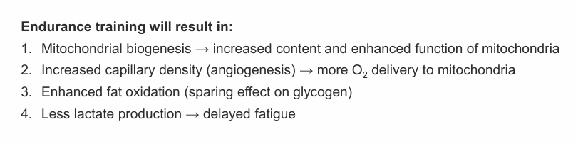
Mitochondrial biogenesis
Different types of exercise provide stimulus for mitochondrial biogenesis (increase in number and size). Through endurance exercise:
# mitochondria increases
Density of cristae increases (more internal SA)
Mitochondrial enzymes and ETC components
# beta oxidation enzymes
Increases mitochondria in long term results in enhances ATP production from fats and carbs, resulting in greater capacity for sustained aerobic energy production
PCG1-a in mitochondrial biogenesis
Key trigger for mitochondrial biogenesis: PGC1-a (peroxisome proliferator-activated receptor gamma coactivator)!
Triggered by: energy stress (AMPK), calcium signaling (Ca2+/CaMK), ROS, mechanical stress, MAPK

Athlete’s paradox
Intramuscular triglycerides (IMTG) are elevated in type 2 diabetes, but also endurance athletes
Athletes have high intermyofibrillar LDs (highly metabolically active that serve as energy reservoir during exercise). They are smaller and more numerous
In obese patients they accumulate subsarcolemmal LDs
Protein synthesis in resistance training
Translational processing and an elevated rate of postexercise protein synthesis results in a modest, same-directional change in protein content. Superimposition of repeated exercise results in gradual accumulation of protein from pulsed increased in mRNA expression.
Long-term adaption to training is due to the cumulative effects of each acute exercise bout leading to a new functional threshold
Resistance training
Exercise mode performed against an external resistance at varying intensities, primarily targeting the neuromuscular system to improve muscular strength, endurance, hypertrophy, and/or power. Short duration with resting intervals in between exercises. (free weights, elastic bands, calisthenics)
Depends heavily on anaerobic metabolism
Myonuclear accretion via satellite cells
Skeletal muscle hypertrophy is accompanied by myonuclear accretion via SC
But hypertrophy can occur without myonuclear accretion, so role of SC on hypertrophy is still controversial
In satellite cells, anabolic signaling promotes:
Cell proliferation
Fusion with myofibers
Several anabolic pathways are activated during and after resistance training, such as ERK/MAPK pathway and mTOR (via dependent and independent of AKT)
Basic mechanism of muscle hypertrophy

Regulation of protein synthesis
Translational efficiency (rate/speed of translation per ribosomes): mediated by anabolic signaling (growth factors, mTORC1, MAPK)
Translational capacity (quantity of ribosomes): mediated by ribosome biogenesis
Transcriptional reserve capacity
The ability myonuclei possess to increase transcriptional rates (increase synthesis of mRNA, rRNA, tRNA, and small RNAs) required to support new demands, such as growth/repair
Role of exercise on muscle protein synthesis (acute and chronic)
Mechanical tension/loading from exercise increases protein synthesis acutely (translational efficiency) and chronically (translational capacity)
Anabolism vs catabolism
Anabolism: synthesis, accumulation
In muscle: building new myofibrillar proteins, resulting in hypertrophy
Requires ATP and sufficient nutrient availability (especially AA)
Catabolism: degradation, breakdown
In muscle: catabolism involves proteolysis, which is physiological and normal, but if chronic may lead to atrophy
Leucine role in protein synthesis
Acts as a nutritional signal that cells can sense. When present at sufficient concentrations, it activates mTORC1 signaling pathway thru intermediates Sestrin2/GATOR2
Inflammation definition
Protective response of organism to stimulation by invading pathogens or endogenous signals such as damaged cells, resulting in elimination of cause of injury, clearance of necrotic cells, and tissue repair
Types of inflammation
Acute: Rapid, short-lived (minutes to days)
Chronic: Persistent, often pathological (weeks to years)
Key mediators of inflammation
Cytokines (pro and anti-inflammatory): TNF-a, IL-1B, IL-6, IL-10
Eicosanoids: Prostaglandins and leukotrienes
Vasoactive amines: Histamine
Cells required to repair/heal muscle tissue
Satellite cells: muscle progenitor cell
Myeloid cells: diverse group of blood and immune cells, such as neutrophils, eosinophils, basophils, monocytes, macrophages, etc.
Phases of acute inflammation in skeletal muscle:
Damage/injury and immune cell recruitment
Clearance of debris
Activation of satellite cells
Repair and remodeling
Normal response to acute inflammatory event
Following muscle injury, neutrophils are rapidly mobilized, increase in circulation and move to the damaged tissue. Initially, M1 macrophage populations dominate. Later M1 macrophages replaced by M2 macrophages which play roles in muscle growth/regen
PUFAs - Polyunsaturated fatty acids
Omega 3: Give rise to anti-inflammatory lipid mediators
Omega 6: Usually pro-inflammatory
Both enzymes can be metabolized by LOX and COX enzymes
LOX and COX
LOX (Lipoxygenases): expressed by leukocytes, eosinophils, platelets, macrophages, etc.
COX (Cyclooxygenases): expressed by endothelial cells, platelets, macrophages, etc
Arachidonic acid
Omega 6 important to inflammatory response
Direct effect on myotube hypertrophy, via COX2 and NSAID-sensitive pathway
Ibuprofen and muscle adaption
Ibuprofen (COX2 inhibitor NSAID) can reduce muscle hypertrophy induced by resistance training. However, in older individuals they promote muscle hypertrophy
Reduce hypertrophy because they delay resolution of inflammation
Muscular dystrophies
Group of rare neuromuscular diseases
Defined as inherited disorders characterized by progressive muscle weakness and degeneration
They can be inherited from 1 autosomal dominant parents or 2 autosomal recessive parents, or they can be sex linked.
Dystrophin
Rod-shaped protein that provides stabilization of the sarcolemma during muscular contraction. It links muscle cytoskeleton and extracellular matrix
DMD
Caused by mutations in DMD gene resulting in complete loss of dystrophin
DMD is X-linked recessive
DMD Treatment
Exon skipping drugs
Gene therapy (Elevidys): delivers shortened micro-dystrophin gene using an adeno-associated viral vector