Week 2 Anatomy Unit 3
 Call Kai
 Learn
 Practice Test
 Spaced Repetition
 Match
 Flashcards
 Knowt Play
1/34

There's no tags or description
Looks like no tags are added yet.
35 Terms
CN I
Olefactory
Cribriform plate
Sensory: Smell
CN II
Optic
Optic canal
Sensory: Vision
CN III
Oculomotor
Superior orbital fissure
Motor: eye movement
CN IV
Trochlear
Superior orbital fissure
Motor: superior oblique eye movement (up and down)
CN V
Trigeminal
V1: Opothalamic (sensory): Superior orbital fissure
V2: Maxillary (sensory): foramen rotundum
V3: Mandibular (both): foramen ovale
Meningeal branch: foramen spinosum
Sensory: chewing and facial sensation; Motor: muscles of mastication
CN VI
Abducens
Superior orbital fissure
Motor: Eye movement (lateral rectus muscle, sideways movement)
CN VII
Facial
Internal acoustic meatus
Motor and sensory: facial expressions, anterior taste, tear production
CN VIII
Vestibulocochlear
Internal acoustic meatus
Sensory: Hearing and balance (semicircular canals)
CN IX
Glossopharyngeal
Jugular foramen
Motor and sensory: swallowing, posterior taste
CN X
Vagus
Jugular foramen
Motor (palate, pharynx, larynx, trachea, bronchial tree, heart, GI tract) and sensory (pharynx, larynx, trachiobronchial tree, lungs, heart, GI)
CN XI
Accessory
Jugular foramen
Motor: Strenocleidomastoid and trapezius
CN XII
Hypoglossal
Hypoglossal canal
Motor: intrinsic and extrinsic muscles of the tongue
Mnemonic for sensory/motor control
Some say money matters, but my brother says big brains matter most
CSF Circulation Steps
Forms in the choroid plexus
Flows into the intraventricular foramen of Monro to get to 3rd ventricle
Goes to the cerebral aqueduct of Sylvius to get to 4th ventricle
Goes through 2lateral apertures (foramen of Luschka) and 1median aperture (foramen of magendie) into the subarachnoid space
At the arachnoid granulations/villi, CSF is reabsorbed into the venous blood of the dural venous sinuses
Diploic veins
Deoxygenated blood travels through the diploe of bone and drains into the dural venous sinuses
Bridges the extracranial venous system to the intracranial venous system
Dural venous sinuses
Valveless channels in the dura mater that collect deoxygenated blood and cerebrospinal fluid (CSF) from the brain and direct it mainly into the internal jugular veins
Veins we’ve studied in class

1
Cribiform plate

2
Optic canal

3
Superior orbital fissure

4
Foramen rotundum (CNV2, maxillary)

5
Foramen ovale (CNV3, Mandibular)

7
Internal acoustic meatus

8
Jugular foramen

9
Hypoglossal canal
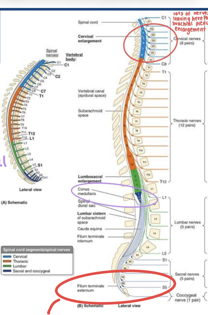
Where does the spinal cord begin and end
Begins at the foramen magnum as a continuation of the medulla, ends around L1/2 in adults
How many spinal nerves are there
8 cervical
C1-C7 exit superiorly from the vertebra; then C8 and future nerves exit inferiorly
12 thoracic
5 lumbar
5 sacral
1 coccygeal
Dermatomes
Somatic sensory innervation of spinal nerve roots
If an injury was before the brachial plexus, we would see dermatome nerve distribution
Myotomes
What muscles are innervated by each spinal nerve root
What is the end of the spinal cord called
Conus medullaris

What structure is below the spinal cord
Cauda equina
L1/L2- rest of the lumbar and sacral spinal canal
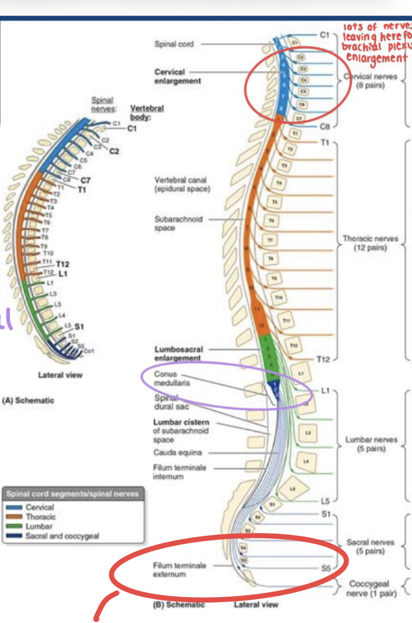
What is the end of the cauda equina called and what does it do
Filum terminale
Anchors the spinal cord to the coccyx
No nerves, just continuation of the dura mater (dural sac terminates at S2)
Anterior and posterior nerve roots
Anterior/ventral: motor (efferent)
Posterior/dorsal: sensory (afferent)
What arises from the spinal cord to become a spinal nerve
Rootlets come off the grey matter (cell bodies)
Become roots
Anterior roots: motor (efferent)
Posterior roots: sensory (afferent)
Become spinal nerve
Branch into rami
Dorsal (posterior) rami
Nerve supply to the skin, deep muscles of back, veretebral column joints
Do not form plexuses
Ventral (anterior) rami
Wrap around to form plexi
Innervate skin and muscles of the trunk and limbs