APEX POCUS
 Call Kai
 Learn
 Practice Test
 Spaced Repetition
 Match
 Flashcards
 Knowt Play
1/29

There's no tags or description
Looks like no tags are added yet.
30 Terms

What are the 3 standard cardiac POCUS imaging windows?
Parasternal → PLAX, PSAX
Apical → A4CH
Subcostal → Subcostal 4CH, IVC

What are the 5 core cardiac POCUS views?
Parasternal long-axis (PLAX)
Parasternal short-axis (PSAX)
Apical 4-chamber (A4CH)
Subcostal 4-chamber
Subcostal IVC

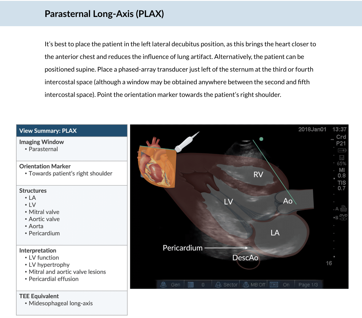
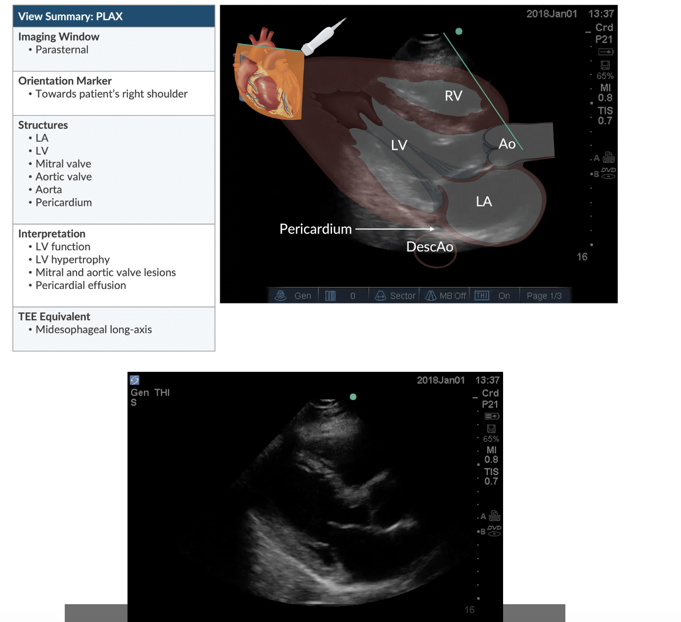
Parasternal Long-Axis (PLAX)
Identify this view. What structures are visible?
View: Parasternal Long-Axis (PLAX)
Structures: LA, LV, RV, mitral valve, aortic valve, aorta, pericardium
Evaluates: LV function/hypertrophy, mitral and aortic valve lesions, pericardial effusion
TEE equivalent: Midesophageal Long-Axis (ME LAX)
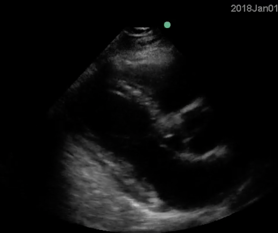

Where do you position the probe for PLAX?
In which direction does the orientation marker point?
Window: Parasternal
Position: Left sternal border, 3rd–4th intercostal space
Marker: Toward patient's right shoulder
Patient: Left lateral decubitus preferred

Parasternal Short-Axis (PSAX)
How to get the PSAX view from the PLAX?
Window: Parasternal
Rotate probe 90° clockwise from PLAX.
Orientation marker: Patient's left shoulder.
TEE equivalent: Transgastric short-axis (TG SAX).


Identify this view. What structures are visible?
View: Parasternal Short-Axis (PSAX)
Structures: LV, RV, pericardium.
Evaluates: LV/RV function and pericardial effusion.
Visual clue: LV appears circular in cross-section, with the RV adjacent to it.


How do you obtain the Apical 4-Chamber view?
Window: Apical
Position: Left lateral decubitus
Probe: Point of maximum impulse (PMI)
Orientation marker: Patient's left side
Ultrasound beam: Toward patient's right shoulder
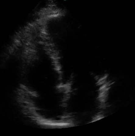
Identify this cardiac POCUS view.
Apical 4-Chamber (A4CH)
Structures: RA, RV, LA, LV, AV valves, pericardium
Evaluates: LV/RV function, AV valve lesions, pericardial effusion
TEE equivalent: Midesophageal 4-chamber (ME 4C)

How do you obtain the Apical 4-Chamber (A4CH) view?
Window: Apical
Patient position: Left lateral decubitus
Probe placement: Point of maximum impulse (PMI)
Men: Just inferolateral to the left nipple
Women: Under the inferolateral quadrant of the left breast
Orientation marker: Toward patient's left side
Ultrasound beam: Toward patient's right shoulder


How do you obtain the Subcostal 4-Chamber view?
Window: Subcostal
Patient position: Supine
Probe placement: Midline, just inferior to the xiphoid process
Orientation marker: Toward patient's left side
If visualization is difficult:
Bend patient's knees to relax abdominal muscles.
Apply additional probe pressure as needed.
Slide probe to patient's right to use the liver as an acoustic window if bowel gas interferes.


Identify this cardiac POCUS view.
Subcostal 4-Chamber (Subcostal 4CH)
Structures: RA, RV, LA, LV, AV valves, pericardial space
Evaluates: RV function and pericardial effusion
TEE equivalent: Midesophageal 4-Chamber (ME 4C)
Visual clue: Liver appears in the near field, with the four cardiac chambers deeper in the image.


Subcostal IVC
How do you obtain the subcostal IVC view?
Window: Subcostal
Starting position: Subcostal 4CH
Probe movement: Rotate transducer 90°
Beam direction: Tilt posteriorly
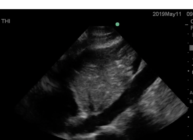

Identify this cardiac POCUS view.
Subcostal IVC
Structures: IVC, RA, liver
Evaluates: Volume status
Key finding: IVC collapse suggests hypovolemia
TEE equivalent: Bicaval view
Visual clue: IVC appears as a long tubular structure entering the RA, with the liver nearby.


Name this view
Parasternal long axis

Name this view
parasternal short axis


Name this view
apical 4 chamber

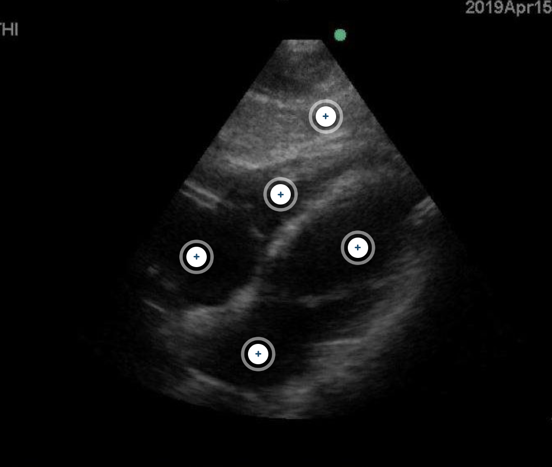
Name this image
Subcostal 4 chamber


Name this view
Subcostal IVC

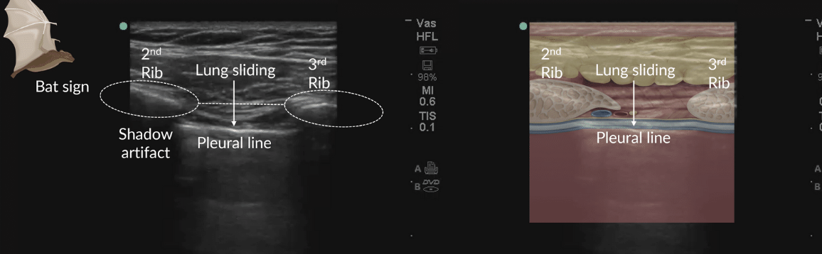
Identify this lung ultrasound sign and its structures.
Bat Sign
Wings: 2nd and 3rd ribs
Body: Hyperechoic pleural line between the ribs
Acoustic shadows: Dark regions deep to the ribs
Key concept: Ribs strongly attenuate ultrasound, producing posterior shadowing.
Memory trick: Ribs = wings; pleural line = body.

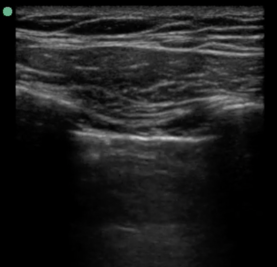
What is lung sliding, and what does its absence suggest?
Lung sliding: Shimmering movement of the pleural line during respiration.
Indicates: Visceral and parietal pleura remain in contact.
Absent lung sliding: May suggest pneumothorax or endobronchial intubation.
Important: Absence alone does not confirm pneumothorax.
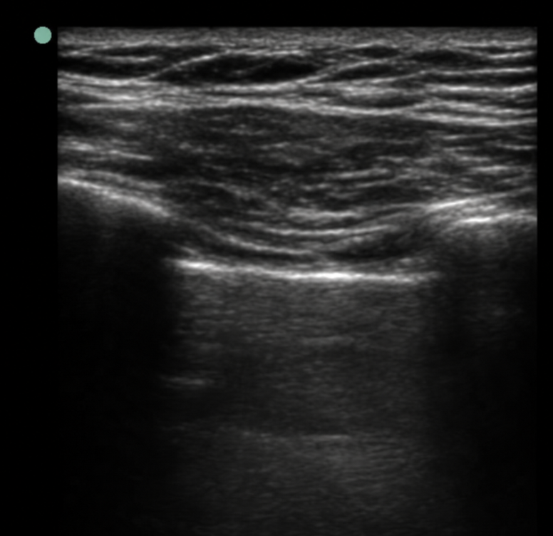

Original APEX image without labels. Identify the artifact.
A-lines = horizontal, parallel to pleura
Caused by reverberation artifact
Usually associated with aerated lung
Memory trick: A = Across / Air


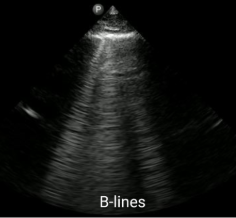
Identify the artifact.
B-lines = vertical, extend from pleura to bottom
Erase A-lines
Multiple B-lines may suggest pulmonary edema
Memory trick: B = Bottom
B BERTICAL (look like the aliens are coming)

How do you perform gastric POCUS?
Patient position: Right lateral decubitus (RLD)
Probe: Curvilinear transducer
Placement: Midline, just inferior to the xiphoid process
Orientation marker: Cephalad (toward the head)
Primary structure: Gastric antrum
Memory trick: Gastric = RIGHT side, BELOW xiphoid, marker UP.
Why is the gastric antrum evaluated in the right lateral decubitus position?
Gravity moves gastric contents toward the pylorus and antrum.
Air rises toward the upper portion of the stomach.
The antrum fills with gastric contents, improving assessment.
Supine positioning may underestimate gastric volume.
Gastric POCUS helps guide decisions about RSI and aspiration risk.
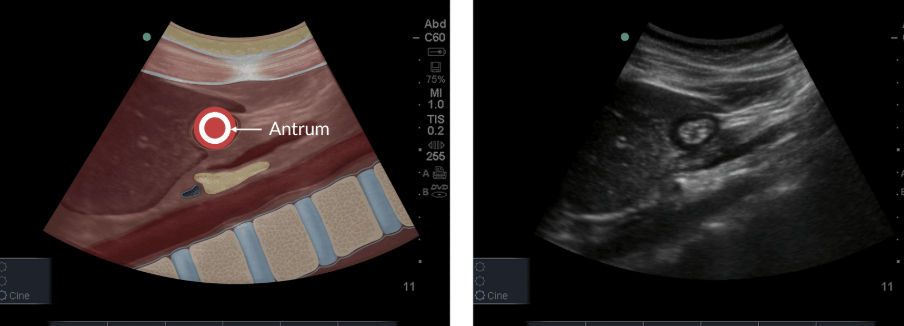
Identify the gastric finding.
Empty Stomach — Bull's Eye Sign
Appearance: Flat or small oval antrum resembling a bull's eye.
Anechoic border: Muscularis propria, NOT fluid.
Anatomical location: Antrum just below the liver.
Pancreas: Deep to the stomach.
Aorta: Deep to the pancreas.
SMA: Sometimes visible.
Quick visual memory trick
Empty stomach = Small bull's eye.
Fluid-filled stomach = Distended antrum.

Identify the gastric finding.
Clear Fluid in the Gastric Antrum
Appearance: Round, distended antrum.
Contents: Anechoic (black).
Why: Clear fluid produces very few internal echoes.
Clinical relevance: Gastric fluid volume helps assess aspiration risk.
»Clear fluid = Big black circle
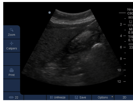
Identify the gastric finding.
Particulate Matter — Non-clear Liquid or Solid Contents
Appearance: Round, distended gastric antrum.
Non-clear liquids (e.g., milk): Hyperechoic.
Solid food: Bright particulate echoes within the antrum.
Clinical significance: Suggests gastric contents that may increase aspiration risk.
» Particulate = Bright speckles
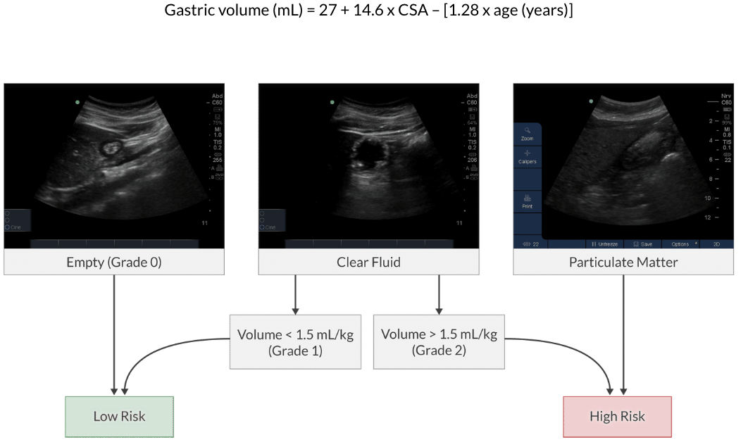
How do you calculate clear gastric fluid volume using POCUS?
Gastric volume: (mL) = 27 + 14.6(CSA) - 1.28 (age)
Position: Right lateral decubitus (RLD)
Measurement: Gastric antrum CSA at the level of the aorta
Accuracy: Average 3 CSA measurements
CSA = cross-sectional area in cm²; age in years.

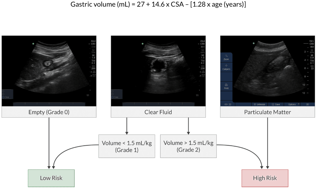
Gastric Grading
Grade 0 = Empty
Grade 1 = Clear fluid below 1.5 mL/kg
Grade 2 = Clear fluid above 1.5 mL/kg
Particulate matter = High risk


APEX - Q - What cardiac view is this
subcostal 4- chamber
bc it shows you the liver
