Deep Back and Spinal Cord
 Call Kai
 Learn
Learn Practice Test
 Spaced Repetition
 Match
Match Flashcards
 Knowt Play
1/27

There's no tags or description
Looks like no tags are added yet.
Name | Mastery | Learn | Test | Matching | Spaced | Call with Kai | Chat |
|---|
No analytics yet
Send a link to your students to track their progress
28 Terms
muscles of the deep back
Erector Spinae
-Act on vertebre; both contract=extension spine, 1 side contract=lateral flexion
Spinalis
Longistimus
Iliocostalis
Deep to Erector Spinae
Multifidus
deep to multifi…
?
Levator Costareum

5 types of vertebrae
-7 servical: neck
-12 thoracic: ribs
-5 lumbar
-(5) sacrum: fused, part of pelvis
-(4) coccyx: tail bone

parts of a typical vertebra
Pictured
-vertebral body
Vertebral Arch (often where muscles attach)
-pedicle: between TP and body
-lamina: between SP and TP
Superior View
-Vertebral foramen: spinal cord
Lateral View
-Superior and inferior vertebral notch: other vertebre articulate

foramens
-intervertebral foramen: exit spinal nerves, R and L side
-inferior vertebral notch and superior vertebral notch
-vertebral foramen: spinal cord


label vertebre

What is the function of the transverse foramina?
vertebral artery
-branch from subclavial a
-to brain

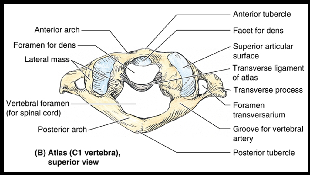
atlas
First cervical vertebra (C 1)
It has no body and leaves a space to accommodate the dens of the axis. Superior articular surfaces articulate with occipital condyles – “YES”movement of the head
*articulates w/ skull
axis
Second cervical vertebra (C 2)
It has a tooth-shaped process, the dens (odontoid process), which articulates with the atlas as a pivot joint – “NO” movement of the head

__has a prominent spinous process
C7
*good for palpation


identify features of a thoracic vertebra
-articulates w/ ribs (facets)
-coronal/verticle plane
-spinous process points inf


identify features of a lumbar vertebra
-very large
-articular process in sagittal plane
-SP ponts post

sacrum and coccyx
-fused together; part of pelvis

Cervical vs. Thoracic vs. Lumbar Vertebrae
CERVICAL
Transverse foramen
Bifid spinous process (most of them)
Articular facets oriented in transverse plane
THORACIC
Costal facets (*ribs articulate)
Articular facets oriented in coronal plane
Spinous process directed inferiorly
LUMBAR
Massive size (*large body, most weight bearing)
Articular facets oriented in para-sagittal plane
Spinous process directed posteriorly

list joints of the vertebral column
1. Joints of the vertebral bodies
a.Intervertebral discs
b.Uncovertebral joints
c.Ligaments – anterior and posterior longitudinal ligaments
2. Joints of the vertebral arches – facet joints (zygapophyseal joints)
3. Craniovertebral joints
a.Atlanto-occipital joints
b.Atlanto-axial joints
4. Accessory ligaments of intervertebral joints
a.Ligamentum flavum
b.Interspinous ligament
c.Supraspinous ligament
d.Ligamentum nuchae (nuchal ligament)
e.Intertransverse ligaments
intervertebral disc
-Annulus fibrosus – outer portion; composed of fibrocartilage and fibrous connective tissue
-Nucleus pulposus – inner portion; semigelatinous fluid with very few, if any, cells (*squisher, gel-like)
-Note: NO intervertebral disc between atlas and axis
-The IV disc is reinforced anteriorly and anterolaterally by the anterior longitudinal ligament.
-The IV disc is reinforced posteriorly by the posterior longitudinal ligament.
-The IV disc is not reinforced posterolaterally.

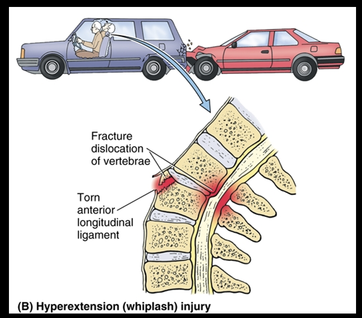
“WHIPLASH” INJURY
-Results from severe hyperextension of the neck, as occurs in during rear-end motor vehicle collisions, especially when the head restraint is too low
-Anterior longitudinal ligament is severely stretched and may be torn
-May injure posterior parts of the vertebral arches and their processes
-Herniation of the nucleus pulposus is almost always in a
___direction
-Practice Question: A 70-year-old female complaining of lower back pain is diagnosed with a herniated disc at the level of L4-5. Which spinal nerve(s) is/are most likely compressed?
-Herniation of the nucleus pulposus is almost always in a
posterolateral direction, passing through a rupture of the annulus fibrosus.
-may lean to opposite side to relieve compression of nerve
*IVD impinge on SN
Practice Question
-L5

Articular facets (zygopophyses)

joints of C1 and C2

ligaments of C1 and C2

Ligaments of intervertebral joints
•Ligamentum flavum: spans the space between neural arches, yellowish color
•Interspinous ligament: between SP
•Supraspinous ligament: spans between post end SP
•Ligamentum nuchae (nuchal ligament): nuchal line cranium → SP cervical vertebra, sagittal articulations, origin trapezius
•Intertransverse ligaments

movements of vertebral column

range of motion in vertebral joints
-atlanto-occipital: not lot rotation
-altanto-axial: most rotation
-other 3: rotation w/ horizantal plane; lumbar not lot bc peri-sagittal orientation

normal CURVATURES OF THE VERTEBRAL COLUMN
-fetus: not lot curves
-4 years: L and S cruves when start to wal
-Lumbar mordosis

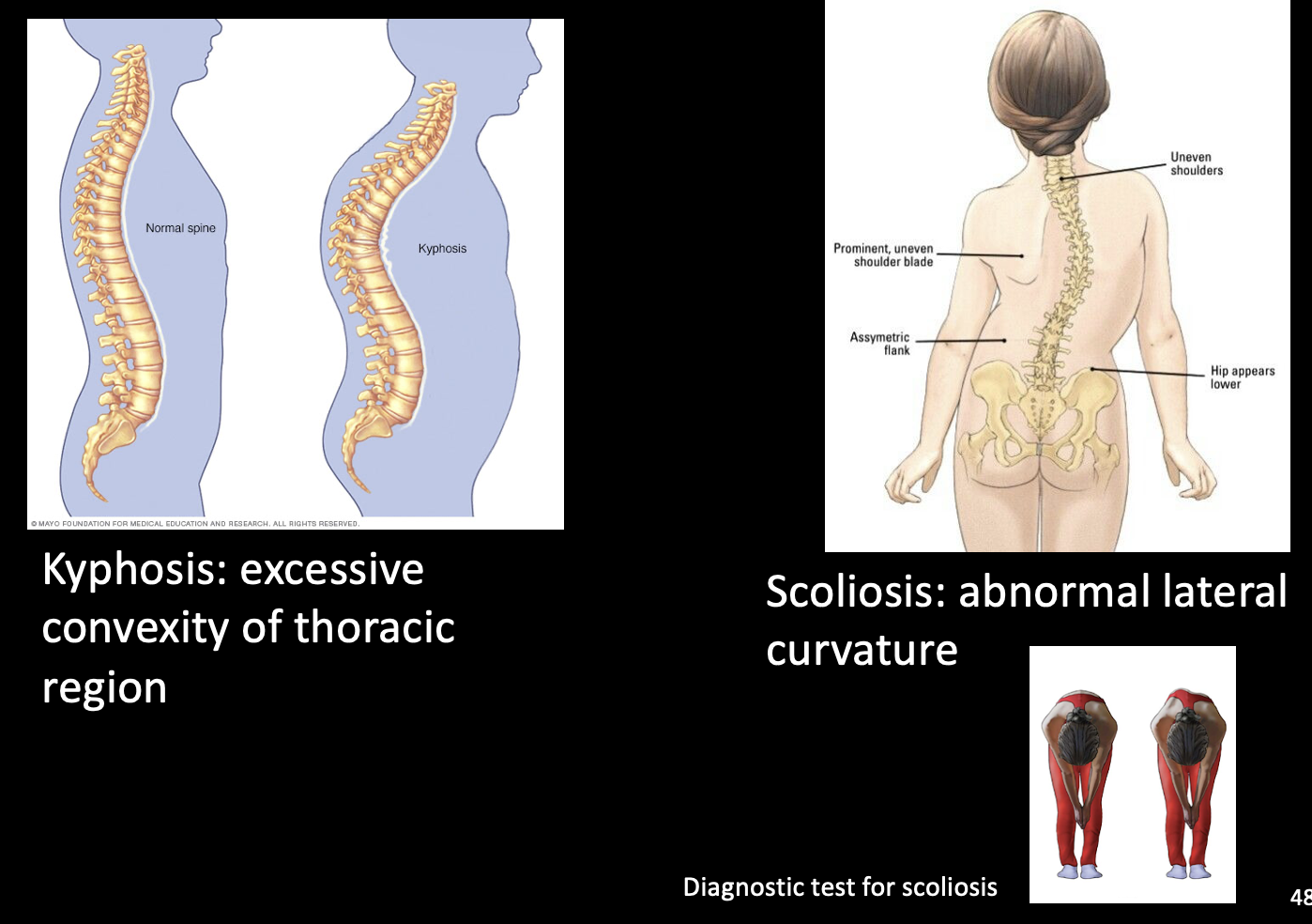
abnormal curvatures of the vertebral column
-Kyphosis: excessive convexity of thoracic region
-Scoliosis: abnormal lateral curvature
*often accompany each other
-(Hyper)lordosis AKA “pelvic tilt”: increased lordotic curve, if pregnant or obese
meninges of spinal cord
+epidural space and subarahnoid space w/ CSF

Spinal cord changes diameter
-Which spinal cord levels contribute to the cervical enlargement of the spinal cord?
-Which spinal cord levels contribute to the lumbar enlargement of the spinal cord?
-C5-T1
-T11-S2

end of the spinal cord
*Cauda Equina=”horses tial”
*Filum terminale: made from pia mater, anchors SC in spinal canal
