Chapter7-bone tissue- sesay emphasized
 Call Kai
 Learn
 Practice Test
 Spaced Repetition
 Match
 Flashcards
 Knowt Play
1/55

There's no tags or description
Looks like no tags are added yet.
56 Terms
Skeletal system
bones, cartilages, ligaments
osteology
study of bone
hyaline cartilage is
forerunner of most bones, and covers joint surfaces.
3x types of cartilage
hylaline
elastic
fibrocartilage
ligament
bone to bone at joints (collagen, a dense regular connective tissue)

tendon
attach muscle to bone
dense regular connective tissue

What do healthy bones do?
1-regulate blood sugar, so unhealthy bones increase risk of diabetes.
bones secrete osteocalcin hormone that affects action of insulin and moderate stresss response.
2- Electrolyte balance- calcium and phosphate levels
3-Acid Base balance- buffers blood against large PH changes by altering phosphate and carboante levels
4- blood formation- red bone marrow is the chief producer of blood cells
healthy bones release
osteocalcin- stimulates pancreas to release insulin
mineralization and calcification
the hardening/stengthening of bone
individual bone consists of
bone tissue,
bone marrow
cartilage
adipose tissue
nervous tissue
fibrous connective tissue
nervous, fibrous, and connective tissues are integrated primarily within the outer protective membranes and the internal channels that run through the mineralized matrix

Compact bone
dense/cortical bone- dense outer shell of bone

compact bone encloses the
marrow/ medullary cavity.

spongy bone/ cancellous
loosely organized bone tissue
Diaphysis
shaft for leverage
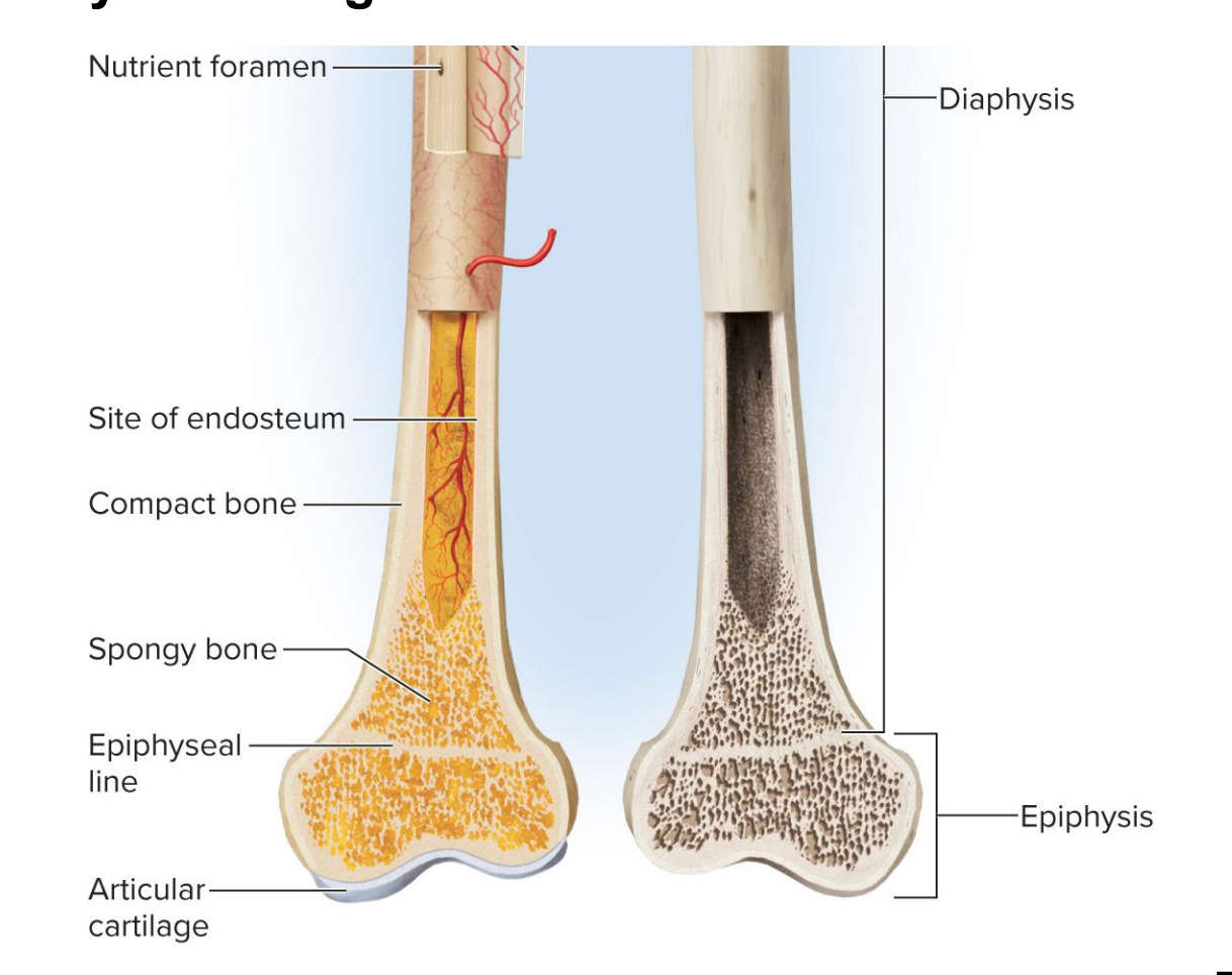
epiphysis
enlarged end of a long bone. strengthens joint, anchors ligaments and tendons.

Epiphyseal line and plate
line- found in adults. remnant of childhood growth zone of hyaline cartilage epiphyseal plate.
plate- found in kids. ontop of epiphysis, contains hyaline cartialge
Osteoblasts
Bone forming cell in endosteum ( The endosteum is a thin, delicate layer of active connective tissue that lines the internal surfaces of all bones.)and inner layer of periosteum(periosteum is a tough, double-layered membrane of fibrous and cellular connective tissue that covers the entire outer surface of all bones, except at the joints where articular cartilage is present.)
osteogenic cell
stem cells found in the endosteum and inner layer of periosteum
Osteocyte
Mature bone cell: former osteoblasts that have become embedded in the matrix they deposit
octeocalcin (not on test)
osteoblasts and osteocytes, under physical stress, secrete osteocalcin. response to stress, inhibits parasympathetic, and allows the sympathetic to work unnopposed. stimulates pancreatic release of insulin, increases insulin sensitivty, promotes energy availability.
Osteoclast
dissolves bone. found on bone surface. performs osteolysis/ resorption or bone breakdown/remodeling.
often reside in pits called resorption bays.
develop from same bone marrow stem cells that give rise to blood cells
Bone matrix
collagen and minerals of bone form a composite. mineral portion allows body to support weight.
Rickets
children with rickets- a disease caused by mineral deficiency results in soft deformed bones.
osteomalacia
vitamin D deficiency in adults that also leads to softening bones and joint pain
Bone marrow
soft tissue occupying marrow cavities of long bones and small spaces of spongy bone
Red bone marrow (myeloid tissue)
contains multiple tissues, including hematopoietic tissue- tissue that produces blood cells
Red bone marrow is hemopoietic (blood-producing) tissue, meaning it is the manufacturing site for all formed elements of the blood. It contains pluripotent stem cells that differentiate not just into erythrocytes (RBCs), but also into all types of leukocytes (white blood cells) and megakaryocytes (which shatter to form platelets).
formed blood elements:
Erythrocytes (Red Blood Cells): Cells responsible for binding and transporting oxygen from the lungs to the body's tissues, and carrying carbon dioxide back to the lungs.
Leukocytes (White Blood Cells): Immune cells
Thrombocytes (Platelets): Cell fragments that play a critical role in blood clotting (hemostasis) to repair damaged blood vessels a
infant bone marrow is
100 percent red in long bones of children
adults bone marrow
70 percent red (In adults, found in skull, vertebrae, ribs, sternum, part of pelvic
girdle, and proximal heads of humerus and femur) and 30 percent yellow.
yellow bone marrow
Fatty marrow that does not produce blood
Can transform back to red marrow in the event of chronic anemia
helps with energy and immunity
ossification or osteogenesis:
Formation of bone:
Occurs in two ways that begin with embryonic mesenchyme:
1-Intramembranous ossification
2- Endochondral ossification
Intramembranous ossification
Produces flat bones of the skull, most of the clavicle, and part of the mandible
Stages (not on test):
1- deposition of osteoid tissue into embryonic mesenchyme
2- calcification of osteoid tissue and entrapment of osteocytes
3-honeycomb of spongy bone with developing periosteum
4- filling of space to form compact bone at surfaces, leaving spongy bone in the middle
key for lifelong thickening and remodeling of long bones
Endochondral ossification
requires hyaline cartilage.
Bone develops from hyaline cartilage: produces most bones of the body (limbs, vertebrae, ribs, sternum, scapula, pelvic girdle.
summary of stages (metacarpal example)
1- Mesenchyme forms early hyaline cartilage model.
2- formation of primary ossification center, bony collar, and periosteum
3- vascular invasion, formation of primary marrow cavity. and appearence of secondary ossification center.
4- Bone at birth contains enlarged marrow cavity and secondary marrow cavity.
5- child has epiphyseal plate
6- adult bone contains single marrow cavity and close epiphyseal plate (becomes epiphyseal line)
*Mesenchyme is a type of loosely organized, undifferentiated embryonic connective tissue. It is made up of star- or spindle-shaped cells floating in a watery, gel-like extracellular matrix
Stages of endochondral ossification

Metaphysis
Epiphyseal plate serves as growth zone. The transitional zone on each side, where cartilage is replaced by bone is called metaphysis.
The structure of metaphysis corresponds to steps of cartilage replacement
steps of metaphysis/ cartilage replacement
Epiphyseal plate serves as growth zone
Transitional zone on each side, where cartilage is replaced by bone is called metaphysis
Steps correspond to steps of cartilage replacement:
1- Zone of reserve cartilage: 100 cells
2- Zone of cell proliferation: 1,000 cells hyaline cartilage
3- Zone of hypertrophy- cells get bigger
4- zone of calcification- cells fill with minerals
5- zone of bone deposition
Zones of metaphysis
1- zone of reserve cartilage: histology of resting hylaine cartilage
2- Zone of cell proliferation: chondrocytes multiplying and lining up in rows of small flattened lanunae
3-zone of cell hypertrophy: cessation of mitosis, enlargment of chondrocytes and thining of lacuna walls
4- Zone of calcification: temporary calcification of cartilage matrix between columns of lacunae
5- zone of deposition: Breakdown of lacuna walls, leaving open channels- death of chondrocytes, bone deposition by osteoblasts, forming spongy bone of trabeculae

Achondroplastic Dwarfism:
Theres a problem with epiphyseal plate
long bones stop growing in childhood; results in normal torso, short limbs
failure of cartilage growth in metaphysis
spontaneous mutation produces mutant dominant allele
pituitary dwarfism
lack growth hormone
normal proportions with short stature
Wolff’s law
the architecture of bone is determined by the forces put on it- the bone adapts to withstand it.
osteoblasts and osteoclasts
if a bone is little used osteoclasts remove matrix
calcium Homestasis
calcium is needed for bone structure, neuron communication, muscle contraction, blood clotting, and exocytosis.
hypocalcemia
calcium deficiency- leads to excessive excitability of nervous system and muscles
hypercalcemia
very rare- excess is sucked into bone. calcium excess. causes nerve and muscle cells to be less excitable than normal.
hypocalcemia causes
low vitamin D, diarrhea, thyroid tumors, underactive parathyroid glands
calcium homestasis is regulated by three hormones
calcitriol
calcitonin
parathyroid hormone
calcitriol
step 1- vitamin D from sun or food… travels to liver and turns into calcidiol
step 2- calcidiol travels to the kidneys where an enzyme converts in into calcitriol- the hormonal active form of Vit D.
necessary for bone deposition- helping provide adequate calcium and phosphate
inadequate calcitriol results in abnormal softness of bones (rickets in children and osteomalacia in adults)
1-It then increases calcium absorption by the small intestine
2- increases calcium resorption from skeleton (stimulates formation of osteoclasts)
3- weakly promotes absorption of calcium from the kidneys (reduced extretion of Ca+)
Calcitonin
If blood calcium concentration goes over 10.4 (9.2-10.4):
The Thyroid senses this- too much calcium in the blood! and releases Calcitonin. calcitonin :Osteoclast inhibition: Rapidly reducing the rate of bone breakdown. Osteoblast stimulation: Promoting calcium deposition into the bone matrix by increasing osteoblast activity (mostly in children). Relevance in the Textbook: Saladin emphasizes that this mechanism is significantly active in children—whose bones are rapidly remodeling and mineralizing—but has a weak or negligible effect in healthy adults
secreted when blood calcium levels are too high.
lowers blood calcium conentration in two ways:
osteoblast inhibition: reducing bone resorption
osteoblst stimulation; to deposit calcium into bone
important in children/weak affect in adults
PTH-Parathyroid Hormone
If too little claclium in blood (under 9.2-10.4):
Parathyroid senses this! it releases PTH, which stimulates osteoclasts in bone to break down osseus tissue and put the calcium back into the blood stream.
Some PTH also goes to the kidneys and said “ speed up activation of vitamin D into Calcitriol. Calcittriol goes to the intestines/gi tract, and gathers calcium to bring into the blood stream.
Direct kidney reabsorption: In addition to speeding up calcitriol production, PTH acts directly on the kidneys, signaling them to stop letting calcium escape in the urine and to reabsorb it straight back into the blood.
The Phosphate Dump: When osteoclasts break down osseous tissue, they release both calcium and phosphate into the blood. If both levels rise together, they will bind and form solid calcium-phosphate crystals (ectopic calcification) instead of staying dissolved as usable ions. To prevent this, PTH forces the kidneys to excrete extra phosphate into the urine.
The Osteoblast Middleman: While PTH does ultimately stimulate osteoclasts, it doesn't bind to them directly. PTH actually binds to osteoblasts (the bone builders). The osteoblasts then release a chemical signal (RANKL) that wakes up the osteoclasts to start resorbing bone.
PTH effect on calcium blood levels:
PTH raises calcium blood level by four mechanisms:
Binds to receptors on osteoblasts, which then stimulate osteoclasts
to resorb bone
Promotes calcium reabsorption by kidneys, so less lost in urine
Promotes the final step of calcitriol synthesis in the kidneys,
enhancing calcium-raising effect of calcitriol
Inhibits collagen synthesis by osteoblasts, inhibiting bone
deposition
calcium intake and excretion image

phosphate homeostais:
calcitriol raises phosphate levels by promoting its absorption by the small intestine
PTH lowers blood phosphate levels, in the blood stream, by promoting its urinary excretion
if phosphate is too high, means kidney has a problem
osteopenia
measurable decline in bone density, may advance to osteoporosis
osteoporosis
severe loss of bone density. bones easily fracture.