Respiratory System
 Call Kai
 Learn
Learn Practice Test
 Spaced Repetition
 Match
Match Flashcards
 Knowt Play
1/132

There's no tags or description
Looks like no tags are added yet.
Name | Mastery | Learn | Test | Matching | Spaced | Call with Kai | Chat |
|---|
No analytics yet
Send a link to your students to track their progress
133 Terms
Path of air in respiratory system
air flows in through mouth and/or nose
Pharynx
oropharynx
nasopharynx
Larynx
Trachea
Bronchi
Bronchioles
Alveoli
Diaphragm
Muscle that controls breathing. Separates thoracic and abdominal cavity
Functions of respiratory system
supply O2 to tissues
get rid of waste like CO2
4 basic steps of respiration/ventilation
pulmonary ventilation (breathing)
external respiration (O2 moves into blood)
transport of gases through the blood
internal respiration (diffusion of gases in/out of tissues)
Respiratory zone
Where respiration actually occurs→alveolar sacs, ducts, and respiratory bronchioles
Conducting zone
“dead space”
air moves in/out→bronchi, bronchioles, trachea
Trachea anatomy
3 layers
goblet cells secrete mucus
cilia beat to elevate trapped particles out of respiratory tract
Alveoli function
Where gas exchange directly occurs
Alveoli structure
sacs
air facing surface is lined with type I alveolar cells and is contiguous with respiratory tract
type II alveolar cells scattered inside alveolus as well
moist inside
Type II alveolar cells
produce surfactant
Surface area of alveoli
Huge surface area, which permits rapid gas exchange
Visceral pleura
Lines lungs
Parietal pleura
Lines chest wall
Intrapleural fluid
Fills the intrapleural space between the parietal and visceral pleura
Ventilation
exchange of air between atmosphere and alveoli
Flow of air equation
F=(Palv-Patm)/R
Pressure-volume relationship
P1V1=P2V2
large volume=less molecules hitting vessel walls=lower pressure
4 lung pressures
intrapulmonary pressure (Palv)
intrapleural pressure (Pip)
transpulmonary pressure (Ptp)
chestwall pressure (Pcw)
Intrapulmonary pressure
pressure inside alveoli
rises and falls with breathing
determines which way air flows
Palv<Patm
air flows in
Palv>Patm
air flows our
Intrapleural pressure
pressure of intrapleural fluid in the intrapleural space
also fluctuates with breathing
always 4 mmHg less than Palv
Palv=Pip
lung collapse
transpulmonary pressure
Ptp=Palv-Pip
pressure across lung wall
governs properties of lungs→drive air in and out
stops lung collapse and chest wall expansion
Chestwall pressure
Pcw=Pip-Patm
pressure across chest wall
Steps of inspiration
Diaphragm and inspiratory intercostal muscles contract
pulls ribcage out and up→thorax expands
Pip becomes more subatmospheric
Ptp increases
lungs expand
Palv becomes subatmospheric
air lows into alveoli
Steps of expiration
Diaphragm and inspiratory intercostal muscles stop contracting
Chest wall recoils inward
Pip moved back up towards preinspiration value
Ptp moved back down towards preinspiration value
lungs recoil back down
air in alveoli is compressured
Air flows out
Are expiration and inspiration passive or active processes?
Inspiration: active
Expiration: passive
What determines lung compliance?
stretchability of lung tissue
surface tension at air water interfaces within alveoli
Greater lung compliance means…
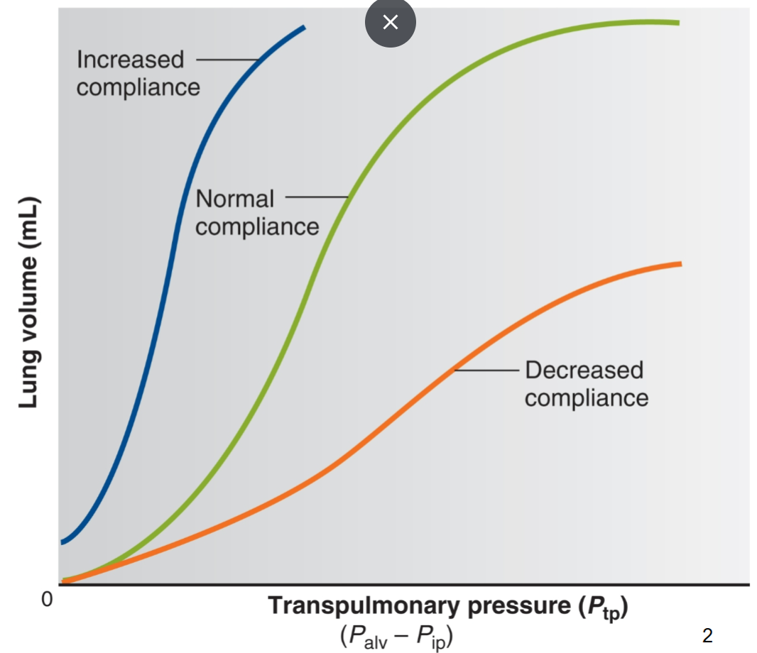
easier to expand lungs at any given Ptp
Lung compliance equation
C=deltaV/deltaPtp
Lung compliance curve
decrease compliance means that less air feels lung even at higher Ptp
both decreased and increased lung compliance are bad!
Effect of surfactant on lung compliance
surfactant reduces cohesive forces between water molecules on alveolar surface, which lowers surface tension and increases lung compliance
Pressure and surface tension relationship equation
P=2T/r
T is surface tension
r is airway radius
What would happens if no surfactant were present?
pressure would be determines by airway radius→air would flow from small to large airways
small airways would collapse
Effect of surfactant on large vs small airways
reduces surface tension in small airways specifically, which allows them to stay open
Premature babies and surfactant
Their type II alveolar cells are not yet mature so they cannot produce surfacant→alveoli collapse and inflate with every breath
Airway resistance
Usually very small, but radius does affect airway resistance
Things that alter airway radius
Physical, neural, chemical factors
Tidal volume
amount of air inhaled/exhaled in one normal breath
500 mL

Residual volume
amount of air remaining in lungs after maximum expiration, which keeps alveoli inflated between breaths
1200 mL

Inspiratory reserve volume
amount of air exhaled in excess when applying maximum effort
3000 mL
Expiratory reserve volume
amount of air exhaled in excess when applying maximum effort
1200 mL
Vital capacity
Amount of air that can be exhaled with maximum effort after maximum inspiration
ERV+TV+IRV
4700 mL
Total lung capacity
Total amount of air that can fit in a lung
RV+VC
5900 mL
Inspiratory capacity
Maximum amount of air that can be inhaled after a normal tidal expiration
TV+IRV
3500 mL
Functional residual capacity
Amount of air remaining in the lungs after a normal expiration
RV+ERV
2400 mL
Minute ventilation
amount of volume breathed in one minute
Minute ventilation equation
TV*RR
5-6 L/min
Alveolar ventilation
Air that enters alveoli for gas exchange
4.2 L/min
Alveolar ventilation equation
(TV-anatomical dead space)(RR)
What is more useful, minute ventilation or alveolar ventilation?
Alveolar ventilation because it does not take into account anatomical dead space where gas exchange does not occur
How much oxygen is introduced into the blood/dropped off at tissues per minute?
250 mL/min
How much CO2 is introduced into the blood/released into the lungs per minute?
200 mL/min
Partial pressures of O2 and CO2 in alveoli
PO2=105 mmHg
PCO2=40 mmHg
Partial pressures of O2 and CO2 in deoxygenated blood
PO2=40 mmHg
PCO2=46 mmHg
Partial pressures of O2 and CO2 in oxygenated blood
PO2=100 mmHg
PCO2=40 mmHg
Partial pressures of O2 and CO2 in the air
PO2=160 mmHg
PCO2=0.3 mmHg
Determinants of alveolar PO2
percent of oxygen in the air
ventilation rate
body’s rate of O2 consumption
Alveolar PO2 equation
(CO2 production)/alveolar ventilation=(O2 consumption)/alveolar ventilation
Hypoventilation
Decreased O2 concentration or decreased alveolar ventilation
increase in the ratio
Hyperventilation
increased O2 or increased alveolar respiration
decrease in the ratio
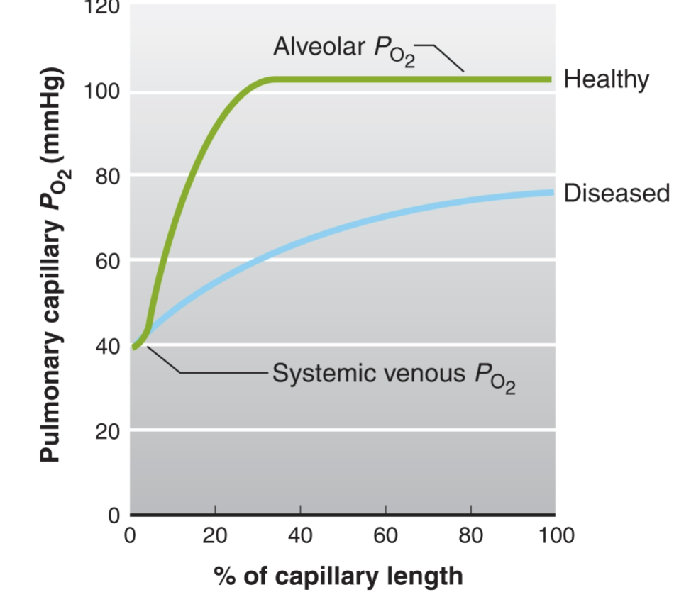
Gas exchange between alveoli and blood pressure curve
Healthy: oxygenation is super rapid
Diseased: oxygenation is slow and not efficient
What is the major reason for insufficiently oxygenated blood?
ventilation-perfusion mismatch
area of lung is receiving sufficient air flow but not blood flow
area of lung receiving sufficient blood flow but not air flow
How the body regulates ventilation-perfusion mismatch
Local controls decrease blood or air flow to the area
if underperfused, body will decrease air saturation to that area
if underventilated, body will decrease perfusion to the area
Transport of gases in hemoglobin-independent manner
CO2 and O2 can be directly dissolved in the plasm
What does solubility of a gas in a liquid depend on?
Directly proportional to its partial pressure (Henry’s law)
Percent of oxygen dissolved directly in plasma
1.5%
Percent of carbon dioxide dissolved directly
10%
Hemoglobin structure
two alpha and two beta polypeptides joined together into a heterotetramer
heme group made up of porphyrin ring and bivalent iron cation
What percent of oxygen is bound to hemoglobin?
95%
How many molecules of O2 can a hemoglobin bind?
4
Hemoglobin saturation curve
sigmoidal curve due to cooperative binding
4 sites are occupied in systemic arterioles
3 sites are occupied in venous arterioles

Steps of oxygen movement from lungs to tissues
O2 is inspired and makes its way to the alveolus
Pressure gradient causes oxygen to move into plasma as dissolved oxygen
As soon as O2 enters blood it binds to Hb due to yet another concentration gradient
RBCs move into systemic circulation
Metabolically active cells whos mitochondria are starved for oxygen receive O2
Reasons Hemoglobin offloads oxygen
metabolically active cells are warm (since heat is generated as a byproduct of respiration), which induces a right shift of the curve
Metabolically active cells are acidic (due to lactic acid and CO2 production), which induces a right shift of the curve
2,3-BPG effect on hemoglobin curve
Shift right
Things that shift Hb curve to the left
cooler temperatures
fetal Hb
Steps of loading CO2 into erythrocytes
metabolically active cells produce CO2
CO2 diffuses into plasma due to concentration gradient
30% of CO2 binds to Hb, 10% stays in plasma, 60% is turned into carbonic acid via carbonic anhydrase
Carbonic acid dissociated into HCO3- and H+
HCO3 exits RBCs while Cl- enters via an antiporter
Steps of unloading CO2 in the lungs
Blood flows past alveolus
CO2 bound to hemoglobin is displaced by plentiful O2
Bicarbonate moves back into RBCs while Cl- moves out→chloride shift
Bicarbonate turned back into carbonic acid
Carbonic anhydrase turns carbonic acid back into CO2 and H2O
Dissolved CO2 and RBC bound CO2 can be moved out through alveolar exchange
Transport of hydrogen ions between tissues and lungs
Hemoglobin can bind free protons in tissues, acting as a buffer against pH changes
Neural regulation of respiration
dorsal and ventral respiratory groups in medulla
Function of dorsal respiratory group in medulla
Regulates contraction of intercostal muscles and diaphragm
Function of ventral respiratory group?
Control intercostals but mainly responsible for respiratory rythmicity/rate
Control of dorsal and ventral respiratory groups
Pneumotaxic and apneustic centres in pons can further fine tune medullary respiratory groups
What happens when one overdoses on a CNS depressant?
Respiratory function compromised
Peripheral chemoreceptors
Neurons located in the body’s periphery that sense O2 and H+ levels
Where are peripheral chemoreceptors located?
Largely in the same spot as arterial baroreceptors: carotid sinus and aortic arch
Graph of chemoreceptors that respond to O2 levels and conclusions
as PO2 decreases, ventilation increases→but it takes quite a large drop in PO2 to trigger a response
These chemoreceptors are not very sensitive

Graph of chemoreceptors that respond to CO2 levels and conclusions
As PCO2 increases, ventilation increases
much more sensitive

Cascade of chemoreceptor and body’s response based on O2 levels
inspired PO2 drops
Alveolar PO2 drops
Chemoreceptor firing increases
Medullary neuron firing increases
Respiratory muscle contraction increases
Ventilation increases
PO2 returns to normal
Cascade of chemoreceptor and body’s response based on acid levels
Increased production of CO2 and non-CO2 acid
increase arterial [H+}
increased firing of chemoreceptors
increased medullary neuron firing
increased respiratory muscle contractions
increased ventilation
decreased alveolar PCO2
decreased arterial PCO2
return of acid levels to normal
Is CO2 directly sensed?
No, H+ levels are sensed by chemoreceptors
Central chemoreceptors
A set of chemoreceptors in the brain that respond to H+ associated with CO2 levels
Central chemoreceptor pathway
increased arterial PCO2
increased brain extracellular fluid PCO2
increase brain extracellular fluid [H+]
same cascade as peripheral chemoreceptors leading to increase ventilation and decrease PCO2
Why does the brain sense H+ associated with CO2 specifically?
Because CO2 can cross the BBB while just protons or other acids cannot
What percentage of CO2-based response is mediated by central vs peripheral chemoreceptors?
70% mediated by central
Acclimatization
Adapting to lower oxygen environments
Hypoxia
inadequate oxygen delivery to tissues
Types of hypoxia
Anemic: not enough RBCs
Ischemic: impaired blood circulation
Histotoxic: impaired O2 metabolism
Hypoxemic: reduced overall oxygen tension in blood
Causes of hypoxemic hypoxia
lack of oxygenated air, lack of ventilation perfusion coupling, pulmonary problems