Liver and pancreas anatomy
 Call Kai
 Learn
 Practice Test
 Spaced Repetition
 Match
 Flashcards
 Knowt Play
1/40

There's no tags or description
Looks like no tags are added yet.
41 Terms
Position of liver in abdomen

Quadrate lobe
Blue structure
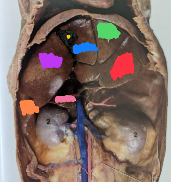
Left medial lobe
Green structure

Left lateral lobe
Red structure

Gallbladder
Yellow structure
Between the quadrate lobe and right medial lobe

Right medial lobe
Purple structure

Caudate lobe
Pink structure

Right lateral lobe
Orange structure

Caudal vena cava
Drains blood from the body wall/pelvic limbs and paired abdominal organs (like the kidneys) to the heart
Hepatic portal vein
Functional blood supply- filters blood from the viscera supplied by the celiac, cranial mesenteric, and caudal mesenteric arteries
Hepatic artery
Nutritional blood supply to liver parenchyma
Right kidney
Makes an impression (renal fossa) in the caudate process of the caudate lobe of the liver
Right lobe
Blue structure

Left lateral lobe
Purple structure

Quadrate lobe
Yellow structure

Caudate lobe
Green structure

Caudate process of the caudate lobe
Red structure

Left medial lobe
Orange structure

Triangular ligaments
Attach the dorsal and lateral aspects of the liver on either side to the diaphragm (f and g)

Falciform ligament/round ligament of the liver
Courses from the liver to the umbilicus and can be identified between the quadrate and left liver lobes (h)

Coronary ligament
Courses between the triangular ligaments around the caudal vena cava. Attaches the liver to the diaphragm
Cecum
Number 4

Descending duodenum
blue asterisk

Base of cecum
black asterisk

Right lobe
Red structure

Caudate process of the caudate lobe
Purple structure

Quadrate lobe
Blue structure

Gallbladder
Green structure

Left lobe
Yellow structure

Caudate lobe
Orange structure

Papillary process of the caudate lobe
White structure

Large animal biopsies
Percutaneous needle biopsies
Right side
Insert needle between the ribs
Goes through the caudal thoracic cavity
This is why the diaphragmatic line of pleural reflection and basal borders of the lung are important
Needle goes through pleural cavity, through the diaphragm, and then immediately into the liver to obtain the simple
Common bile duct
Drains into duodenum along with pancreas secretions
Major duodenal papilla
Where the bile duct and pancreatic duct both open
Minor duodenal papilla
Where the accessory pancreatic duct opens
Right lobe of the pancreas
This body structure lies near the duodenum
Left lobe of the pancreas
This is located near the stomach
Body of the pancreas
In the middle and near the hepatic portal vein
Canine and Equine major duodenal papilla
Where bile duct and pancreatic duct (main duct for pancreatic secretions in equine) empty
Minor duodenal papilla
Where the accessory pancreatic duct empties
The accessory pancreatic duct is the primary route for pancreatic secretions in bovine and canine
Often small or even absent in equine
Bovine major duodenal papilla
Where the bile duct empties
Pancreatic duct not usually present