Spiny Dogfish Shark Practice
 Call Kai
 Learn
 Practice Test
 Spaced Repetition
 Match
 Flashcards
 Knowt Play
 Podcast
1/53

There's no tags or description
Looks like no tags are added yet.
54 Terms
Nares (singular: naris)
These are a pair of openings on each side of the rostrum, anterior to the eyes. Water is taken in and then immediately expelled, indicating this is a "blind pit" and not connected by a pathway to the oral cavity/pharynx. Inside the pit, the water passes by a sensory membrane allowing the shark to detect specific chemicals in the water.
Spiracles
These are small openings caudal to the eyes. These openings allow water to pass through the gills even when the sharks mouth is closed.
Oral Cavity/Mouth
Although the eating function is obvious, this cavity is also used for the intake of water that passes through the gills.
External Gills
Five vertical slits which allow water to exit after passing over the gills. They are located caudally from the jaws.
Lateral Line System
A pale line that extends noticeably from the pectoral fin past the the pelvic fin. This is actually a group of small pores which open into the underlying lateral line canal, a sensory organ that detects water movements and changes in pressure.
Cloaca
This is the common exit from the urinary and digestive tract in both male and female sharks, combined with being the opening for the reproductive tract In females. This area lies between the pelvic fins.
Clasper
Found on male sharks only, these are finger-like extensions of each pelvic fin. They may have a single spine associated with each clasper. These structures aid in sperm transfer during mating.
Anterior Dorsal Fin
Located on the dorsal side of the fish along the spine and closest to the head. Used to help stabilize the body and prevent rotation while moving through water. In addition, helps in species identification.
Posterior Dorsal Fin
Located on the dorsal side of the fish along the spine and closest to the caudal fin. Used to help stabilize the body and prevent rotation while moving through water. In addition, helps in species identification.
Rostrum
This is the pointed area at the anterior end of the head.
Dorsal Spines
Just cranial to each dorsal fin and is used defensively by the shark. Has a poison gland associated with it.
Esophagus
The connection between the pharynx and the stomach. In the shark this part of the alimentary canal is very short and wide and contains many papillae to help pull food into the stomach.
Stomach
This J-shaped Organ is composed of cardiac portion which lies near to the head and pyloric portion which is after the primary bend. The organ ends at the pyloric sphincter - a muscular ring which opens or closes to allow food to pass into the intestine. The pyloric sphincter can be felt if you choose to find it.
Duodenum
This is a short section of the alimentary canal immediately caudal from the stomach. It receives liver secretions known as bile from bile duct and several types of pancreatic digestive juices.
Liver
This organ is composed of three lobes, two large and one smaller. The gall bladder is located alongside/within the smaller lobe and stores the bile created by this organ. In a shark, this organ also helps with maintaining buoyancy.
Pancreas
Divided into two lobes: the ventral lobe, which is easily viewed on the ventral surface of the duodenum and dorsal lobe, which is long and thin located behind the duodenum and extends causally toward the spleen.
Valvular Intestine
Located caudally from the duodenum and distinguished by the extensive network of arteries and veins over its surface. Site of absorption of nutrients into the bloodstream.
Spiral Valve
Interior structure of valvular intestine that allows for increase of surface area for digestion and absorption of nutrients in shark.
Colon/Rectum
This is the short, end portion of the digestive tract between the intestine and the cloaca. Solid waste is passed through here before exiting the body and reabsorption of water into the bloodstream occurs.
Spleen
Located just caudal to the first curve of the stomach. This organ is not part of the digestive tract, but is associated with the circulatory system.
Sinus Venosus
Dorsal to the ventricle, this is a thin walled, non-muscular sac which acts as a collecting place for deoxygenated blood.
Atrium
The first true chamber that blood enters when flowing through the heart.
Conus Arteriosus
A muscular reservoir and accessory chamber that empties after the ventricle contracts. It connects to the ventral aorta.
Ventricle
The main contracting chamber of the heart.
Teeth
These are derived from the scales which cover the shark's body! They have been adapted to function as cutting structures. They are replaced regularly as they wear out.
Pharynx
The area of the oral cavity just posterior to the spiracles and anterior to the esophagus.
Internal Gill Slits
These opening are located on either side of the pharynx. They are protected by gill rakers and supported by gill arches.
Gill Rakers
Cartilaginous protrusions which prevent large particles of food from entering the gills from the oral cavity.
Tongue
Located in the oral cavity, this structure is attached and immobile. In a human, ours is unattached!
Kidney
The shark has two dark-colored, elongated organs on either side of the midline in the pleuroperitoneal cavity. The shark regulates its urinary system in a way unique compared to most other vertebrates. This organ extracts urea from urine and returns the urea to the blood. In this way, the water pressure of the shark's body fluids are maintained as high as that of sea water.
Rectal Gland
A finger-like extension of the rectum. This gland controls the salt concentration within the body and is thus an osmoregulator. Excess salt is secreted into the gland tubule. Via the central gland cavity, salt is released into the colon for expulsion.
Testes (singular: testis/testicle)
These are oval shape and are dorsal to the liver. This organ is where male gametes are produced.
Ovary
These are oval shape and are dorsal to the liver. This organ is where female gametes are produced.
Vas Deferens (aka Ductus deferens)
A highly coiled tube that carries sperm to the clasper.
Oviducts
Elongated tubes that lay dorsal and lateral along the body cavity. These structures can be very prominent in mature female sharks. Both of these tubes share a common opening to the body cavity called the ostium where eggs released by the ovaries will enter.
Uterus
The enlarged caudal end of the oviduct. It is here that fertilized eggs develop into shark pups.
Placoid Scales
Present in cartilaginous fishes and project from surface with a spine extending posteriorly. These structures are toothlike and have a sandpaper-like texture.

Pectoral Fins
A pair of fins, situated just posterior to the head in fishes, that help to control the direction of movement during locomotion
Caudal Fin
The most posterior of the fins. This structure propels the fish.
Urinary Papilla
Lump in the center of this area highlighted by the picture.

Gill Filaments/Lamellae
These feathery and filamentous structures are part of the gills and contain capillary beds necessary for respiration/gas exchange.

Afferent Branchial Arteries
These carry blood away from heart to gills.
Pelvic Fin
which fin?

Pores of Ampullae of Lorenzini
Sensory organs which are sensitive to changes in temperature, water pressure, electrical fields and salinity

Pericardial Cavity
Cavity that contains the heart.

Coelom
Larger, all-encompassing ventral cavity.
Conus Arteriosus
What is D?
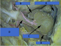
Ventricle
What is C?

Atrium
What is B?
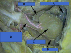
Rugae
What is this internal structure?

Gall Bladder
Sac located alongside the medial lobe of the liver.
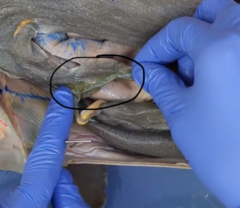
Gill Pouch
Open space where water flows between the internal gill slits and external gill slits where the gill lamellae can be located.

Dorsal aorta
Carries oxygenated blood from the gills to the body.

Efferent Branchial Arteries
Conduct blood from the gills to the dorsal aorta. Can be seen in the roof of the oral cavity once skin removed.
