Stem Cells and Gametogenesis
 Call Kai
 Learn
 Practice Test
 Spaced Repetition
 Match
 Flashcards
 Knowt Play
1/114

Earn XP
Description and Tags
115 Terms
Stem Cells
cells that have clonogenic and self-renewing capabilities and differentiate into multiple cell lineages
History Mid of 1800
the discovery that some cell could generate other cells
History early 1900
the first stem cells were discovered to generate blood cells
History in 1998
James Thomson and coworker derived the first human embryonic stem cell
History in 2005
Scientists at Kingston University in England were purported to have found another category of stem cell.
History 2004-2005
Hwang Wol-suk claimed to have created several human embryonic stem cell lines from unfertilised human oocytes by cloning
History 2006
Scientists at Newcastle University create the first ever artificial liver cells using umbilical cord
History 2007
Dr. Anthony Atala claimed that a new type of stem cells had been isolated in amniotic fluid
Mario Capecchi, Martin Evans, and Oliver Smithies won the 2007 Nobel prize for physiology or medicine
History 2009
Found a way to improve chemotherapy of mouse muscle stem cells
History 2010
Human embryonic stem cells have been cultured under chemically controlled conditions without the use of animal substances
Classification of stem cells
on the basis of Potency
on the basis of their sources
on the basis of Potency
totipotent
pluripotent
multipotent
unipotent
oligopotent
on the basis of their sources
embryonic stem cells
adult stem cell
induced pluripotent
Totipotent
the ability to differentiate into all possible cell types
Totipotent examples
Zygote formed at egg fertilization and the first few cells that result from the division
- gensis
formation
Pluripotent
differentiate into almost all cell types
Pluripotent example
embryonic stem cells and cell that are derived from the mesoderm, endoderm, and ectoderm germ layers
Multipotent
differentiate into a closely related family of cells
Multipotent example
include Hemopoietic stem cells that can become red and white blood cells
Oligopotent
ability to differentiate into few cells
Oligopotent
differentiate into few cells
Oligopotent example
include lymphoid or myeloid stem cells
Unipotent
only produce their own type
Unipotent
Include muscles stem cells
Early or Embryonic
found in the inner cell mass of a blastocyte after approximately five days of development and include totipotent or pluripotent (typically 4-5 days old and hollow microscopic ball of cell called blastocytes)
Mature or Adult
found in specific mature body tissue as well as the umbilical cord and placenta after birth and includes multipotent, oligopotent and unipotent cells
Induced pluripotent stem cells (iPS)
Lab made. they are reprogramming somatic cells back to pulripotent state
goal of stem cell therapies
to promote cell replacement in organs that are damaged and do not have the ability for self repair
PGCs
Promordial germ cells
gametogensis
the differentiation of the germ cells into gametes: eggs and sperms
Spermatogenesis
the development pathway from germ cell to mature sperm
male gametes are formed in
the testis within the seminal tubules
Testicular tissue consists of
tunica capsule surrounding a large number of long, tubular convoluted seminal tubes that form sperms
seminal tubules are linked to each other by
connective tissue rich in blood vessels known as tubular connective
Primoridial germ cells
initiate the formation of spermatogonia that move during its transformation into sperm to the inner region
Ledge cells
secrete the hormone the hormone testosterone
Stages of spermatogenesis
Multiplication
Growth phase
Maturation
Differentiation
Multiplication
primary germ cells divide several consecutive divisions by indirect mitosis method, producing a large number of Spermatogenic cells, each containing (2n)
Growth phase
The spermatogenic cells stop dividing and grow and increase in size, so the nucleus enlarges and this moves towards the center of the tubule and forms the primary spermatocytes cells
Maturation phase
primary spermatocytes divide by meiosis
Maturation phase include
Meiosis 1st
Meiosis 2nd
meiosis 1st
leads to the formation of two cells of equal size and whose nucleus contain an odd number of chromosomes (secondary spermatocytes)
meiosis 2nd
division of each two secondary spermatogenic cells divides into two spermatids each containing (n) thus 4 spermatids are formed
Differentiation
each spermatids will start to differentiate into sperms
each spermatids is a cell that contains
a round nucleus is relatively small in size
chromatin bodies
cytoplasm contains the Golgi apparatus near the nucleus
two centrioles near the periphery of the cell
chromatid body, along with other organelles
Golgi Phase
perforating body
acrosome
Cap phase
gap that contains the acrosomal loses the fluids in it and relaxes over the top of the sperm and becomes what is known as the head cover
nucleus shrinks gradually and the chromosomes are confined to a narrow space, which helps reduce the size and weight of the sperm
Centeral bodies move toward the posterior pole of the nucleus
capitis
head cover
Anterior Central Body
one of the two bodies take a position close to the nucleus
Posterior Central Body
the bodies that take a distant position and is known as the posterior central body, which rests at the base of the tail that began to elongate
Acrosomal Phase
the nucleus moves to the edge and elongate or flattens.
most of cytoplasm moves to the back end of nucleus and then surrounds the front part of tail to which the mitrichondria migrate (mid piece)
the chromatid body moves towards distal central body (tail)
Maturation
transform into a sperm is complete
nucleus and the apex take the final form
annular centering body moves away in the direction of the tail
mitochondria are concentrated in the middle segment
a large number of organelles are destroyed by the sertoli cells
what shape does a mouse nucleus take
sickle
Function of the Sertoli cells
protection through tight junction
remove the cytoplasmic bodies that excrete them sperm
secretion of fluid flowing from the cavities of the seminal tubules toward the genital canals
helping in the release of the sperms
provides nutrients
secrete ABP
ABP
Androgen-Binding protein
Sperms Head
contains the nucleus, and the enzyme acrosome
Acrosome
works to penetrate the outer membrane of the egg with the enzyme it secretes
middle piece of the sperm
contains the mitochondria and two centrioles
Sperms tail
helps the sperm move forward due to the presence of fine fibers in the form of tubules.
Oogenesis
multiplication
growth phase
maturation
Ovum structure
composed of plasma membrane, nucleus, and cytoplasm that contain extra structures such as yolk, colored granules, cortical granules, and glycoproteins
oocyte poles
animal pole
vegetative pole
animal pole
upper half containing the nuclues
vegetative pole
lower half containing the amount of yolk
Ovum membranes
primary
secondary
Primary membrane
formed while the egg cell is inside the ovary
Primary membrane contains
plasma membrane
radial layer
transport membrane
granular layer
vitelline membrane are
the transport membrane of amphibian and birds
chorion are
the transport membrane of fish
Secondary membrane
they are the membranes secreted by the oviducts, uterus, or some secondary glans
secondary glans are
albumin
double shell membrane
hard calcareous crust
classification of oocytes
isolecithal
mesolecithal
telolecithal
centrolecithal
Isolecithal
very little yolk like in placental mammals
Mesolecithal
Moderate amount of yolk like in amphibian and fish eggs
Telolecithal
Lots of yolk at vegetal pole like in reptiles and birds egg
Centrolecithal
lots of yolk centrally located like in insects
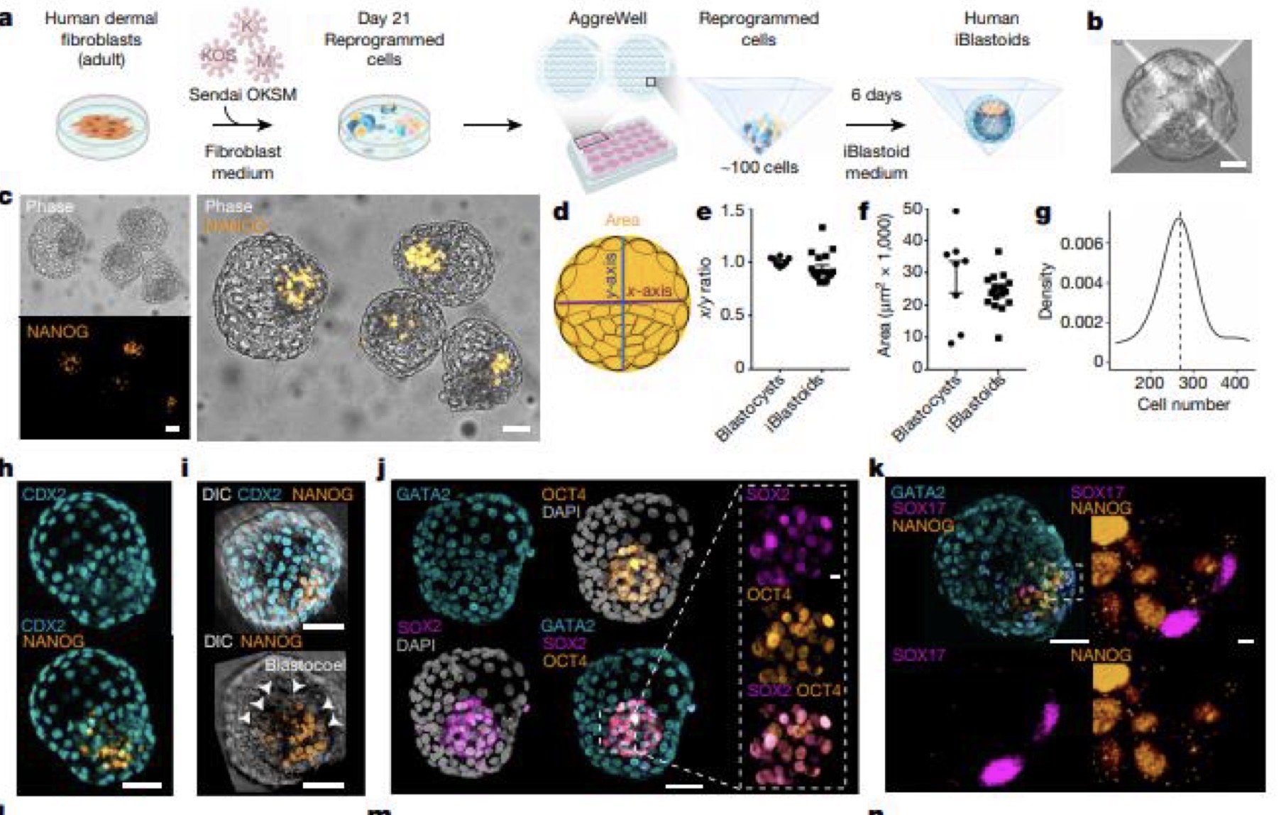
Generation of human iBlastoids

Blastocyst

Embryonic Stem Cell Colony

Mature / Adult cells

Reprogrammed Somatic cells

Induced Pluripotent stem cell

Spermatogenesis

Golgi phase

Cap phase

Acrosomal phase

Maturation phase

Mature Sperm
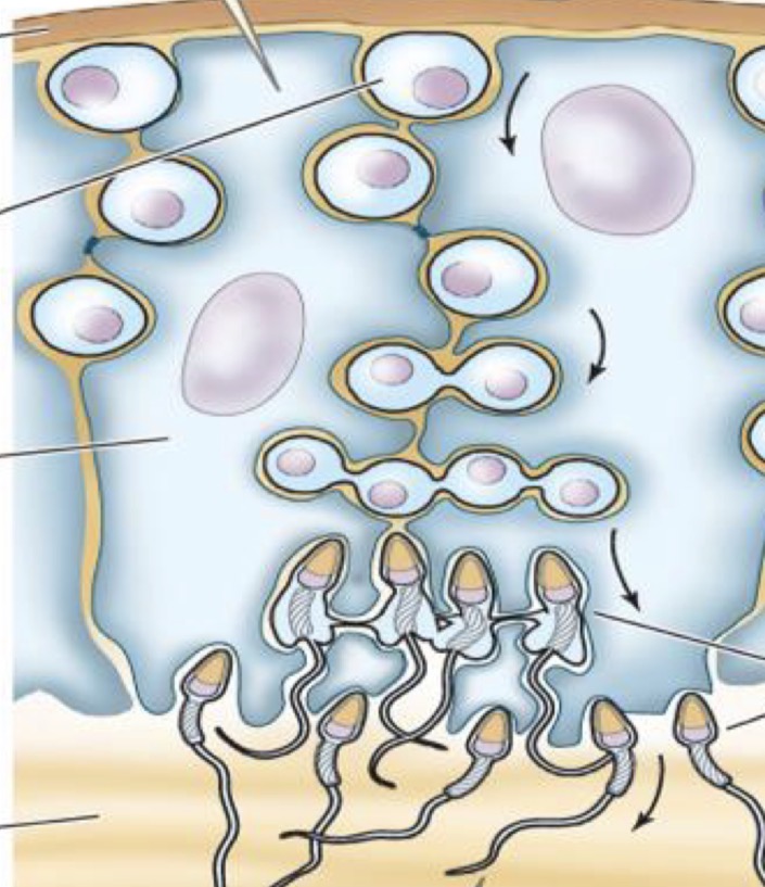
Sertoli cells

Oogenesis phases

Structure of an ovum

Vitelline membrane

Plasma membrane

Transport membrane

Radical layer

Granular layer

Air cell

Thick Albumen