Ch. 9 : Organization of the Motor System
 Call Kai
 Learn
 Practice Test
 Spaced Repetition
 Match
 Flashcards
 Knowt Play
1/22

There's no tags or description
Looks like no tags are added yet.
23 Terms
The Motor System
→ initiating & producing movement requires interaction of info from senses & the entire brain
Motor System:
Motor cortex = plans & initiates movement
Basal Ganglia & cerebellum = coordinate movement
Spinal Cord = conducts info to the muscles
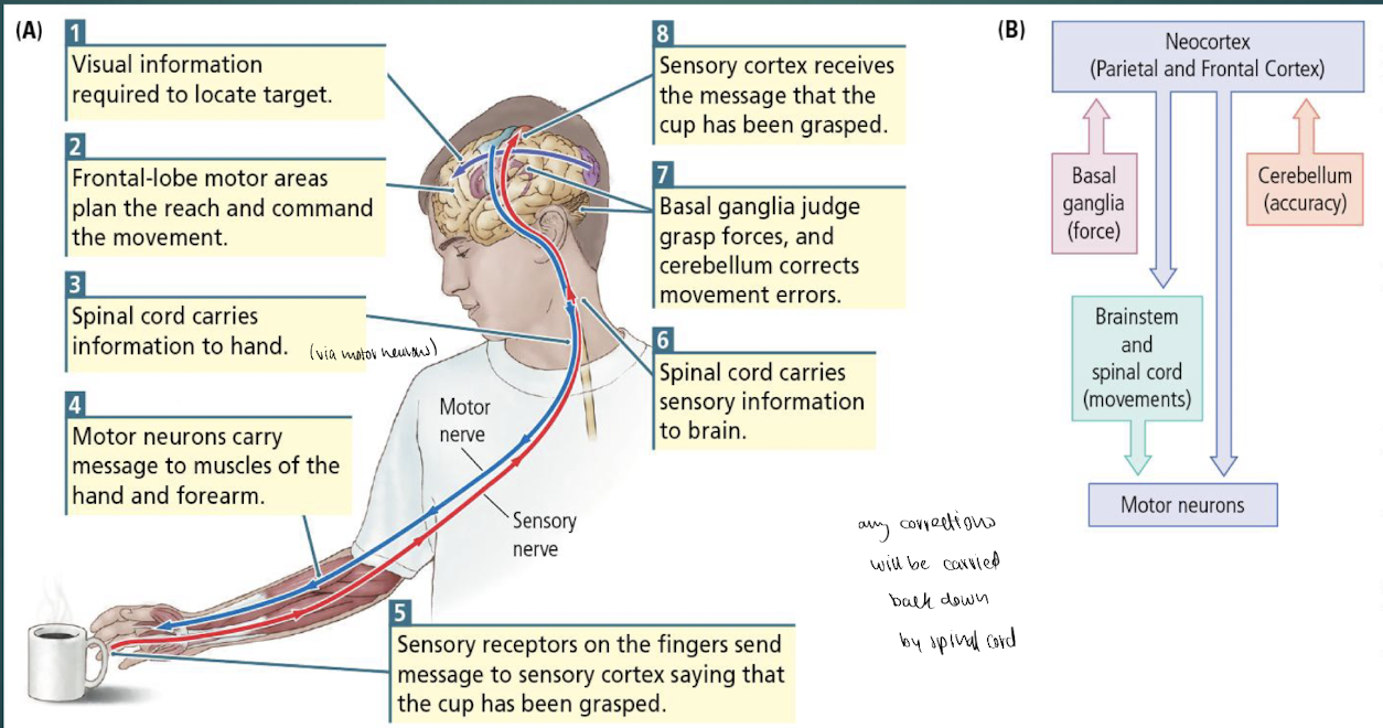
Sequential Production of Movement
visual info locates target
frontal-lobe motor areas plan the reach & command the movement
spinal cord carries info to hand/forearm muscles via motor nuerons
sensory receptors in fingers send message to sensory cortex to let it know the cup was grasped
spinal cord carries sensory info to brain
basal ganglia judges grasp forces & cerebellum corrects movement errors
sensory cortex receives message that the cup has been grasped
note: any corrections will be sent back down the spinal cord
Neocortex : Initiating Movement
→ initiating movement starts in the neocortex
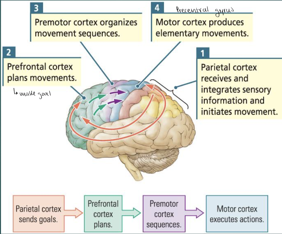
Parietal cortex : integrates info from senses & initiates movements based on that info
Prefrontal cortex : plans movements based on experiences, goals & sensory input
makes goal
ex. want to get water, thus need to know where water is
Premotor Cortex = coordinates whole-body movements & organizes action sequences
→ goal is sent here to plan sequence
most active BEFORE the movement occurs (planning)
Primary Motor Cortex (precentral gyrus) = produces simple movements
looks at plan & produces simple movements based on what muscles will need to do
last step of movement control in the cortex
primary is last here bc it does simple things, unlike the primaries for other systems (auditory, visual,..)
damage to primary motor cortex results in largest impact to movement (cant move)
primary cortex sends it outside of cortex, to thalamus → spinal cord
Hierarchical Control of Movement
→ theres a hierarchical control of movement : as movement complexity increases, more brain areas are recruited to coordinate it
ex. when someone taps their finger : blood flow increases in primary motor cortex & adjacent sensory cortex
if parietal lobe is damaged (sensory cortex) : may not be able to tell when tapping ur finger
ex. when someone performs a movement sequence: blood flow increases in the premotor cortex
if premotor region is damaged:
can still tap & feel fingers tap
CANT plan (use premotor) to move fingers
ex. when someone uses a finger to navigate a maze : blood flow increases in the prefrontal, temporal & parietal cortices

Mapping the Motor Cortex
electrical stimulation of the primary motor cortex (precentral gyrus) triggered movements of diff parts of the body
electrical stimulation of the premotor cortex (anterior to primary) also elicited movement
Motor Homunculus = similar to the somatosensory homunculus, BUT the body is represented upside down
the motor cortex is organized/mapped based on the control of diff body parts:
feet in the central fissure (top of brain)
thus, damage to top of head is more likely to impact moot control of legs/feet
head near the inferior lateral surface (bottom of brain)
has larger representation for hands & tongue - areas of fine motor control
note: modern techniques using microelectrodes that penetrate to layer 5 of the cortex (one of the motor layers) suggest ~10 DIFF homunculi for the motor cortex
not all of these representations are mapped the same way
Ethological Movements
→ brief electrical stimulation of the motor cortex produce movement in a specific body part (ex. leg twitch)
→ LONGER electrical stimulation of motor cortex results in ethological behaviours (ex. defensive postures, reaching movements, climbing & leaping postures)
note: stimulation of primary motor cortex produces less specific ethological behaviours than the premotor motor cortex
ex. longer stim. of primary→ hand in lower space, chewing/licking
ex. longer stim. of premotor → defence, hand to mouth, reach to grasp
Ethological Movements = innate, stereotyped & species-specific sequences of behaviour that run to completion once triggered
behaviours an animal may use in everyday activities
triggered by longer electrical stimulation of motor cortex
also triggered by stimulation of parietal cortex
movements : include the parts of the body to be moved, the destination to which the movement is directed & the function of the movement
→ types of ethological movements seem to be clustered in parts of the motor cortex - bc brain regions for the muscles used are closer
ethological movements are also somewhat organized by the homunculus:
ex. hand movements are ventral
ex. movements of hand to mouth are most ventral
ex. complex, whole-body movements are closer to e/o in the premotor area
note: premotor section of the homunculus is based on types of movement, thus not specific movements
Motor in the Parietal Lobe : Visual-Parietal-Motor Connections
→ ethological movements can ALSO be elicited from stimulation of the parietal cortex
map in a similar way to those stimulated in motor cortex of frontal lobe
ex. stepping movements are triggered by dorsal (top) parietal stim.
ex. hand movements are triggered by ventral (side) parietal stim.
ex. mouth movements are triggered by most ventral (side) parietal stim.
→ sensory info abt nature & location of an object is needed to successfully reach it
→ somatosensory info about body position indicates where the movement is starting
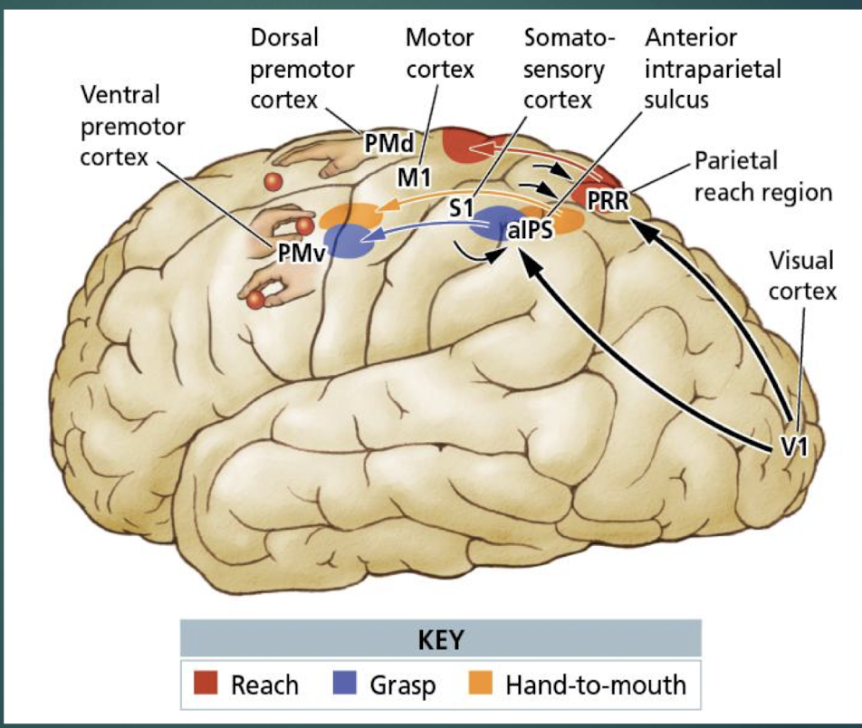
Sensory-to-Motor Channels for Reaching (comparing paths to reach, grasp, and hand-to-mouth)
ex. Path of hand-to-mouth movement:
visual cortex (V1)
ventral path
plan in motor cortex
execute in motor cortex
Movement Lexicon
Movement Lexicon = set movement patters that we know and use to build behaviours
depends on experience & changes constantly
ex. Pincer grip = a developmental milestone in first few weeks of life that gets refined overtime
at birth do the whole hand grasp → learn to use two finger to pick up things at ~3 weeks and continue to improve at it

→ damage to cortical motor areas (of a muscle) impairs the ENTIRE movement, not just the one body part/muscle that correspond to the damaged cortical region
idea that if you’d cant use a muscle, then u must not be able to do the entire movement that involves the muscle
ex. ppl getting physiotherapy to retrain their body to do movements w/o a damaged muscle
impaired movements are replaced by movements that are controlled by an intact part of the cortex
ex. areas that once did the hand grasp in babies are now used for pincer grip
→ stroke studies suggest that movements are encoded in multiple places in the cortex
Specific Damage to Primary motor cortex vs Premotor cortex
damage to primary motor cortex : produces muscle weakness + impairs individual movements
MOST signif damage
damage to premotor cortex : impairs complex movements involving multiple body parts (ex. reach, grasp)
→ movements coordinated by motor planning areas integrate & refine basic movements (walking or climbing movements)
role of neocortex & motor planning areas seems to involve blending these relearned moments
ex.
normal animal : can coordinated 2 movements
animal 5 months after lesion to premotor cortex : CANT coordinate movements

Corticomotor-Neuron activity : Role of Neurons in the Motor Cortex for planning & executing movements
motor cortex = premotor & primary motor cortex ?
study: wanted to understand the role of neurons in the motor cortex for planning & executing movements → tracked neuronal activity of monkeys electrode implanted in wrist area of PRIMARY motor cortex before & during movement with different weights
tracking neuronal activity of wrist area in primary motor cortex
findings:
increased neuronal activity in primary motor when planning movement (before movement)
further increased activity of primary motor during actual movement
primary motor cortex fires MOST during movement
even higher neuronal activity with increased weight on bar
→ primary motor activity increases w more weight

some neurons fired during flexion movements but not during extension → THUS, neurons encode direction of movement
→ diff neurons need diff movements of the wrist to activate

→ movement is encoded by a population of neurons, NOT a single cell
Mirroring Movement : Motor Mirror Neurons
→ many moments are learned & modified based on our interactions with other (learn from others)
Motor Mirror Neurons = active when someones performing a movement & when they observe someone else making that same movement
found in the ventral premotor area (planning & complex movement areas)
allow us to mirror movement (help w learning & emotions)
1st discovered in monkeys & we DONT know how they’re distributed
types:
some neurons are specific to size of the target object
some neurons discharge ONLY when the object in within reach
CORE mirror neurons are less specific & respond to broad classes of movement
→ THUS, we have motor mirror neurons for general movement & specific
note:
additional mirror neurons have been found in the temporal & parietal lobes
believed to be for visual movement & to help us understand others (empathy bc found in emotional areas)
mirror neuron system:

Subcortical (below cortex) Motor Control : 1. Basal Ganglia
Subcortical motor control: basal ganglia, cerebellum & brainstem are important for control of movement
motor cortex sends input to the basal ganglia & cerebellum
Basal Ganglia : receives input form the motor cortex & substantia nigra in the midbrain → sends projections back to these areas
path: substantia nigra → caudate (basal ganglia) → thalamus → cortex → movement
includes : caudate, putamen, globus pallidus
substantia nigra is FUNCTIONALLY part of the basal ganglia
symptoms of movement disorders suggest the role of the basal ganglia is to modulate movements (NOT planning or initiating, just refining)
Subcortical (below cortex) Motor Control : 1. Basal Ganglia - Movement Disorders
→ study of movement disorders allowed us to understand fxn of motor areas
categorized based on types of symptoms
Hyperkinetic Symptoms = Increased movement & Increased unwanted movement
caused by damage to caudate & putamen (basal ganglia)
ex. huntington disease
ex. Tourette syndrome
Hypokinetic Symptoms = decreased voluntary movement & CANT inhibit smaller movements
caused by loss of dopamine input from the substantia nigra to the basal ganglia
associated w difficulty making voluntary movements ( struggle to start or inhibit movement)
ex. parkinson’s disease ( tremors are a lack of inhibiting bc of low dopamine)
→ combination of symptoms suggests the basal ganglia role is to modulate movement - refine it, NOT plan or initiate movement
Subcortical (below cortex) Motor Control : 1. Basal Ganglia - Hypothesis of Movement Force
→ hypothesis : that the basal ganglia controls movement force (ex. how hard we grip), THUS movement disorders are issues w too little/much force
ex. hyperkinetic symptoms would be due to too much force, while hypo kinetic symptoms are too little force
Regulating Movement Force
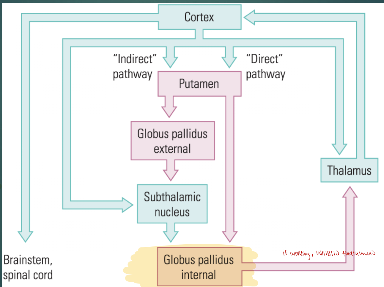
cortex sends excitatory glutamate signal to putamen (in both pathways)
interior globus pallidus sends output to thalamus & motor cortex for movement to occur
if the thalamus is inhibited by the internal globus → not sent to motor cortex → movement DOESNT occur
→ cortex sends info to Basal Ganglia in 2 pathways:
Direct pathway = INCREASES movement by inhibiting internal globus pallidus
ACTIVATED by dopamine - thus w increased dopamine → increased movement
Indirect pathway = DECREASES movement by activating internal globus pallidus
INHIBITED by dopamine - thus w decreased dopamine → decreased movement
→ dopamine levels determine what path occurs:
high dopamine = increased movement
low dopamine = decreased movement

Subcortical (below cortex) Motor Control : 2. Cerebellum
Cerebellum = important for acquiring (learning movement) & maintaining motor skills
organized topographically in a homunculus
receives copy of motor plan from primary motor cortex
contains:
4x as many neurons as the entire cortex
3 sections: 2 hemispheres + a small horizontal lobe (flocculus)
Homunculus → the 3 sections of the cerebellum are involved in diff aspects of motor control
Flocculus = receives info from vestibular system + is involved in balance
Midline areas = control the midline of body (ex. waist, internal organs)
lateral areas = involved in movements of limbs & hands

Subcortical (below cortex) Motor Control : 2. Cerebellum - Role in Movement
Roles of cerebellum in movement:
important in timing of movements
damage to cerebellum impairs ability to move at a regular rhythm & ability to accurately perceive time
important for monitoring movement accuracy
plays role in adjustment
→ pts with intact cerebellum: adjust overtime to distorted glasses and can throw accurately, however when they’re removed they’ll throw incorrectly bc they have to readjust
→ whereas, pts w a damaged cerebellum: UNable to adjust their movements to hit a target, but after removing the distorted goggles they threw accurately, bc they NEVER adjusted in the first place (due to damaged cerebellum) - no adjustment to return to their baseline

Subcortical (below cortex) Motor Control : 2. Cerebellum - Motor Feedback
when a movement DOESNT reach the intended target: adjustment Is needed
cortex sends motor command to spinal cord + a copy to cerebellum
sensory info from muscles & visual system is sent to cerebellum (feedback)
cerebellum compares the intention (motor output) w the results (sensory input) → generates a correction
THUS, cerebellum corrects movements & learns for future movements. whereas the basal ganglia only refines before movements

note: cognitive process (like language) use a smiler error correction strategy based in the cerebellum
Subcortical (below cortex) Motor Control : 3. Brainstem
Brainstem : the main motor control pathway originates in the motor cortex, BUT splits into multiple pathways in the brainstem
spinal cord pathways provide input abt balance & posture to refine movements commanded by the cortex
movements specified by the brainstem tend to be coarse movements of the ENTIRE body (ex. fully body reflex/spasms)
→ electrical stimulation of brainstem areas in a cat: produce arched back & erect hair behaviours + walking or running behaviours
timing of these behaviours depended on the timing of stimulation & intensity was determined by magnitude of the stimulation → led to question of whether its bc ur stimulating the brainstem or bc it communicates
Communicating with the Spinal Cord
→ the spinal cord is the FINAL pathway to convert motor plans into movements
corticospinal & corticobulbar tracts project from cortex → spinal cord
corticobulbar tracts = control facial movements
corticospinal tracts = influence movement of limbs, digits & body
originate in somatosensory areas, motor cortex & premotor cortex
input from somatosensory areas modulates afferent sensory info (sensory info twos the CNS)
input from motor areas → motor neurons of brain stem & spinal cord to influence movement
Spinal Cord Pathways
→ corticospinal & corticobulbar tracts originate in layer 6 (V) of the motor cortex → project twd spinal cord
note: layer 6 sends motor info to the thalamus
Corticospinal-Tract Pathway
95% of descending motor pathways (from cortex) decussate (join axons) on the VENTRAL surface of the brainstem → results in a bump called the pyramids
lateral corticospinal tract = decussates (joins axons) & influences the movements of limbs & digits contralateral (opposite) to the hemisphere from the motor command side
anterior corticospinal tract = DOESNT decussate + controls movement of the trunk
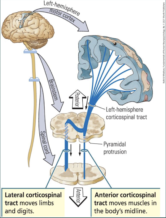
ex. L-hemis motor cortex sends commands down L-hemisphere corticospinal tract:
some commands are sent down the R-body’s lateral cortiscopinal tract → move the limbs & digits
some commands are sent down the L-body’s anterior corticopsinal tract → moves midline muscles (trunk)
Spinal Motor Neurons
→ motor neurons of the spinal cord provide the output connection b/w the nervous system & muscles
neurons from both cortiospinal tracts (lateral & anterior) synapse on interneurons AND motor neurons
lateral tract : synapses w interneurons & motor neurons that innervate muscles of limbs & digits
anterior tract : synapses w interneurons & motor neurons that innervate the trunk (midline of body)
interneurons in spinal cord project to motor neurons → motor neurons projects to muscles of body
spinal motor neurons & interneurons are arranged in a homunculus
lateral motor neurons: influence fingers & hand
intermediate motor neurons: control limbs
medial motor neurons: control trunk
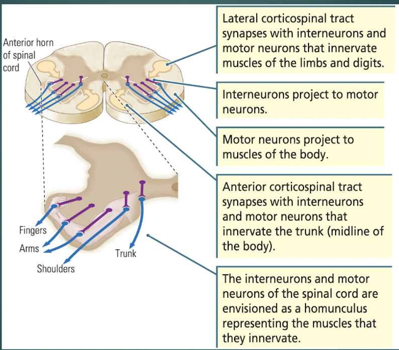
Muscular coordination in the Spinal cord
muscles that control limbs are arranged in pairs (anatgonist muscles)
extensor muscles = move limb AWAY from trunk
flexor muscles = move limb twd trunk
→ spinal circuits cause one muscle of the pair to relax while the other muscles is excited → allows precise movements & prevents muscle fatigue
