1062 FINAL EXAM Topics I,II,III

0.0(0)
Studied by 0 people
call kaiCall Kai
learnLearn
examPractice Test
spaced repetitionSpaced Repetition
heart puzzleMatch
flashcardsFlashcards
GameKnowt Play
Card Sorting

1/231

encourage image

There's no tags or description

Looks like no tags are added yet.

232 Terms

1
New cards

What is DNA?

  • Stores hereditary information (is a polymer of nucleotide monomers)

  • The sequence of nucleotides gives rise to unique sequences that are called genes, and they code for specific proteins

  • Double stranded

  • 4 Nucleotides:

    • Adenine (A), Guanine (G) → Purines (2 aromatic rings = double carbon/nitrogen ring)

    • Cytosine (C) and Thymine (T) → Pyrimidines (1 aromatic ring = 1 carbon/nitrogen ring) + Uracil

    • Complementary base pairing: A <-> T (2 H-bonds), G <-> C (3 H-Bonds)

  • General note: Standard notation is always in the 5’ to 3’ direction, the question is always given in the 5’ to 3’

    • E.g., Determine the probe needed to identify the sequence AUGGGAU

      • 5’ AUGGGAU 3’

      • 3’ TACCCTA 3’

      • ANSWER: 5’ ATCCCAT 3’

      • Also note probes for DNA can be made from DNA or RNA


<ul><li><p>Stores hereditary information (is a polymer of nucleotide monomers)</p></li><li><p>The sequence of nucleotides gives rise to unique sequences that are called genes, and they code for specific proteins</p></li><li><p>Double stranded</p></li><li><p>4 Nucleotides:</p><ul><li><p>Adenine (A), Guanine (G) → Purines (2 aromatic rings = double carbon/nitrogen ring)</p></li><li><p>Cytosine (C) and Thymine (T) → Pyrimidines (1 aromatic ring = 1 carbon/nitrogen ring) + Uracil</p></li><li><p>Complementary base pairing: A &lt;-&gt; T (2 H-bonds), G &lt;-&gt; C (3 H-Bonds)</p></li></ul></li><li><p><span style="color: rgb(157, 248, 227);">General note: Standard notation is always in the 5’ to 3’ direction, the question is always given in the 5’ to 3’ </span></p><ul><li><p>E.g., Determine the probe needed to identify the sequence AUGGGAU </p><ul><li><p>5’ AUGGGAU 3’</p></li><li><p>3’ <span style="color: rgb(137, 218, 88);">TACCCTA </span>3’ </p></li><li><p>ANSWER: 5’ <span style="color: rgb(126, 211, 80);">ATCCCAT </span>3’</p></li><li><p>Also note probes for DNA can be made from DNA or RNA </p></li></ul></li></ul></li></ul><p></p>
2
New cards
<p>PCR Primers: Forward and Reverse </p>

PCR Primers: Forward and Reverse

  • Add a huge excess of both forward and reverse primers into the reaction mix

    • This is important because every cycle, every available template strand needs a primer to bind to it (quick exponential growth)

  • The forward primer is designed to match a sequence on the bottom strand, and it binds there, ready to extend left-to-right (5'→3') along the top strand's sequence

  • The reverse primer is designed to match a sequence on the top strand, and it binds there, ready to extend right-to-left, but still synthesizing 5'→3' (i.e. it goes in the opposite physical direction)

  • The primer doesn't detach or get removed. It becomes covalently bonded, permanently, into the new strand


How does removing one primer lead to a change in length?

  • Product length = the distance between where the forward primer starts and where the reverse primer starts

  • The reverse primer acts as a stop signal, once synthesis reaches the reverse primer's binding site, that's exactly where the new strand ends

  • Without the reverse primer, there's no defined stopping point anywhere, it keeps going until it physically runs out of template to copy

  • That "end of the template" is essentially arbitrary

  • Thus you get Long, run-off single-stranded products of variable, unpredictable lengths


3
New cards

PCR Temperature

If temperatures are too high:

  • Denaturation too hot (e.g., way above 95–98°C): The DNA polymerase itself can start to denature and lose activity. Taq polymerase is heat-stable but not indestructible — repeated cycles at excessive heat degrade it over the course of the reaction, so amplification efficiency drops off in later cycles

  • Annealing too hot: The primers can't form stable hydrogen bonds with the template DNA (their melting temperature is exceeded), so they fail to bind at all. This gives you little or no product, since primers can't initiate synthesis

  • Extension too hot: This pushes past the polymerase's optimal working temperature, slowing it down or damaging it, reducing how much DNA gets synthesized per cycle


If temperatures are too low:

  • Denaturation too cold: The double-stranded DNA doesn't fully separate into single strands. Since primers need single-stranded template to bind, incomplete denaturation means incomplete or failed amplification, and it gets worse each cycle as unseparated strands accumulate.

  • Annealing too cold: Primers bind too loosely and non-specifically — they'll stick to sequences that are only a partial match, not just the exact target. This causes non-specific amplification (extra bands on a gel, primer-dimers, off-target products) rather than a clean single product.

  • Extension too cold: The polymerase works, but more slowly and less efficiently, so you may not get full-length products within the cycle time — especially a problem for longer amplicons

.

4
New cards
<p>Why the lagging strand loses its 3′‑OH (in relation to telomerase) </p>

Why the lagging strand loses its 3′‑OH (in relation to telomerase)

  • DNA polymerase can only add new nucleotides to a free 3′‑OH group on an existing strand

  • Every time DNA polymerase extends a strand, it’s doing this reaction:

    (3′‑OH of existing strand)+(incoming dNTP)→phosphodiester bond

  • Without that 3′‑OH, polymerase literally has nowhere to attach the next base


On the lagging strand, replication happens in short Okazaki fragments, each starting with an RNA primer.
When the final primer at the very end is removed (RNase):

  • There’s no upstream fragment providing a 3′‑OH for polymerase to extend from

  • The template still has a short unreplicated region, but polymerase can’t fill it

  • → That small gap remains unfilled → the daughter chromosome is slightly shorter

So it’s not that the primer “can’t fit” — it’s that after the primer is gone, there’s no 3′‑OH left to continue synthesis.


<ul><li><p><span>DNA polymerase can only add new nucleotides to a <strong>free 3′‑OH group</strong> on an existing strand</span></p></li><li><p><span>Every time DNA polymerase extends a strand, it’s doing this reaction:</span></p><p><span>(3′‑OH&nbsp;of&nbsp;existing&nbsp;strand)+(incoming&nbsp;dNTP)→phosphodiester&nbsp;bond</span></p></li><li><p><span>Without that 3′‑OH, polymerase literally has nowhere to attach the next base</span></p></li></ul><p></p><p><span>On the lagging strand, replication happens in short <strong>Okazaki fragments</strong>, each starting with an <strong>RNA primer</strong>.<br>When the final primer at the very end is removed (RNase):</span></p><ul><li><p><span>There’s <strong>no upstream fragment</strong> providing a 3′‑OH for polymerase to extend from</span></p></li><li><p><span>The template still has a short unreplicated region, but polymerase can’t fill it</span></p></li><li><p><span>→ That small gap remains unfilled → the daughter chromosome is slightly shorter</span></p></li></ul><p><span>So it’s not that the primer “can’t fit” — it’s that <strong>after the primer is gone, there’s no 3′‑OH left to continue synthesis</strong>.</span></p><p></p>
5
New cards
<p>How to use a gel electrophoresis to calculate allele frequency and genotype frequency?</p>

How to use a gel electrophoresis to calculate allele frequency and genotype frequency?

This includes calculating the allele frequency - that is, the frequency of each of the I and D alleles in the population. We can also calculate the genotype frequency - the frequency of each of the three genotypes II, ID and DD in the population

<p><span>This includes calculating the </span><strong>allele frequency</strong><span> - that is, the frequency of each of the I and D alleles in the population. We can also calculate the </span><strong>genotype frequency</strong><span> - the frequency of each of the three genotypes II, ID and DD in the population</span></p>
6
New cards
<p>QS: The image above depicts a polyacrylamide gel obtained following electrophoresis of fragments generated via amplifcation of a template fragment of DNA with chain-terminating nucleotides:&nbsp;Sanger sequencing.</p><p>Based on the gel, the sequence of the original DNA fragment is: 5’________ 3’</p>

QS: The image above depicts a polyacrylamide gel obtained following electrophoresis of fragments generated via amplifcation of a template fragment of DNA with chain-terminating nucleotides: Sanger sequencing.

Based on the gel, the sequence of the original DNA fragment is: 5’________ 3’

  1. First find out the sequence of the DNA on the gel electrophoresis - 5’ ATGCCAGTA 3’

  2. Now find its complementary sequence - 3’ TACGGTCAT 5’

  3. The question asks for the 5’ → 3’ direction, thus flip the sequence from step 2


*ALWAYS NOTE THAT THE GEL GIVES IT IN THE 5’ TO 3’ DIRECTION


7
New cards
<p>What are some Molecular biology techniques </p>

What are some Molecular biology techniques

  • Note when to use them (e.g. do you need to identify a gene, do you need to count a gene)

  • Also note you should preferably use DNA if you need to identify something


  • Visualise the regions in a tissue where a specific gene is highly expressed → FISH

  • Compare expression of a large set of genes involved in antiviral response before and after monkeypox infection

    • Microarray can be quantitative and allows comparison of two conditions (before/after, healthy/disease...)

  • Compare expression of a single gene involved in antiviral response before and after monkeypox infection → RT-qPCR

    • The reverse transcription (RT) allows synthesis of DNA from the mRNA pool that reflects levels of expression. The amplification allows quantitative comparison of the mRNA from the two conditions (qPCR)

  • Identify mutated genes in a cancerous cell → NGS

    • NGS allows the identification of all mutations present in the cancerous cells without prior knowledge of the specific gene(s)

  • Identify over-expressed genes in a cancerous cell → Microarray

    • Best tool to allow quantitative comparison of expression levels between cancerous and healthy cell

  • Identify the molecular basis of a disease involving a deficient DNA helicase → Sanger sequencing and functional analysis

    • The gene can be sequenced to identify the mutation(s) in this gene

    • It should then be cloned and expressed to investigate the functionality of the mutant (gain of function, loss of function, specificity change...)


  • Sanger sequencing is qualitative (and semi‑quantitative at best) — meaning it tells you what the sequence is, which variants are present, and whether something is there or not, but not how much of each variant is present

<ul><li><p>Note when to use them (e.g. do you need to identify a gene, do you need to count a gene)</p></li><li><p>Also note you should preferably use DNA if you need to identify something</p></li></ul><p></p><div data-type="horizontalRule"><hr></div><p></p><ul><li><p>Visualise the regions in a tissue where a specific gene is highly expressed → FISH</p></li><li><p>Compare expression of a large set of genes involved in antiviral response before and after monkeypox infection</p><ul><li><p><span style="color: yellow;">Microarray can be quantitative and allows <strong>comparison </strong>of two conditions (before/after, healthy/disease...)</span></p></li></ul></li><li><p>Compare expression of a <em>single gene</em> involved in antiviral response before and after monkeypox infection → RT-qPCR</p><ul><li><p>The reverse transcription (RT) allows synthesis of DNA from the mRNA pool that reflects levels of expression. The amplification allows <em>quantitative </em>comparison of the mRNA from the two conditions (qPCR)</p></li></ul></li><li><p>Identify mutated gene<u>s</u> in a cancerous cell → NGS</p><ul><li><p>NGS allows the identification of <em>all mutations</em> present in the cancerous cells <strong>without prior knowledge</strong> of the specific gene(s)</p></li></ul></li><li><p>Identify over-expressed genes in a cancerous cell → Microarray</p><ul><li><p>Best tool to allow quantitative <strong>comparison </strong>of expression levels between cancerous and healthy cell</p></li></ul></li><li><p>Identify the molecular basis of a disease involving a deficient DNA helicase → Sanger sequencing and functional analysis</p><ul><li><p>The gene can be sequenced to identify the mutation(s) in this gene</p></li><li><p>It should then be cloned and expressed to investigate the functionality of the mutant (gain of function, loss of function, specificity change...)</p></li></ul></li></ul><p></p><p></p><ul><li><p>Sanger sequencing is qualitative (and semi‑quantitative at best) — meaning it tells you what the sequence is, which variants are present, and whether something is there or not, but not how much of each variant is present</p></li></ul><p></p>
8
New cards
<p>Complementary Base Pairing </p>

Complementary Base Pairing

  • Nitrogenous bases united by hydrogen bonds

    • A purine on one backbone with a pyrimidine on the other

    • A–T two hydrogen bonds (weaker)

    • C–G three hydrogen bonds (stronger cuz of the 3 H-bonds + due to better stacking)

  • DNA base pairing → A–T and C–G

  • Law of complementary base pairing

    • One strand determines base sequence of other

  • Base complementarity allows DNA to be precisely copied and to be read for gene expression

    • The weak bonds b/w strands is also an important factor that enables DNA to be copied, as the DNA helix has to actually be able to unwind for RNA polymerase to DO the copying!


<ul><li><p>Nitrogenous bases united by hydrogen bonds</p><ul><li><p>A purine on one backbone with a pyrimidine on the other</p></li><li><p>A–T two hydrogen bonds (weaker)</p></li><li><p>C–G three hydrogen bonds (stronger cuz of the 3 H-bonds + due to better stacking) </p></li></ul></li><li><p>DNA base pairing → A–T and C–G</p></li><li><p>Law of complementary base pairing</p><ul><li><p>One strand determines base sequence of other</p></li></ul></li><li><p>Base complementarity allows DNA to be precisely copied and to be read for gene expression</p><ul><li><p>The weak bonds b/w strands is also an important factor that enables DNA to be copied, as the DNA helix has to actually be able to unwind for RNA polymerase to DO the copying!</p></li></ul></li></ul><p></p>
9
New cards

What do the bases look like?

  • Look at the blue circles, these are what differentiates between the pyrimidines and purines


<ul><li><p>Look at the blue circles, these are what differentiates between the pyrimidines and purines </p></li></ul><p></p>
10
New cards

What is the central dogma of Molecular Biology?

  • Information flows FORWARDS (not backwards) DNA → RNA → Polypeptide

  • Not ALWAYS forwards, there are times backwards
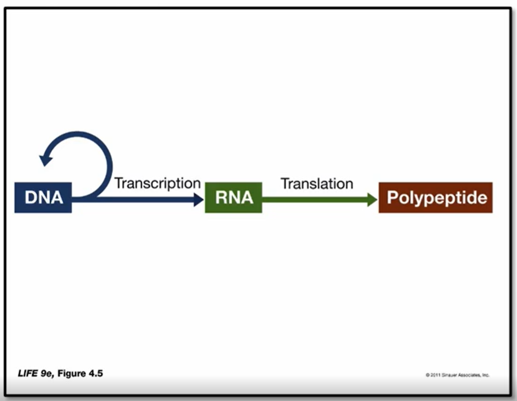

<ul><li><p>Information flows FORWARDS (not backwards) DNA → RNA → Polypeptide </p></li><li><p>Not ALWAYS forwards, there are times backwards </p></li></ul><p></p>
11
New cards
<p>Difference between RNA and DNA</p>

Difference between RNA and DNA

  • RNA - ribose sugar, DNA - Deoxyribose sugar (2’ has no oxygen attached)


<ul><li><p>RNA - ribose sugar, DNA - Deoxyribose sugar (2’ has no oxygen attached)</p></li></ul><p></p>
12
New cards

Nucleoside

  • The phosphate is connected to the 5’ position of the sugar → nucleotide

  • If no phosphate, only sugar → nucleoside


<ul><li><p>The phosphate is connected to the 5’ position of the sugar → nucleotide</p></li><li><p>If no phosphate, only sugar → nucleoside </p></li></ul><p></p>
13
New cards

What are the different outcomes for selection via blue/white screening?

  • When grown in the presence of X-gal/Bluo-Gal, bacterial colonies that express b-galactosidase turn blue (Lac Z gene encodes b-galactosidase)

  • There are 3 different outcome: Plasmid WITH inserted gene of interest in bacteria, plasmid WITHOUT inserted gene of interest in bacteria, no plasmid in bacteria


<ul><li><p>When grown in the presence of X-gal/Bluo-Gal, bacterial colonies that express b-galactosidase turn blue (Lac Z gene encodes b-galactosidase) </p></li><li><p>There are 3 different outcome: Plasmid WITH inserted gene of interest in bacteria, plasmid WITHOUT inserted gene of interest in bacteria, no plasmid in bacteria </p></li></ul><p></p>
14
New cards

What does DNA/RNA stand for?

  • Acidic due to the phosphate molarity


<ul><li><p>Acidic due to the phosphate molarity </p></li></ul><p></p>
15
New cards

How does nucleotides link (sugar-phosphate backbone)?

  • Polarity: 5’ phosphate (C5) – 3’ OH (C3)

    • The 3’ holds the phosphodiester bond

  • Phosphodiester bond (covalent)

  • Catalysed by a polymerase (DNA ligase)

  • Forms linear polynucleotide


<ul><li><p>Polarity: 5’ phosphate (C5) – 3’ OH (C3)</p><ul><li><p>The 3’ holds the phosphodiester bond</p></li></ul></li><li><p>Phosphodiester bond (covalent)</p></li><li><p>Catalysed by a polymerase (DNA ligase)</p></li><li><p>Forms linear polynucleotide</p></li></ul><p></p>
16
New cards

What are the features that define the DNA structure?

  • Minor and major grooves

    • Major → proteins can bind more readily here (wider)

    • Minor → More narrow, can find some molecules such as dye and drugs

  • Uniform diameter of 2 nm in its most common B-form

  • Right-handed twist with 10-10.5 base pairs per turn (~3.4nm)

  • Antiparallel

  • The double helical structure of DNA is essential to its functions:

    • Storage of an organism’s genetic information; order of bases

    • It must be precisely copied in the cell division cycle; base complementarity means that one strand of bases defines the other

    • It must be readable to express genetic information; the weak interactions involved in connecting the two DNA strands means that they can be dissociated and transcribed into RNA

    • Susceptible to change (mutation); change of specific bases


<ul><li><p>Minor and major grooves</p><ul><li><p>Major → proteins can bind more readily here (wider)</p></li><li><p>Minor → More narrow, can find some molecules such as dye and drugs</p></li></ul></li><li><p>Uniform diameter of 2 nm in its most common B-form</p></li><li><p>Right-handed twist with 10-10.5 base pairs per turn (~3.4nm)</p></li><li><p>Antiparallel</p></li><li><p>The double helical structure of DNA is essential to its functions:</p><ul><li><p>Storage of an organism’s genetic information; order of bases</p></li><li><p>It must be precisely copied in the cell division cycle; base complementarity means that one strand of bases defines the other</p></li><li><p>It must be readable to express genetic information; the weak interactions involved in connecting the two DNA strands means that they can be dissociated and transcribed into RNA</p></li><li><p>Susceptible to change (mutation); change of specific bases</p></li></ul></li></ul><p></p>
17
New cards

General features of RNA

  • 4 bases - A,C,G,URACIL

  • Only one nucleotide strand (single strand)

  • For coding RNAs, the base sequence form a message … but not all RNAs are coding

  • Much shorter than DNA

    • tRNA (transfer), 70 - 90 bases

    • mRNA (messenger), variable length often <10 kb (kb = kilo bases)

    • microRNAs, 21-25 bases

    • rRNAs (ribosomal), several kb

  • Migrates to the site of protein synthesis

  • Directs protein synthetic machinery

  • RNA is transient (short-period of time)


18
New cards
<p>What are the major species of RNA</p>

What are the major species of RNA

  • Functional RNA species are: mRNA (no major 2nd structure), tRNA and rRNA

    • rRNA forms a very large 3D secondary structure


<ul><li><p>Functional RNA species are: mRNA (no major 2nd structure), tRNA and rRNA</p><ul><li><p>rRNA forms a very large 3D secondary structure </p></li></ul></li></ul><p></p>
19
New cards

Secondary Structures of RNA

knowt flashcard image
20
New cards

Conformation of DNA in Prokaryotes

  • Prokaryotic DNA is generally circular, supercoiled and complexed with an RNA protein core


<ul><li><p>Prokaryotic DNA is generally <strong><u>circular</u></strong>, supercoiled and complexed with an RNA protein core</p></li></ul><p></p>
21
New cards

What are the structural properties of DNA that influence its physical properties?

  • Double stranded helix

  • Complementary base pairing b/w strands (via H-bonds) - WEAK

  • Length/Diameter (e.g. in plasmids)

  • Acidity due to negatively charged phosphate group

    • Can be used to determine the purity of DNA


22
New cards

DNA: Solubility

DNA is polyanionic due to its high phosphate content (negatively charged and slightly acidic). Thus it is:

  • Soluble in aqueous (high polarity) / low salt conditions

    • In water (+) the negative charges in DNA repel each other strongly → soluble

  • A salt of Na+, Mg2+ etc. at neutral pH

    • Cations shield the negative charge, therefore a little shielding makes it more comfortable in solution → increases solubility

  • Insoluble in alcohols

    • Alcohol is much less polar than water. In water, salts help DNA stay dissolved by shielding its negative charges. In alcohol, salts don’t work well → DNA becomes sticky → DNA precipitates (white)


<p>DNA is <strong>polyanionic </strong>due to its high phosphate content (negatively charged and slightly acidic). Thus it is:</p><ul><li><p><span style="color: rgb(178, 217, 89);"><strong>Soluble </strong></span>in aqueous (high polarity) / low salt conditions</p><ul><li><p>In water (+) the negative charges in DNA repel each other strongly → soluble </p></li></ul></li><li><p>A salt of Na+, Mg2+ etc. at neutral pH</p><ul><li><p>Cations shield the negative charge, therefore a little shielding makes it <em>more comfortable</em> in solution → increases solubility </p></li></ul></li><li><p>Insoluble in alcohols</p><ul><li><p>Alcohol is much less polar than water. In water, salts help DNA stay dissolved by shielding its negative charges. In alcohol, salts don’t work well → DNA becomes sticky → DNA precipitates (white)</p></li></ul></li></ul><p></p><p></p>
23
New cards

DNA: Viscosity

DNA is naturally very viscous (resists flow, thick and clinging like egg white)

  • Long/large, rigid, rod-like shape

  • Large hydrodynamic volume - attracts a very large shell of water (due to the phosphate groups)

  • Disruption of H-bonding decreases viscosity (dsDNA → (thru heating) ssDNA)

  • Mechanical shearing decreases viscosity

    • This is breaking down the length/ breaking it down into smaller pieces - still dsDNA

  • Can be used to determine the purity of DNA


24
New cards

DNA: UV light Absorption and Fluorescence

The nitrogenous bases of DNA strongly absorb UV light maximally at 260nm

  • UV absorption is due to the aromatic nature of the nitrogenous bases which can absorb energy from the UV light

  • Used to measure concentration

  • ssDNA absorption > dsDNA

    • Double helix shields bases

  • Dye bound to minor groove emits blue light when illuminated with UV light (i.e. fluoresces)

  • RNA can absorb UV better than DNA because nitrogenous are exposed and can absorb more

  • Proteins absorb UV light maximally at 280nm due to Trp and Tyr amino acids. This information can be used to determine the purity of a DNA sample (e.g. see if there are proteins in the DNA sample, thus a higher UV max abs lvl)

  • Can also be used to quantify the amount of DNA?


<p>The nitrogenous bases of DNA strongly <span style="color: rgb(255, 222, 78);">absorb UV light maximally at 260nm</span></p><ul><li><p>UV absorption is due to the aromatic nature of the nitrogenous bases which can absorb energy from the UV light</p></li><li><p>Used to measure concentration</p></li><li><p>ssDNA absorption &gt; dsDNA</p><ul><li><p>Double helix shields bases</p></li></ul></li><li><p>Dye bound to minor groove emits blue light when illuminated with UV light (i.e. fluoresces)</p></li><li><p>RNA can absorb UV better than DNA because nitrogenous are exposed and can absorb more</p></li><li><p><span style="color: rgb(160, 151, 151);">Proteins absorb UV light maximally at 280nm due to Trp and Tyr amino acids. This information can be used to determine the purity of a DNA sample (e.g. see if there are proteins in the DNA sample, thus a higher UV max abs lvl)</span></p></li><li><p><span style="color: rgb(160, 151, 151);">Can also be used to quantify the amount of DNA?</span></p></li></ul><p></p>
25
New cards
<p>DNA: Denaturation </p>

DNA: Denaturation

  • H-bonding is disrupted

  • Caused by INCREASE in­ temperature, BOTH extremes of pH

  • On denaturation viscosity decreases and UV absorption increases , fluorescence decreases (as no more minor grooves)

    • Due to the single separated strands

  • Can be used to determine the purity of DNA

  • In the picture above, the “bubbles” shown are the single stranded DNA, in yellow are the dsDNA

    • Happens during transcription (if mediated by enzymes, not temperature)


<ul><li><p>H-bonding is disrupted</p></li><li><p>Caused by INCREASE in­ temperature, BOTH extremes of pH</p></li><li><p>On denaturation viscosity decreases and UV absorption increases , fluorescence decreases (as no more minor grooves)</p><ul><li><p>Due to the single separated strands</p></li></ul></li><li><p>Can be used to determine the purity of DNA</p></li><li><p>In the picture above, the “bubbles” shown are the single stranded DNA, in yellow are the dsDNA</p><ul><li><p>Happens during transcription (if mediated by enzymes, not temperature) </p></li></ul></li></ul><p></p>
26
New cards
<p>DNA Melting curve </p>

DNA Melting curve

(Pic above)

  • Increasing temperature (X-axis) leads to denaturation of double stranded DNA into single stranded DNA

  • This leads to an increased absorbance of UV light as indicated by the Y-axis

  • Tm → melting temperature/melting point, this is the temperature at which half (50%) the strands of DNA are denatured and the other half in a double strand (dsDNA) form at equilibrium

    • Depends on:

      • The size of the molecule

      • Its GC content where… Tm is proportional to G + C (more GC, higher mp, line shifts to the right)

      • The pH and ionic strength

        • High pH → alters the ionisation state of H-bonds → more denaturation → lower Tm

        • High Salt → stabilise dsDNA → Higher Tm


(Pic below)

Steepness/GRADIENT of transition depends on the complexity of DNA

  • Steeper — melts over a narrower temperature range and is less complex (red is less complex than blue)

  • Complex DNA tends to have many different sequences, whereas simple DNA is usually repeated sequences

  • BUT IT DOES NOT CHANGE THE Tm → this is going to be based on the number of GC content or the length of the the molecule


Higher pH = destabilisation = Lower Tm

  • Fun fact, low pH can destroy/denature DNA way worse than high (alkaline) pH can, therefore high pH is used more than low pH (cuz otherwise there’s no DNA left to work with!)

Higher ionic strength = stabilisation = higher Tm

<p>(Pic above)</p><ul><li><p>Increasing temperature (X-axis) leads to denaturation of double stranded DNA into single stranded DNA</p></li><li><p>This leads to an increased absorbance of UV light as indicated by the Y-axis</p></li><li><p>T<sub>m</sub> → melting temperature/melting point, this is the temperature at which half (50%) the strands of DNA are denatured and the other half in a double strand (dsDNA) form at <u>equilibrium</u></p><ul><li><p><span style="color: rgb(226, 159, 159);">Depends on</span>:</p><ul><li><p>The size of the molecule</p></li><li><p>Its GC content where… T<sub>m</sub> is proportional to G + C (more GC, higher mp, line shifts to the right)</p></li><li><p>The pH and ionic strength</p><ul><li><p>High pH → alters the ionisation state of H-bonds →  more denaturation → lower Tm</p></li><li><p>High Salt → stabilise dsDNA → Higher Tm</p></li></ul></li></ul></li></ul></li></ul><div data-type="horizontalRule"><hr></div><p>(Pic below)</p><p>Steepness/GRADIENT of transition depends on the <u>complexity</u> of DNA</p><ul><li><p>Steeper — melts over a narrower temperature range and is less complex (red is less complex than blue)</p></li><li><p>Complex DNA tends to have many different sequences, whereas simple DNA is usually repeated sequences</p></li><li><p>BUT IT DOES NOT CHANGE THE Tm → this is going to be based on the number of GC content or the length of the the molecule</p></li></ul><p></p><div data-type="horizontalRule"><hr></div><p>Higher pH = destabilisation = Lower Tm</p><ul><li><p>Fun fact, low pH can destroy/denature DNA way worse than high (alkaline) pH can, therefore high pH is used more than low pH (cuz otherwise there’s no DNA left to work with!)</p></li></ul><p>Higher ionic strength = stabilisation = higher Tm</p>
27
New cards

Renaturation/Annealing

  • Easier for smaller molecules

  • larger molecules will find it harder to renature, and instead these separated strands can form clumps of precipitate which can then be removed - this process (where you go from high pH and back to neutral pH) can help to remove complex/long DNA as smaller DNA will anneal while the rest precipitate (the large DNA won’t dissolve as they may form weak interactions which makes them insoluble)


<ul><li><p>Easier for smaller molecules </p></li><li><p>larger molecules will find it harder to renature, and instead these separated strands can form clumps of precipitate which can then be removed - this process (where you go from high pH and back to neutral pH) can help to remove complex/long DNA as smaller DNA will anneal while the rest precipitate (the large DNA won’t dissolve as they may form weak interactions which makes them insoluble) </p></li></ul><p></p>
28
New cards

Hybridisation

  • Nucleic acids from different species can form hybrids through complementary base pairing’

  • Two DNA species are heat denatured, mixed and cooled. In addition to renaturation, hybrid molecules can form where the

    species have complementary sequences

  • Hybrids may also involve RNA species e.g. DNA-RNA

    • BUT THE DNA must be single stranded DNA


<ul><li><p>Nucleic acids from different species can form hybrids through complementary base pairing’</p></li><li><p>Two DNA species are heat denatured, mixed and cooled. In addition to renaturation, hybrid molecules can form where the</p><p>species have complementary sequences</p></li><li><p>Hybrids may also involve RNA species e.g. DNA-RNA</p><ul><li><p>BUT THE DNA must be single stranded DNA</p></li></ul></li></ul><p></p>
29
New cards
<p>Probes</p>

Probes

  • Labelled DNA or RNA fragments are used to detect specific nucleic acid sequences by hybridisation

  • Probes are among the most useful of molecular tools

    • Can be labelled with fluorescent tags

    • Can be used on membranes, cells, tissues…
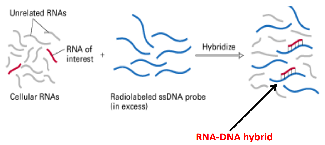

<ul><li><p>Labelled DNA or RNA fragments are used to detect specific nucleic acid sequences by <em>hybridisation</em></p></li><li><p>Probes are among the most useful of molecular tools</p><ul><li><p>Can be labelled with fluorescent tags</p></li><li><p>Can be used on membranes, cells, tissues…</p></li></ul></li></ul><p></p>
30
New cards

Separation of DNA by Gel Electrophoresis - stains

  • DNA is negatively charged

  • DNA bands are invisible

  • Needs to be stained in some way

  • Gel is emersed in buffer solution

  • Ethidium bromide binds to DNA and fluoresces under UV light

  • Other stains visible under blue light (SyBr safe stain)

  • Or no light- eGel


<ul><li><p>DNA is negatively charged</p></li><li><p>DNA bands are <em>invisible</em></p></li><li><p>Needs to be stained in some way</p></li><li><p>Gel is emersed in buffer solution </p></li><li><p><span style="color: rgb(0, 125, 204);">Ethidium bromide </span>binds to DNA and fluoresces under UV light</p></li><li><p>Other stains visible under blue light (<span style="color: rgb(0, 125, 204);">SyBr safe stain</span>)</p></li><li><p>Or no light- eGel</p></li></ul><p></p>
31
New cards

Agarose Gels

  • Polysaccharide extracted from seaweed

    • Coarse pore size – adjustable

    • Used to separate fragments in the size range ~ 0.6 - 25 kb

    • Discrimination of ~1% of length

      • This means for DNA fragment of1000 bp, the gel can tell apart fragments that differ by at least 10 bp. If two fragments differ by less than that, they’ll run almost together and look like one band.

  • Detection of bands:

    • Autoradiography, or

    • Staining e.g. with ethidium bromide and viewed under UV illumination (as here).


<ul><li><p><strong>Polysaccharide extracted from seaweed</strong></p><ul><li><p>Coarse pore size – adjustable</p></li><li><p>Used to separate fragments in the size range ~ 0.6 - 25 kb</p></li><li><p>Discrimination of ~1% of length</p><ul><li><p>This means for DNA fragment of1000 bp, the gel can tell apart fragments that differ by at least 10 bp. If two fragments differ by less than that, they’ll run almost together and look like one band.</p></li></ul></li></ul></li><li><p>Detection of bands:</p><ul><li><p>Autoradiography, or</p></li><li><p>Staining e.g. with ethidium bromide and viewed under UV illumination (as here).</p></li></ul></li></ul><p></p>
32
New cards

Separating DNA — Polyacrylamide gel electrophoresis

  • Vertical gel to separate smaller fragments

  • Much more accurate and precise than horizontal

  • Made from polyacrylamide

  • Very thin


Polyacrylamide Gels

  • Synthetic

  • A neurotoxin in its normal form, therefore care needs to be taken when polymerising it into a gel

  • Finer pore size - variable

  • Can separate fragments differing by a single nucleotide - used for sequencing

  • Used over size range 1-250b (up to ~1kb)

  • Illustration of autoradiographic band detection
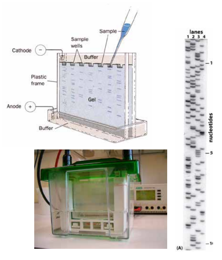

<ul><li><p><span style="color: rgb(187, 241, 210);"><strong>Vertical </strong></span>gel to separate smaller fragments</p></li><li><p>Much more accurate and precise than horizontal </p></li><li><p>Made from <span style="color: rgb(187, 241, 210);">polyacrylamide</span></p></li><li><p>Very <span style="color: rgb(187, 241, 210);">thin</span></p></li></ul><p></p><p>Polyacrylamide Gels</p><ul><li><p>Synthetic</p></li><li><p>A neurotoxin in its normal form, therefore care needs to be taken when polymerising it into a gel</p></li><li><p><em>Finer </em>pore size - variable</p></li><li><p>Can separate fragments <span style="color: rgb(224, 219, 127);">differing by a single nucleotide</span><span style="color: rgb(219, 230, 126);"> </span>- used for sequencing</p></li><li><p>Used over size range 1-250b (up to ~1kb)</p></li><li><p>Illustration of autoradiographic band detection</p></li></ul><p></p>
33
New cards
<p>Measurement of Fragment size/<span style="color: yellow;">length </span>and the influence of <span style="color: yellow;">shape</span></p>

Measurement of Fragment size/length and the influence of shape

  • DNA must be Linear to measure the size

  • The image above shows how you can run an agarose gel electrophoresis with a standard ladder and then form a graph, which can then be used to figure out the size (bp) of an unknown DNA fragment since you have the distance migrated

  • Supercoiled DNA can travel further/faster (lighter), whereas linear DNA (human) cannot travel as far and as fast

    • Supercoiled DNA structure is much more compact and can migrate through the pores in the gel much faster. Only linear DNA moves through at a predictable rate that allows us to determine its fragment size

  • Supercoiled DNA can also be human, id does not matter the origin of the DNA (?)


<ul><li><p>DNA must be <span style="color: rgb(243, 92, 92);"><strong>Linear </strong></span>to measure the size</p></li><li><p>The image above shows how you can run an agarose gel electrophoresis with a standard ladder and then form a graph, which can then be used to figure out the size (bp) of an unknown DNA fragment since you have the distance migrated</p></li><li><p>Supercoiled DNA can travel further/faster (lighter), whereas linear DNA (human) cannot travel as far and as fast</p><ul><li><p>Supercoiled DNA structure is much more <strong>compact </strong>and can migrate through the pores in the gel much faster. Only linear DNA moves through at a predictable rate that allows us to determine its fragment size</p></li></ul></li><li><p>Supercoiled DNA can also be human, id does not matter the origin of the DNA (?)</p></li></ul><p></p>
34
New cards
<p>Overview of DNA Replication</p>

Overview of DNA Replication

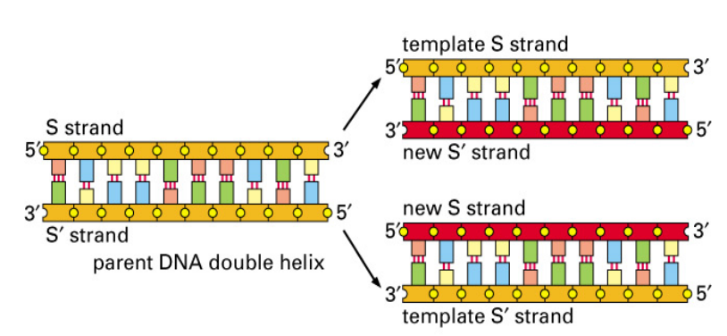
  • All cells replicate their DNA by templated polymerisation — where the existing double stranded DNA is used as a template for making copies of itself

    • The substrates, deoxyribonucleoside triphosphates (dNTPs), are hydrolysed as added, providing the energy required for DNA synthesis

  • An enzyme called helicase separates (unwinds/denatures) double stranded DNA starting at the origin of replication, giving rise to two template strands

    • Single-strand binding proteins bind to the unwound strands to keep them apart

  • A second enzyme call DNA polymerase binds to the template strand and is responsible for adding free nucleotides to the new strand, complementary to the templates strand

    • DNA polymerase catalyzes the formation of the phosphodiester bond between:

      • the 3′‑OH of the growing DNA strand

      • the 5′‑phosphate of the incoming dNTP

  • DNA replication occurs prior to cellular division, ensuring each daughter cell receives a copy of this hereditary information

  • Each new daughter strand of DNA is synthesized slightly differently based on the antiparallel nature of DNA, that is one parental/template strand runs 5’ to 3’ and the other runs 3’ to 5’. This is important as DNA polymerase can only synthesize new DNA in the 5’ to 3’ direction - nucleotides are only added to the 3’ end of the growing strand


*DNA ligase does not ADD new nucleotides, it forms phosphodiester bonds to seal the gaps after polymerase

<ul><li><p>All cells replicate their DNA by <u>templated polymerisation</u> — where the existing double stranded DNA is used as a template for making copies of itself</p><ul><li><p>The substrates, deoxyribonucleoside <strong><u>tri</u></strong>phosphates (dNTPs), are hydrolysed as added, providing the energy required for DNA synthesis</p></li></ul></li><li><p>An enzyme called <span style="color: rgb(255, 185, 0);"><strong>helicase </strong></span>separates (unwinds/denatures) double stranded DNA starting at the origin of replication, giving rise to two template strands</p><ul><li><p>Single-strand binding proteins bind to the unwound strands to keep them apart</p></li></ul></li><li><p>A second enzyme call <span style="color: rgb(255, 185, 0);"><strong>DNA polymerase</strong></span> binds to the template strand and is responsible for adding free nucleotides to the new strand, complementary to the templates strand</p><ul><li><p><span>DNA polymerase catalyzes the formation of the <strong>phosphodiester bond</strong> between:</span></p><ul><li><p><span>the <strong>3′‑OH</strong> of the growing DNA strand</span></p></li><li><p><span>the <strong>5′‑phosphate</strong> of the incoming dNTP</span></p></li></ul></li></ul></li><li><p>DNA replication occurs prior to cellular division, ensuring each daughter cell receives a copy of this hereditary information</p></li><li><p><span style="font-size: medium;">Each new daughter strand of DNA is synthesized slightly differently based on the <u>antiparallel</u> nature of DNA, that is one parental/template strand runs 5’ to 3’ and the other runs 3’ to 5’. This is important as </span><span style="font-size: medium; color: rgb(252, 191, 191);"><em>DNA polymerase can only synthesize new DNA in the 5’ to 3’ direction - nucleotides are only added to the 3’ end of the growing strand</em></span></p></li></ul><p></p><p><span style="color: rgb(155, 154, 154);"><em>*DNA ligase does not ADD new nucleotides, it forms phosphodiester bonds to seal the gaps after polymerase</em></span></p>
35
New cards

What is the origin of replication?

  • Process of DNA replication begins with special initiator proteins that bind to double stranded DNA to pry the two strands apart, breaking the hydrogen bonds between the bases

  • Positions where DNA helix is first opened are called replication origins

  • These sites have short sequences to attract the initiator proteins and stretches of DNA that are easy to open

  • The small circular chromosomes of prokaryotes have a single origin of replication (500-1000 nt/s)

    • Highly regulated, Only initiated when sufficient nutrients, Blocked until A’s are methylated

  • Large, linear DNAs have many origins (50 nt/s)

    • Eukaryote chromosomes can have hundreds of origins of replication, and replication occurs at many different sites simultaneously, this increases SPEED and EFFICANCY (so many genes can be replicated at the same time)

    • Also you have more than one site of origin, so just in case that place mutates, you still have back-up origins for replication!


<ul><li><p>Process of DNA replication begins with special initiator proteins that bind to double stranded DNA to pry the two strands apart, breaking the hydrogen bonds between the bases</p></li><li><p>Positions where DNA helix is first opened are called <span style="color: rgb(234, 144, 144);">replication origins</span></p></li><li><p>These sites have short sequences to attract the initiator proteins and stretches of DNA that are easy to open</p></li><li><p>The small circular chromosomes of prokaryotes have a <span style="color: yellow;"><em>single </em></span>origin of replication (500-1000 nt/s)</p><ul><li><p>Highly regulated, Only initiated when sufficient nutrients, Blocked until A’s are methylated</p></li></ul></li><li><p>Large, linear DNAs have many origins (50 nt/s)</p><ul><li><p>Eukaryote chromosomes can have <span style="color: yellow;">hundreds </span>of origins of replication, and replication occurs at many different sites <u>simultaneously</u>, this increases SPEED and EFFICANCY (so many genes can be replicated at the same time)</p></li><li><p>Also you have more than one site of origin, so just in case that place mutates, you still have back-up origins for replication!</p></li></ul></li></ul><p></p>
36
New cards

What are the ingredient needed for DNA replication?

  • Essential ingredients:

    • ATP, TTP, GTP, CTP (deoxynucleotides)

    • DNA polymerase

    • Primer

  • Additional proteins: increase efficiency and allow bidirectionality

  • Begins at a specific point, called the origin, and proceeds bidirectionally at the replication forks


37
New cards
<p><span style="font-size: medium;">Replication Bubble and Replication Forks - DNA Replication </span></p>

Replication Bubble and Replication Forks - DNA Replication

  • When double stranded DNA is opened up at a point called the origin, a replication bubble is formed, giving rise to two replication forks, one at each end of the bubble

  • To begin with however, we will consider just one. At the replication fork, helicase continues to open up the double stranded DNA and replication progresses

  • Once single stranded DNA is exposed, RNA primers are synthesised on each strand

  • DNA polymerase binds to the RNA primer and synthesises the addition of nucleotides to the 3’ OH (free) end of the primer and continues to add nucleotides from the 3’ end only


<ul><li><p><span style="font-size: medium;">When double stranded DNA is opened up at a point called the origin, a&nbsp;<strong><u>replication bubble</u></strong> is formed, giving rise to two <strong><u>replication forks</u></strong>, one at each end of the bubble</span></p></li><li><p><span style="font-size: medium;">To begin with however, we will consider just one. At the replication fork, helicase continues to open up the double stranded DNA and replication progresses</span></p></li><li><p><span style="font-size: medium;">Once single stranded DNA is exposed, RNA primers are synthesised on each strand</span></p></li><li><p><span style="font-size: medium;">DNA polymerase binds to the RNA primer and synthesises the addition of nucleotides to the 3’ OH (free) end of the primer and continues to add nucleotides from the</span><span style="font-size: medium; color: rgb(255, 82, 82);"><strong> 3’ end only</strong></span></p><p><br></p></li></ul><p></p>
38
New cards
<p>Steps in DNA replication </p>

Steps in DNA replication

  1. DNA helix opened up by helicase – Enzyme that breaks hydrogen bonds holding strands together

  • DNA helicase hydrolyses ATP and propel rapidly along a DNA single strand, prying apart the helix at rates up to 1000 nucleotide pairs per second

  • This created a replication fork + bubble


  1. The unwound DNA is stabilized by single strand binding protein (SSB)

  • SSB proteins bind tightly and cooperatively to exposed single stranded DNA without covering the bases

  • This ensure the DNA does not fold up again before it can be replicated


  1. A DNA primase catalyses the synthesis of short RNA primers, to which nucleotides are added

  • Since DNA polymerase cannot start a strand without a primer


  1. Clamp assembled with the clamp loader next to the 3’ end of the growing chain. The Clamp loader dissociates once clamp assembled (this requires ATP)

  • DNA polymerase tends to fall of the template (lol) so a new DNA polymerase needs to be recruited every time - this decreases efficiency, so we need the sliding clamp and clamp loader to help


  1. DNA polymerase catalyses nucleotide polymerisation

  • This can ONLY happen in the direction of the 5’ → 3’ (this mean new nucleotides can only be added to the 3’ end)

  • We then have the emergence of the leading and the lagging strands


Tidying up


  1. DNA topoisomerases relieve helical winding (when helicase pulls the strands apart, the DNA ahead of the fork becomes overwound, therefore DNA topoisomerases is Infront of the helicase)

  1. Once DNA polymerase has finished, RNA primers are removed by enzymes called exonucleases (RNase H) (for both strands)

  2. DNA polymerase fills in these gaps with deoxyribonucleotides

  3. DNA ligase joins up the fragments by forming a covalent phosphodiester bond between the free 3' and 5' ends of adjacent fragments


<ol><li><p>DNA helix opened up by <span style="color: rgb(255, 185, 0);"><strong>helicase </strong></span>– Enzyme that breaks hydrogen bonds holding strands together</p></li></ol><ul><li><p>DNA helicase hydrolyses ATP and propel rapidly along a DNA single strand, prying apart the helix at rates up to 1000 nucleotide pairs per second</p></li><li><p>This created a replication fork + bubble</p></li></ul><p></p><ol start="2"><li><p>The unwound DNA is stabilized by single strand binding protein (SSB)</p></li></ol><ul><li><p>SSB proteins bind tightly and cooperatively to exposed single stranded DNA without covering the bases</p></li><li><p>This ensure the DNA does not fold up again before it can be replicated</p></li></ul><p></p><ol start="3"><li><p>A DNA primase catalyses the synthesis of short RNA <em>primers</em>, to which nucleotides are added</p></li></ol><ul><li><p>Since DNA polymerase cannot start a strand without a primer</p></li></ul><p></p><ol start="4"><li><p>Clamp assembled with the clamp loader next to the 3’ end of the growing chain. The Clamp loader dissociates once clamp assembled (this requires ATP)</p></li></ol><ul><li><p>DNA polymerase tends to fall of the template (lol) so a new DNA polymerase needs to be recruited every time - this decreases efficiency, so we need the sliding clamp and clamp loader to help</p></li></ul><p></p><ol start="5"><li><p>DNA polymerase catalyses nucleotide polymerisation</p></li></ol><ul><li><p>This can ONLY happen in the direction of the 5’ → 3’ (this mean new nucleotides can only be added to the 3’ end)</p></li><li><p>We then have the emergence of the leading and the lagging strands</p></li></ul><p></p><p>Tidying up</p><p></p><ol start="6"><li><p>DNA topoisomerases relieve helical winding (when helicase pulls the strands apart, the DNA <strong>ahead </strong>of the fork becomes overwound, therefore DNA topoisomerases is Infront of the helicase)</p></li></ol><ol start="7"><li><p><span style="font-size: medium;">Once DNA polymerase has finished, RNA primers are removed by enzymes called <u>exonucleases</u> (RNase H) (for both strands)</span></p></li><li><p><span style="font-size: medium;">DNA polymerase fills in these gaps with deoxyribonucleotides</span></p></li><li><p><span style="font-size: medium;"><u>DNA ligase</u> joins up the fragments by forming a <u>covalent</u> phosphodiester bond between the free 3' and 5' ends of adjacent fragments</span></p></li></ol><p></p>
39
New cards

Primase and the RNA Primer - In DNA replication

  • While DNA polymerase is responsible for the synthesis of new daughter strands, it cannot initiate this process on its own

  • A primer is required to allow DNA polymerase to extend the new strand from

  • The primer is a short piece of RNA, complementary to the template strand, extending around 10-12 nucleotides in length

  • An enzyme called primase is responsible for synthesising the RNA primer

  • From the primer, DNA polymerase is then able to continue adding nucleotides complementary to the template strand, extending the new daughter strand


40
New cards
<p>Leading and Lagging Strands - DNA replication</p>

Leading and Lagging Strands - DNA replication

Leading Strand

  • For one strand, as DNA polymerase adds nucleotides to the 3’ end of the new strand, it can extend and elongate continuously, following behind helicase as it continues to open up the double stranded DNA

  • This strand is referred to as the leading strand and is synthesised with its 3’ end running away from the origin of replication (right ward in the figure)


Lagging Strand

  • The same is not the case for the other daughter strand running antiparallel, referred to as the lagging strand

  • The lagging strand must be synthesized in chunks, running towards the origin of replication

  • Multiple RNA primers bind to this template strand and DNA polymerase synthesizes short pieces of the new strand in the 5’ to 3’ direction (leftward in the figure), until it reaches the next RNA primer

  • New primers are added to the template strand as it opens up and the strand is synthesised in a ‘back-stitching’ manner

  • These short fragments are called Okazaki fragments, named after the researcher who first described them in the 1960’s

  • As this strand is synthesised in pieces, it is synthesised discontinuously


*Each fork has one leading and one lagging strand (look at top image, 2nd image)

<p>Leading Strand</p><ul><li><p><span style="font-size: medium;">For one strand, as DNA polymerase adds nucleotides to the 3’ end of the new strand, it can extend and elongate <u>continuously</u>, <em>following behind helicase</em> as it continues to open up the double stranded DNA</span></p></li><li><p><span style="font-size: medium;">This strand is referred to as the <u>leading strand</u> and is synthesised with its 3’ end running </span><span style="font-size: medium; color: rgb(248, 178, 178);"><strong>away </strong></span><span style="font-size: medium;">from the origin of replication (right ward in the figure)</span></p></li></ul><p></p><p><span style="font-size: medium;">Lagging Strand</span></p><ul><li><p><span style="font-size: medium;">The same is not the case for the other daughter strand running antiparallel, referred to as the <u>lagging strand</u></span></p></li><li><p><span style="font-size: medium;">The lagging strand must be synthesized in chunks, running </span><span style="font-size: medium; color: rgb(248, 178, 178);"><strong>towards </strong></span><span style="font-size: medium;">the origin of replication</span></p></li><li><p><span style="font-size: medium;"><u>Multiple RNA primers</u> bind to this template strand and DNA polymerase synthesizes short pieces of the new strand in the 5’ to 3’ direction (leftward in the figure), until it reaches the next RNA primer</span></p></li><li><p><span style="font-size: medium;">New primers are added to the template strand as it opens up and the strand is synthesised in a ‘back-stitching’ manner</span></p></li><li><p><span style="font-size: medium;">These short fragments are called <u>Okazaki fragments</u>, named after the researcher who first described them in the 1960’s</span></p></li><li><p><span style="font-size: medium;">As this strand is synthesised in pieces, it is synthesised <u>discontinuously</u></span></p></li></ul><p></p><p>*Each fork has <strong>one leading</strong> and <strong>one lagging</strong> strand (look at top image, 2nd image)</p>
41
New cards


1. Helicase (blue) unwinds the DNA

→ exposes two single strands.

2. Primase (purple) lays down RNA primers

→ gives polymerase a starting point.

3. DNA polymerase (green) builds new DNA

  • Leading strand: continuous (bottom)

  • Lagging strand: Okazaki fragments (top)

    • You can see the green molecule (DNA polymerase coming and going continuously + you can also see the loading clamp)

4. New DNA (white) grows as polymerase adds nucleotides

→ complementary to the parental DNA (red).

42
New cards
<p>G0 phase </p>

G0 phase

  • Phase where the cell is undergoing its functions and is not cell dividing


43
New cards
<p>Mitosis: Interphase </p>

Mitosis: Interphase

G1 Phase — 46 chromosomes, 46 chromatids

  • The cell is growing and doing its normal job

  • Chromosomes are not replicated (single‑chromatid)

  • Cell increases size, makes proteins, organelles, and enzymes needed for DNA replication

    • The cell also prepares for the initiation of DNA replication - where proteins identify origins of replication and load Helicases onto these sites

  • A major checkpoint ensures the cell is healthy enough to replicate DNA

    • Check for DNA damage

    • Enough nutrients for cell division

    • Verify cell is big enough to divide

  • There is a point of no return between, where at this point the cell commits to entering the S phase


S Phase — 46 chromosomes, 92 chromatids, 4C DNA content

  • Cell activates the previously loaded DNA helicases

  • DNA replication happens here

  • Each chromosome is copied → forming two sister chromatids

    • Chromosome number stays 46, but chromatids double to 92

    • Centrosomes also duplicate (important for spindle formation later)


G2 Phase — 46 chromosomes, 92 chromatids

  • Final prep before mitosis

    • Cell checks for DNA replication errors (makes sure S phase has been correctly completed, that is chromosomes have completely and accurately replicated)

    • Produces proteins for mitosis (e.g., tubulin for spindle fibres)

    • Chromosomes are still duplicated (two chromatids each)


44
New cards
term image
  • PCR primers must perfectly base-pair with the target DNA, especially at the 3′ end, because DNA polymerase extends from that end

  • If the 3′ base is mismatched, the polymerase cannot easily start synthesis, leading to poor amplification

  • Hence the second primer performs less well because its 3′ end (G) does not correctly pair with the template’s T

  • DNA polymerase requires a correctly paired 3′ end to initiate replication, a mismatch here triggers proofreading activity, slowing or preventing extension


*remember the primer is given in 5’ to 3’ direction - standard notation

45
New cards

Mitosis

  • Interphase (S phase completed) — 46 chromosomes, 92 chromatids

    • DNA has already replicated. Each chromosome now consists of two sister chromatids joined at a centromere.

  • Prophase — 46 chromosomes, 92 chromatids

    • Chromosomes condense and become visible. Each still consists of two sister chromatids. Nuclear envelope breaks down

  • Metaphase — 46 chromosomes, 92 chromatids

    • Mitotic spindle forms. Microtubules grow from the mitotic spindle and attach on both sides at the centromere of the chromosome. They pull, such that the chromosomes line up at the metaphase plate.

  • Anaphase — 92 chromosomes, 92 chromatids

    • Sister chromatids separate and are pulled to opposite sides of the cell by the mitotic spindle. Even though the cell still has the same amount of DNA, the number of chromosomes doubles because chromatids are now counted as individual chromosomes

  • Telophase — 46 chromosomes per nucleus, 46 chromatids per nucleus

    • Chromosomes reach opposite poles and begin to decondense. Nuclear envelopes reform. Each daughter nucleus receives 46 chromosomes (each now a single chromatid).

  • Cytokinesis — two identical daughter cells (not a phase of mitosis)

    • Each daughter cell ends with:

      • 46 chromosomes

      • 46 chromatids
        Genetically identical to the parent cell (2C)


<ul><li><p><strong>Interphase </strong>(S phase completed) — 46 chromosomes, 92 chromatids</p><ul><li><p>DNA has already replicated. Each chromosome now consists of <strong>two sister chromatids</strong> joined at a centromere.</p></li></ul></li><li><p><strong>Prophase </strong>— 46 chromosomes, 92 chromatids</p><ul><li><p>Chromosomes condense and become visible. Each still consists of <strong>two sister chromatids</strong>. Nuclear envelope breaks down</p></li></ul></li><li><p><strong>Metaphase </strong>— 46 chromosomes, 92 chromatids</p><ul><li><p>Mitotic spindle forms. Microtubules grow from the mitotic spindle and attach on both sides at the centromere of the chromosome. They pull, such that the chromosomes line up at the metaphase plate.</p></li></ul></li><li><p><strong>Anaphase </strong>— 92 chromosomes, 92 chromatids</p><ul><li><p>Sister chromatids separate and are pulled to opposite sides of the cell by the mitotic spindle. Even though the cell still has the same <em>amount</em> of DNA, the number of <strong>chromosomes doubles</strong> because chromatids are now counted as individual chromosomes</p></li></ul></li><li><p><strong>Telophase </strong>— 46 chromosomes per nucleus, 46 chromatids per nucleus</p><ul><li><p>Chromosomes reach opposite poles and begin to decondense. Nuclear envelopes reform. Each daughter nucleus receives <strong>46 chromosomes</strong> (each now a single chromatid).</p></li></ul></li><li><p><strong>Cytokinesis </strong>— two identical daughter cells (not a phase of mitosis) </p><ul><li><p>Each daughter cell ends with:</p><ul><li><p><strong>46 chromosomes</strong></p></li><li><p><strong>46 chromatids</strong> <br>Genetically identical to the parent cell (2C)</p></li></ul></li></ul></li></ul><p></p>
46
New cards
<p>How to inhibit DNA replication?</p>

How to inhibit DNA replication?

  • The use of nucleobase/nucleoside

    • Acyclovir (after phosphorylation) mimics guanosine and competitively inhibits viral DNA polymerases analogues

  • Mutagens

    • Induce mutation, they can be incorporated into the DNA but are not correct

  • Modified nucleotides used as drugs

    • E.g. change the 3’ so that no more bonding can occur


47
New cards

How did Meselson-Stahl Experiment work?

  • E. coli were grown for many generation in heavy nitrogen (15N), this means their DNA became fully heavy

    • This means when they were spun, heavy DNA forms a lower band (since its denser)

    • Therefore before replication: DNA = all heavy strands

  • Now they transferred the bacteria to a light nitrogen medium (14N) and allowed them to replicate once

    • After 1 round of replication, they centrifuged the DNA again. The results now showed a single intermediate band appeared in between (heavy + light)

  • After 2 generations: one intermediate band + one light band (2 bands)

    • Half the molecules: heavy–light

    • Half the molecules: light–light

  • Probably used UV absorbance to see these bands


*DNA bases (A, T, G, C) contain nitrogen atoms. When bacteria grow in heavy N medium, they build new nucleotides using ^15N → so their DNA becomes “heavy.” When you move them to ^14N medium, the cell now builds new nucleotides using ^14N → “light” DNA

*Note you can move them to either light → heavy or heavy → light medium - read question carefully!
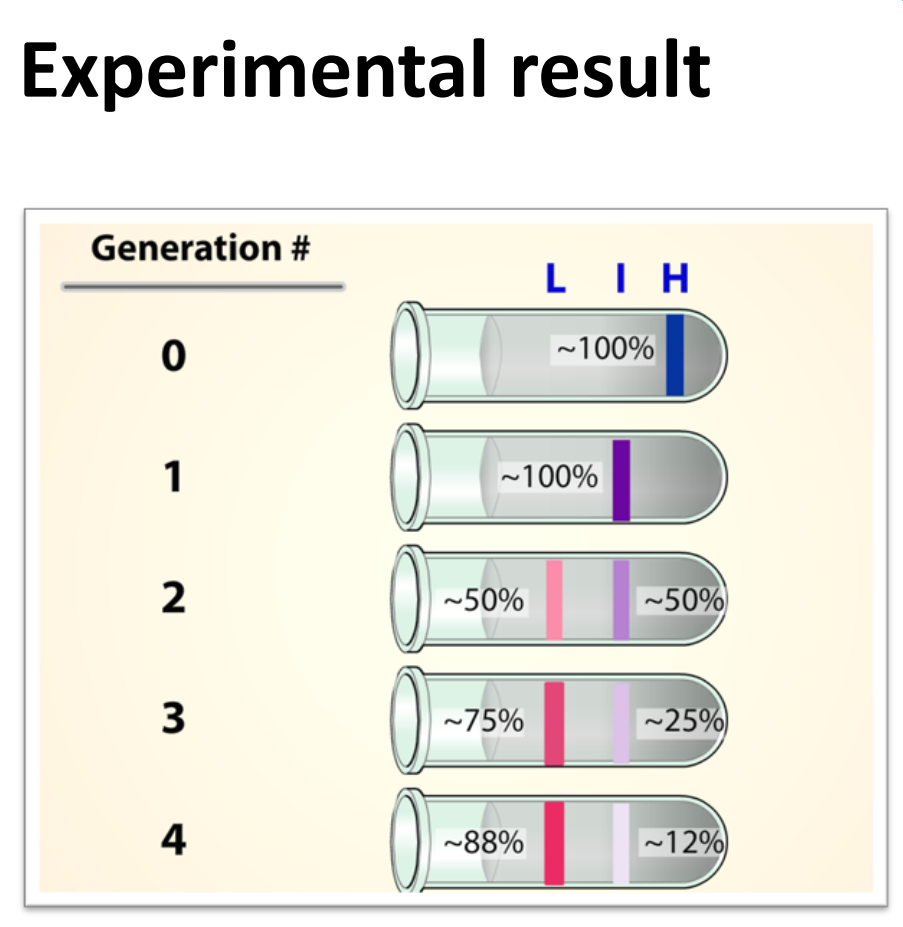
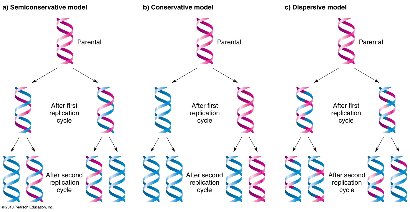
<ul><li><p>E. coli were grown for many generation in heavy nitrogen (<sup>15</sup>N), this means their DNA became fully heavy</p><ul><li><p>This means when they were spun, heavy DNA forms a lower band (since its denser)</p></li><li><p>Therefore before replication: DNA = all heavy strands</p></li></ul></li><li><p>Now they transferred the bacteria to a light nitrogen medium (<sup>14</sup>N) and allowed them to replicate once</p><ul><li><p>After 1 round of replication, they centrifuged the DNA again. The results now showed a single <em>intermediate </em>band appeared in between (heavy + light)</p></li></ul></li><li><p>After 2 generations: <strong>one intermediate band + one light band (2 bands)</strong></p><ul><li><p>Half the molecules: heavy–light</p></li><li><p>Half the molecules: light–light</p></li></ul></li><li><p>Probably used UV absorbance to see these bands</p></li></ul><p></p><p><span style="color: rgb(187, 184, 184);"><em>*DNA bases (A, T, G, C) contain </em><strong><em>nitrogen atoms</em></strong><em>. When bacteria grow in heavy N medium, they </em><strong><em>build new nucleotides using ^15N</em></strong><em> → so their DNA becomes “heavy.” When you move them to ^14N medium, the cell now builds </em><strong><em>new nucleotides using ^14N</em></strong><em> → “light” DNA</em></span></p><p><span style="color: rgb(187, 184, 184);"><em>*Note you can move them to either light → heavy or heavy → light medium - read question carefully!</em></span></p>
48
New cards
<p>What did Meselson-Stahl Experiment prove about the type of replication theory? </p>

What did Meselson-Stahl Experiment prove about the type of replication theory?

  • The Conservative replication was rejected (B)

    • After 1 generation, you should see two bands:

      • One heavy-heavy molecule

      • One light-light molecule

      • But Meselson–Stahl saw only one intermediate band in 1st gen, not two

      • Also the original amount/density of the HH remains the same


  • The Dispersive replication was rejected (C)

    • Model prediction:

      • After 1 generation: one intermediate band (same as semi-conservative)

      • After 2 generations: DNA should become even lighter, but still only one band, because every strand is a mix of old + new DNA

      What Meselson–Stahl actually saw after 2 generations as 2 band (one light band (LL) and one intermediate (LH)), whereas Dispersive replication predicts only one band, so it is wrong.


  • The Semi-conservative replication (SUPPORTED) (A)

    • Model prediction:

      • After 1 generation: one intermediate band

        • Each daughter DNA molecule has one heavy strand + one light strand

      • After 2 generations: one intermediate band + one light band

        • Half the molecules: heavy–light

        • Half the molecules: light–light

      This is exactly what Meselson–Stahl observed


<ul><li><p>The <span style="color: rgb(200, 169, 107);"><strong>Conservative replication</strong></span> was rejected (B)</p><ul><li><p>After 1 generation, you should see <strong>two bands</strong>:</p><ul><li><p>One <strong>heavy-heavy</strong> molecule</p></li><li><p>One <strong>light-light</strong> molecule</p></li><li><p>But Meselson–Stahl saw <strong>only one intermediate band in 1st gen</strong>, not two</p></li><li><p>Also the original amount/density of the HH remains the same</p></li></ul></li></ul></li></ul><p></p><ul><li><p>The <span style="color: rgb(200, 169, 107);"><strong>Dispersive replication</strong></span> was rejected (C)</p><ul><li><p>Model prediction:</p><ul><li><p>After 1 generation: <strong>one intermediate band</strong> (same as semi-conservative)</p></li><li><p>After 2 generations: DNA should become <strong>even lighter</strong>, but <strong>still only one band</strong>, because every strand is a mix of old + new DNA</p></li></ul><p>What Meselson–Stahl actually saw after 2 generations as 2 band (one light band (LL) and one intermediate (LH)), whereas Dispersive replication predicts <strong>only <em>one </em>band</strong>, so it is <strong>wrong</strong>.</p></li></ul></li></ul><p></p><ul><li><p>The <span style="color: rgb(200, 169, 107);"><strong>Semi-conservative replication</strong></span> (SUPPORTED) (A)</p><ul><li><p>Model prediction:</p><ul><li><p>After 1 generation: <strong>one intermediate band</strong></p><ul><li><p>Each daughter DNA molecule has <strong>one heavy strand + one light strand</strong></p></li></ul></li><li><p>After 2 generations: <strong>one intermediate band + one light band</strong></p><ul><li><p>Half the molecules: heavy–light</p></li><li><p>Half the molecules: light–light</p></li></ul></li></ul><p>This is <strong>exactly</strong> what Meselson–Stahl observed</p></li></ul></li></ul><p></p>
49
New cards

Why are RNA primers rather than DNA primers used to initiate replication?

  • Primase is error-prone (hence you need to get rid of them later?)

  • The cell has enzymes that can remove the RNA primers (it’d be difficult to distinguish DNA primers from normal dsDNA)

  • Cells use RNA primers instead of DNA primers because RNA can be made quickly, cheaply, and without needing a pre‑existing 3′‑OH end, which solves the fundamental problem that DNA polymerase cannot start a new strand on its own.
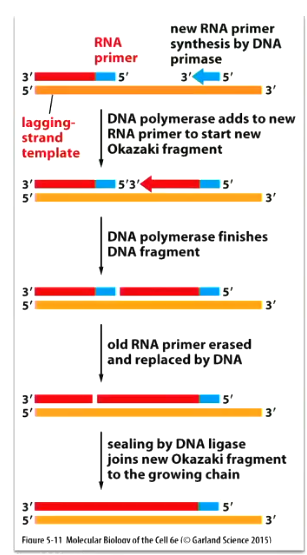

<ul><li><p>Primase is error-prone (hence you need to get rid of them later?)</p></li><li><p>The cell has enzymes that can remove the RNA primers (it’d be difficult to distinguish DNA primers from normal dsDNA)</p></li><li><p>Cells use RNA primers instead of DNA primers because RNA can be made quickly, cheaply, and without needing a pre‑existing 3′‑OH end, which solves the fundamental problem that DNA polymerase cannot start a new strand on its own.</p></li></ul><p></p>
50
New cards
<p>High Fidelity DNA synthesis</p>

High Fidelity DNA synthesis

  • DNA fidelity refers to the accuracy with which a cell duplicates its genetic code. It represents the ability of enzymes like DNA polymerase to avoid and correct errors while copying a template strand. High fidelity prevents mutations and ensures genetic integrity across cell generations

  • Only about 1 mistake in every 1010 nucleotides copied

  • Amazing accuracy – much higher than expected from accuracy of complementary base-pairing

  • Sometimes errors can occur

    • With small changes in geometry, two hydrogen bonds can form between G and T

    • Rare tautomeric forms of DNA bases occur transiently causing incorrect pairing


<ul><li><p>DNA fidelity refers to the accuracy with which a cell duplicates its genetic code. It represents the ability of enzymes like DNA polymerase to avoid and correct errors while copying a template strand. High fidelity prevents mutations and ensures genetic integrity across cell generations</p></li><li><p>Only about 1 mistake in every 10<sup>10</sup> nucleotides copied</p></li><li><p>Amazing accuracy – much higher than expected from accuracy of complementary base-pairing</p></li><li><p>Sometimes errors can occur</p><ul><li><p>With small changes in geometry, two hydrogen bonds can form between G and T</p></li><li><p>Rare tautomeric forms of DNA bases occur transiently causing incorrect pairing</p></li></ul></li></ul><p></p>
51
New cards
<p>Tautomeric Forms </p>

Tautomeric Forms

  • Sometimes errors can occur

    • With small changes in geometry, two hydrogen bonds can form between G and T

    • Rare tautomeric forms of DNA bases occur transiently causing incorrect pairing

  • In the rare tautomeric form, C can pair with A instead of G, G can pair with T etc. → but they also rapidly return to their normal form

  • DNA Polymerase to the rescue!

    • Tautomeric shifts cause mispairing during DNA replication, not transcription

    • The enzyme responsible for replication fidelity and proofreading is DNA polymerase, which ensures correct base pairing and detects mismatches caused by rare tautomeric forms

    • Correct nucleotide has a higher affinity for polymerase (more energetically favourable)


<ul><li><p>Sometimes errors can occur</p><ul><li><p>With small changes in geometry, two hydrogen bonds can form between G and T</p></li><li><p>Rare tautomeric forms of DNA bases occur transiently causing incorrect pairing</p></li></ul></li><li><p>In the rare tautomeric form, C can pair with A instead of G, G can pair with T etc. → but they also rapidly return to their normal form</p></li><li><p>DNA Polymerase to the rescue!</p><ul><li><p>Tautomeric shifts cause mispairing during <u>DNA replication</u>, not transcription</p></li><li><p>The enzyme responsible for replication fidelity and proofreading is DNA polymerase, which ensures correct base pairing and detects mismatches caused by rare tautomeric forms</p></li><li><p>Correct nucleotide has a <span style="color: rgb(120, 170, 235);"><strong>higher affinity</strong></span> for polymerase (more energetically favourable)</p></li></ul></li></ul><p></p>
52
New cards
<p>How does DNA polymerase help maintain/improve fidelity in DNA synthesis</p>

How does DNA polymerase help maintain/improve fidelity in DNA synthesis

DNA polymerase before covalent binding (above)

  • Correct nucleotides have greater affinity for the DNA polymerase active site and form the correct base-pair geometry

  • This allows the polymerase to undergo a conformational change and close around the nucleotide

  • This tightening only happens easily when the base pairs correctly (A–T, G–C)

  • If the base is wrong, the enzyme struggles to close, which slows the reaction and gives the polymerase a chance to reject the incorrect nucleotide


DNA polymerase after covalent binding (below) - PROOFREADING

  • DNA molecules with mismatched nucleotide at 3’ OH end not effective

  • If a wrong nucleotide does get added, polymerase has a second safety mechanism: exonucleolytic proofreading

  • 3’-to-5’ proofreading exonuclease clips off any unpaired residues from the 3’ end of the newly synthesised strand

  • DNA polymerase functions as a self correcting enzyme with 5’–3’ DNA synthesis activity and 3’–5’ exonuclease activity


<p>DNA polymerase <u>before</u> covalent binding (above)</p><ul><li><p>Correct nucleotides have greater affinity for the DNA polymerase active site and form the correct base-pair geometry</p></li><li><p>This allows the polymerase to undergo a <span style="color: rgb(142, 244, 195);"><strong>conformational </strong></span>change and close around the nucleotide</p></li><li><p>This tightening only happens <strong>easily</strong> when the base pairs correctly (A–T, G–C)</p></li><li><p>If the base is wrong, the enzyme struggles to close, which slows the reaction and gives the polymerase a chance to reject the incorrect nucleotide</p></li></ul><p></p><p>DNA polymerase <u>after</u> covalent binding (below) - PROOFREADING</p><ul><li><p>DNA molecules with mismatched nucleotide at 3’ OH end not effective</p></li><li><p>If a wrong nucleotide <em>does</em> get added, polymerase has a second safety mechanism: exonucleolytic proofreading</p></li><li><p><u>3’-to-5’</u> proofreading exonuclease clips off any unpaired residues from the 3’ end of the newly synthesised strand </p></li><li><p>DNA polymerase functions as a self correcting enzyme with 5’–3’ DNA synthesis activity and 3’–5’ exonuclease activity</p></li></ul><p></p>
53
New cards

A need for proofreading may explain the 5’ to 3’ direction of DNA chain growth

  • Growth in 5’-to-3’ direction allows chain to continue to be elongated when a mistake has been removed by exonucleolytic proofreading

  • Exonucleolytic proofreading in hypothetical 3’-to-5’ direction would block further chain elongation

  • Not assessable


<ul><li><p>Growth in 5’-to-3’ direction allows chain to continue to be elongated when a mistake has been removed by exonucleolytic proofreading</p></li><li><p>Exonucleolytic proofreading in hypothetical 3’-to-5’ direction would block further chain elongation</p></li><li><p>Not assessable </p></li></ul><p></p>
54
New cards

Strand-directed Mismatch Repair in Prokaryotes

  • In E.coli, DNA methylation (DNA adenine methyltransferase) adds methyl groups to all A nucleotides in the sequence GATC, but not immediately during replication

  • Therefore, GATC sequences that have not yet been methylated are in the new strands just behind the replication fork (tells the cell that the error is on the newly made strand not on the template strand)

  • Three step process

    • Recognition of a mismatch

    • Excision of the segment of DNA with mismatch

    • Resynthesis of the excised segment using old strand as template

  • Reduces number of errors made by factor of 100-1,000


55
New cards

Strand distinction in Eukaryotes

  • In eukaryotes, mechanism for distinguishing newly synthesized strand from parental template does not depend on DNA methylation

  • Newly synthesized lagging-strand transiently contains nicks which provides the signal that directs the mismatch proof reading system

  • However, this also requires the newly synthesized DNA on the leading strand to be transiently nicked – how this occurs is uncertain


56
New cards

Model for strand-directed mismatch repair in eukaryotes

  • MutS detects and binds specifically to mismatched base pair

    • MutS detects the physical bulge in the helix caused by a mismatched base pair.

  • MutL scans nearby DNA for a nick, triggers degradation of nicked strand all the way back through the mismatch

    • That nick tells the system which strand is the newly synthesized one


<ul><li><p>MutS detects and binds specifically to mismatched base pair</p><ul><li><p>MutS detects the physical bulge in the helix caused by a mismatched base pair.</p></li></ul></li><li><p>MutL scans nearby DNA for a nick, triggers degradation of nicked strand all the way back through the mismatch</p><ul><li><p><span>That nick tells the system which strand is the newly synthesized one</span></p></li></ul></li></ul><p></p>
57
New cards
<p>Eukaryotic chromosomes</p>

Eukaryotic chromosomes

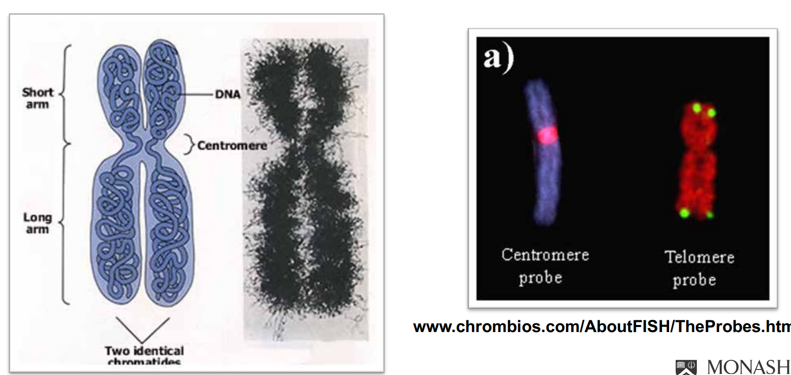
  • DNA is organised as fibre like-structures called chromosomes

    • This like 50/50 proteins + DNA

    • DNA: Protein is 1:3 if including all DNA bond proteins

  • Each chromosome consists of a single linear DNA molecule – No. of chromosomes varies between different species

  • Eukaryotic organisms generally diploid (two copies of each chromosome 2N) – Prokaryotes are haploid (one copy per cell)

  • Green image is a karyotype


Number of chromosomes in a haploid (sperm, ovum) cell = N

In Humans N = 23

Fruit fly N = 4

Dogs N = 39

*Somatic cells are diploid (2N), they have 2 (slightly different) homologous chromosomes

Human 2N = 46

<ul><li><p>DNA is organised as fibre like-structures called chromosomes</p><ul><li><p>This like 50/50 proteins + DNA</p></li><li><p>DNA: Protein is 1:3 if including all DNA bond proteins </p></li></ul></li><li><p>Each chromosome consists of a single linear DNA molecule – No. of chromosomes varies between different species</p></li><li><p>Eukaryotic organisms generally diploid (two copies of each chromosome 2N) – Prokaryotes are haploid (one copy per cell)</p></li><li><p>Green image is a karyotype </p></li></ul><p></p><p>Number of chromosomes in a haploid (sperm, ovum) cell = N</p><p>In Humans N = 23</p><p>Fruit fly N = 4</p><p>Dogs N = 39</p><p>*Somatic cells are diploid (2N), they have 2 (slightly different) homologous chromosomes</p><p>Human 2N = 46</p>
58
New cards
<p>How is DNA Organised </p>

How is DNA Organised

  • Nucleosomes: 147 bp of DNA wraps around 8 histone proteins (2x H2a, H2b, H3, H4), there is one full tuner for every nucleosome

    • The colourful stuff in the 2nd picture, inside the DNA are the histone proteins!


<ul><li><p>Nucleosomes: 147 bp of DNA wraps around 8 histone proteins (2x H2a, H2b, H3, H4), there is one full tuner for every nucleosome </p><ul><li><p>The colourful stuff in the 2nd picture, <em>inside </em>the DNA are the histone proteins!</p></li></ul></li></ul><p></p>
59
New cards

Chromosome structure: nucleosomes

  • The nucleosome is the basic structure of chromatin

  • Invariant component of euchromatin (lightly packed) and heterochromatin (densely packed)

  • Nucleosome is composed of

    • A core region comprising two copies each of the histone proteins H2A, H2B, H3 and H4 → altogether 8 proteins

      • Histones are small basic proteins (positive charge, rich in Lys and Arg)

        • DNA has a negatively charged phosphate backbone. Positively charged lysine and arginine residues in histones are attracted to it

    • DNA wound around the core

      • 146 bp of DNA/octomer of histones plus ~50 bp linker DNA

      • Linker DNA is like the strong and the nucleosomes are like the beads

  • Wrapped as slightly less than 2 turns around the core

  • An associated histone H1 protein binds the linker DNA and is involved in/regulates compaction


<ul><li><p>The nucleosome is the basic structure of chromatin</p></li><li><p>Invariant component of euchromatin (lightly packed) and heterochromatin (densely packed)</p></li><li><p>Nucleosome is composed of</p><ul><li><p>A <span style="color: rgb(255, 173, 238);"><strong>core region</strong></span> comprising two copies each of the histone proteins H2A, H2B, H3 and H4 → altogether 8 proteins</p><ul><li><p>Histones are small basic proteins (positive charge, rich in Lys and Arg)</p><ul><li><p><span style="color: yellow;">DNA has a negatively charged phosphate backbone. Positively charged lysine and arginine residues in histones are attracted to it</span></p></li></ul></li></ul></li><li><p><span style="color: rgb(255, 173, 238);"><strong>DNA wound around the core</strong></span></p><ul><li><p>146 bp of DNA/octomer of histones plus ~50 bp linker DNA</p></li><li><p>Linker DNA is like the strong and the nucleosomes are like the beads</p></li></ul></li></ul></li><li><p>Wrapped as slightly less than 2 turns around the core</p></li><li><p>An <span style="color: rgb(255, 173, 238);"><strong>associated histone H1</strong></span> protein binds the linker DNA and is involved in/regulates compaction</p></li></ul><p></p>
60
New cards

What is Chromatin?

  • The human genome is about 3m long, the average diameter of the nucleus is 6 micrometres, therefore we must be able to fit the DNA in there (this is where chromatin comes in)

  • Density of chromatin packaging is flexible

    • Alters during different phases of cell cycle

  • During mitosis and meiosis becomes 5-10 times more densely packed

    • Only time you can visualise the chromosomes

  • Less tightly packed between mitotic cycles


<ul><li><p>The human genome is about 3m long, the average diameter of the nucleus is 6 micrometres, therefore we must be able to fit the DNA in there (this is where chromatin comes in)</p></li><li><p>Density of chromatin packaging is flexible</p><ul><li><p>Alters during different phases of cell cycle</p></li></ul></li><li><p>During mitosis and meiosis becomes 5-10 times more densely packed</p><ul><li><p>Only time you can visualise the chromosomes</p></li></ul></li><li><p>Less tightly packed between mitotic cycles</p></li></ul><p></p>
61
New cards
<p>Chromosome structure: centromeres</p>

Chromosome structure: centromeres

  • Centromeres situated near the centre

  • One centromere per chromosome

  • Directs chromosome segregation during mitosis and meiosis

    • Contains site at which sister chromatids are paired before segregation

    • Section that pulls chromosomes to either pole

    • Interacts with proteins (kinetochore) and microtubules

  • Contains short, highly-repetitive sequences (satellite DNA)

    • In humans short 170 bp repeats

    • Repeated 2,000 to 30,000 times


<ul><li><p>Centromeres situated near the centre</p></li><li><p>One centromere per chromosome </p></li><li><p>Directs chromosome segregation during mitosis and meiosis</p><ul><li><p>Contains site at which sister chromatids are paired before segregation</p></li><li><p>Section that pulls chromosomes to either pole</p></li><li><p>Interacts with proteins (kinetochore) and microtubules </p></li></ul></li><li><p>Contains short, highly-repetitive sequences (satellite DNA)</p><ul><li><p>In humans short 170 bp repeats </p></li><li><p>Repeated 2,000 to 30,000 times</p></li></ul></li></ul><p></p>
62
New cards
<p>Problem replicating the end of chromosomes</p>

Problem replicating the end of chromosomes

  • Problem replicating end of chromosomes in eukaryotes

    • Telomeres

    • Eukaryotic chromosomes linear not circular

  • Leading strand

    • DNA polymerase can synthesize continuously all the way to the end

    • No problem here — the leading strand reaches the chromosome tip

  • Lagging strand

    • DNA polymerase synthesizes Okazaki fragments, each requiring an RNA primer

    • At the very end of the chromosome, there is no place to put the final RNA primer

    • Without that primer, the last fragment cannot be synthesized

  • Result

    • The newly made daughter strand ends up with a short 3′ overhang of the paternal strand

    • This means the daughter chromosome is shorter than the parent

    • With each round of replication, chromosomes would keep getting shorter


<ul><li><p>Problem replicating end of chromosomes in eukaryotes</p><ul><li><p>Telomeres</p></li><li><p>Eukaryotic chromosomes linear not circular</p></li></ul></li><li><p><strong>Leading strand</strong></p><ul><li><p>DNA polymerase can synthesize <strong>continuously</strong> all the way to the end</p></li><li><p>No problem here — the leading strand reaches the chromosome tip</p></li></ul></li><li><p><strong>Lagging strand</strong></p><ul><li><p>DNA polymerase synthesizes Okazaki fragments, each requiring an RNA primer</p></li><li><p>At the very end of the chromosome, there is no place to put the final RNA primer</p></li><li><p>Without that primer, the last fragment cannot be synthesized</p></li></ul></li><li><p><strong>Result</strong></p><ul><li><p>The newly made daughter strand ends up with a <strong><em>short 3′ overhang of the paternal strand</em></strong></p></li><li><p>This means the daughter chromosome is <strong>shorter</strong> than the parent</p></li><li><p>With each round of replication, chromosomes would keep getting shorter</p></li></ul></li></ul><p></p>
63
New cards

Chromosome structure: Telomeres

  • Telomeres form protective cap at ends of chromosomes

  • They contain a unique DNA sequence which is repeated several times

  • A protein complex called shelterin sequesters the 3’ overhang

    • Prevents the 3’ overhand from being mistaken as double strand break and dealt with by DNA repair machinery


<ul><li><p>Telomeres form protective cap at ends of chromosomes</p></li><li><p>They contain a unique DNA sequence which is repeated several times </p></li><li><p>A protein complex called <span style="color: rgb(230, 163, 163);"><strong>shelterin </strong></span>sequesters the 3’ overhang </p><ul><li><p>Prevents the 3’ overhand from being mistaken as double strand break and dealt with by DNA repair machinery</p></li></ul></li></ul><p></p>
64
New cards

Solution to shortening

  • Put multiple copies of non-coding tandem repeat sequence at ends

  • Nonsense DNA later added back after replication by telomerase


65
New cards

How does telomere DNA replicate?

  1. The paternal strand ends with a 3′ overhang - Lagging strand can’t finish (due to no 3’ OH when primer is removed)

  2. Telomeres associate with Shelterin protein DNA complex which protects telomeres from being treated as damaged DNA by sequestering the 3’ overhang

  3. Telomere DNA repeats are recognised by Telomerase

  4. Telomerase binds to that 3′ overhang

  5. It aligns its RNA template with the very end of the DNA

  6. Telomerase uses its RNA to add DNA (transcriptase activity) → It literally reads its own RNA and synthesizes new DNA repeats: extended by RNA-templated DNA synthesis

  7. Telomerase slides forward and repeats → It adds multiple copies of TTAGGG, making the telomere longer

  8. Now the lagging strand can finish → DNA polymerase can now place primers and complete the lagging strand using the newly extended region as a template


<ol><li><p>The paternal strand ends with a 3′ overhang - Lagging strand can’t finish (due to no 3’ OH when primer is removed) </p></li><li><p>Telomeres associate with Shelterin protein DNA complex which protects telomeres from being treated as damaged DNA by sequestering the 3’ overhang</p></li><li><p>Telomere DNA repeats are recognised by Telomerase</p></li><li><p>Telomerase binds to that 3′ overhang</p></li><li><p>It aligns its RNA template with the very end of the DNA</p></li><li><p>Telomerase uses its RNA to add DNA  (transcriptase activity) → It literally reads its own RNA and synthesizes new DNA repeats: extended by RNA-templated DNA synthesis</p></li><li><p>Telomerase slides forward and repeats → It adds multiple copies of TTAGGG, making the telomere longer</p></li><li><p>Now the lagging strand can finish → DNA polymerase can now place primers and complete the lagging strand using the newly extended region as a template</p></li></ol><p></p>
66
New cards
<p>What is the Telomerase enzyme </p>

What is the Telomerase enzyme

  • Telomere DNA sequences are recognized by sequence-specific DNA binding proteins that attract an enzyme called telomerase

  • Telomerase recognizes tip of existing telomere DNA repeat sequence and elongates it in the 5’ to 3’ direction using an RNA template that is a component of the enzyme itself

  • The enzymatic portion of telomerase has a reverse transcriptase activity, i.e. synthesizes DNA using an RNA template

  • This extends the parental DNA strand so that replication of the lagging strand at the chromosome end can be completed by DNA polymerase


<ul><li><p>Telomere DNA sequences are recognized by sequence-specific DNA binding proteins that attract an enzyme called telomerase </p></li><li><p>Telomerase recognizes tip of existing telomere DNA repeat sequence and elongates it in the 5’ to 3’ direction using an RNA template that is a component of the enzyme itself </p></li><li><p>The enzymatic portion of telomerase has a reverse transcriptase activity, i.e. synthesizes DNA using an RNA template </p></li><li><p>This extends the parental DNA strand so that replication of the lagging strand at the chromosome end can be completed by DNA polymerase</p></li></ul><p></p>
67
New cards

Telomere length as a measuring stick

  • Without telomerase activity, cells can lose 100-200 nucleotides from each telomere every division (cell replicates the genome every 8 hours)

    • After some time the telomere becomes shorter and shorter, and will then undergo apoptosis and the cell will then create a new telomere (?)

  • After many generations, the descendent cells will inherit defective chromosomes and ultimately cease dividing – replicative cell senescence

    • Counting mechanism in somatic cells

  • This safeguards against uncontrolled cell proliferation


68
New cards

Telomeres and Medicine

  • Werner syndrome is a premature aging disease

  • Begins in adolescence or early adulthood

  • Results in old appearance by 30-40 years

  • Physical characteristics:

    • Short stature (common from childhood on)

    • Bird-like features

    • Baldness, cataracts, muscular atrophy

  • Inherited, autosomal, recessive trait

  • Cells from WS patients have shorter telomeres

  • Mutation in WRN gene (codes for the helicase part of Telomeric cap structure), which promotes telomere instability

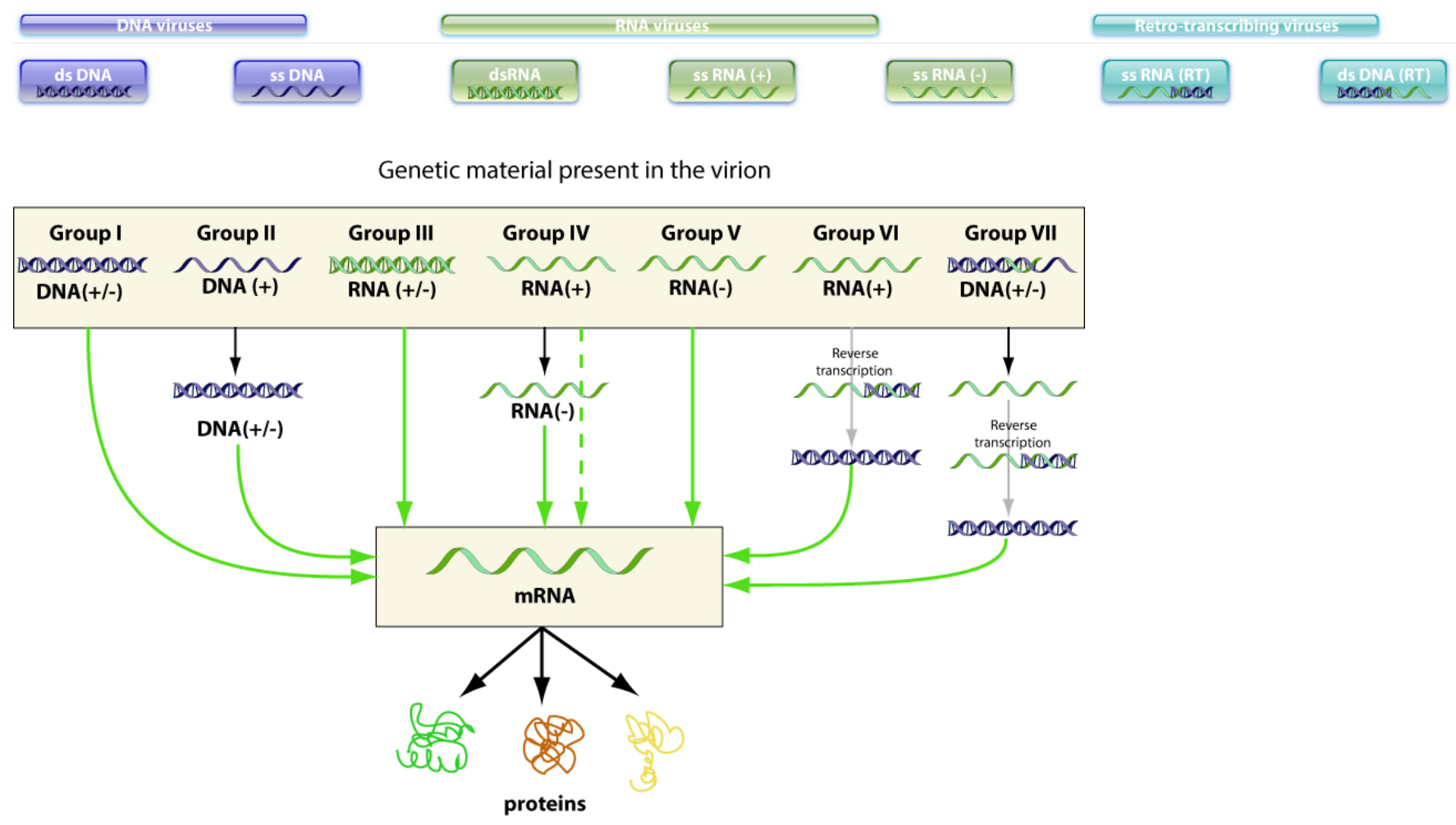
<ul><li><p>Werner syndrome is a premature aging disease </p></li><li><p>Begins in adolescence or early adulthood</p></li><li><p>Results in old appearance by 30-40 years </p></li><li><p>Physical characteristics: </p><ul><li><p>Short stature (common from childhood on)</p></li><li><p>Bird-like features </p></li><li><p>Baldness, cataracts, muscular atrophy</p></li></ul></li><li><p>Inherited, autosomal, recessive trait </p></li><li><p>Cells from WS patients have shorter telomeres </p></li><li><p>Mutation in WRN gene (codes for the helicase part of Telomeric cap structure), which promotes telomere instability</p></li></ul><p></p>
69
New cards
<p>What is the Baltimore classification of viruses?</p>

What is the Baltimore classification of viruses?

ds = double-stranded; ss = single-stranded; +/- refer to whether the RNA is the coding strand (+) or the non-coding strand (-); RT = reverse-transcribed

  • Group I — dsDNA viruses
    Use host RNA polymerase to transcribe DNA → mRNA
    Examples: Herpesviruses, Adenoviruses

  • Group II — ssDNA viruses
    Convert ssDNA → dsDNA → mRNA
    Examples: Parvoviruses

  • Group III — dsRNA viruses
    Viral RNA‑dependent RNA polymerase (RdRp) transcribes dsRNA → mRNA
    Examples: Reoviruses

  • Group IV — (+)-sense ssRNA viruses
    Genome acts directly as mRNA
    Examples: Picornaviruses, Coronaviruses

  • Group V — (–)-sense ssRNA viruses
    Must synthesize complementary (+)RNA → mRNA
    Examples: Orthomyxoviruses, Rhabdoviruses

  • Group VI — ssRNA-RT viruses
    (+)RNA → DNA intermediate → mRNA (reverse transcription)
    Examples: Retroviruses

  • Group VII — dsDNA-RT viruses
    dsDNA genome with RNA intermediate; use reverse transcription
    Example: Hepadnaviruses (e.g., Hepatitis B)

  • SOME CAN BE CIRCULAR

*They are not included in the tree of life!

70
New cards
<p>What are the main classes of organisms?</p>

What are the main classes of organisms?

  • Animals (multicellular)

  • Bacteria (single-celled)

  • Viruses (acellular) infectious particles, not considered alive outside a host)


<ul><li><p>Animals (multicellular)</p></li><li><p>Bacteria (<span style="color: yellow;">single-celled</span>)</p></li><li><p>Viruses (acellular) infectious particles, not considered alive outside a host)</p></li></ul><p></p>
71
New cards

What features does the organism class ANIMALS have?

  • Multicellular organisms with specialized tissues and organs

  • Genome: Multiple double stranded and linear DNA

  • Eukaryotic cells (have nucleus, mitochondria, etc.)

  • Living — perform metabolism, grow, reproduce sexually or asexually

  • Large (millimetres to metres)

  • Reproduction: internal cellular machinery; do not require a host cell

  • Examples: humans, insects, fish


72
New cards

What features does the organism class BACTERIA have?

  • Single‑celled prokaryotes — no nucleus; DNA floats freely

  • Genome: Typically a single circular, double‑stranded DNA chromosome + plasmids, starts from 1000+ genes

  • Living organisms with active metabolism

  • Size: ~0.2–10 µm

  • Reproduction: binary fission (independent)

  • Structure: cell wall (usually peptidoglycan), membrane, cytoplasm, ribosomes

  • Treatment: antibiotics often effective

  • Examples: E. coli, Streptococcus


73
New cards

What features does the organism class VIRUS have?

  • Acellular particles — not cells

  • Genome: Baltimore class (could be single-stranded DNA or RNA), very few genes 10s of gene (less than 300)

  • Non‑living outside host (no metabolism)

  • Size: 20–300 nm (much smaller than bacteria)

  • Structure: DNA or RNA + protein capsid; sometimes lipid envelope

  • Reproduction: must infect a host cell and use host machinery

  • Treatment: antivirals or vaccines; antibiotics do not work

  • Examples: influenza virus, HIV


74
New cards

Genomic Diversity (Virus, Bacteria, yeast, plasmodium, human)

Plasmodium → 3 genomes

Coding for 28 proteins is roughly equal to having 28 genes → Virus

Hundreds of genes are essential to sustain bacterial life (currently estimated at 256 genes)

<p>Plasmodium → 3 genomes</p><p>Coding for 28 proteins is roughly equal to having 28 genes → Virus </p><p><span>Hundreds of genes are essential to sustain bacterial life (currently estimated at 256 genes)</span></p>
75
New cards
<p>What are some human pathogens? Place them in order of complexity </p>

What are some human pathogens? Place them in order of complexity

knowt flashcard image
76
New cards
<p>Compare the genome sizes of Viruses, Prokaryotes and Eukaryotes? </p>

Compare the genome sizes of Viruses, Prokaryotes and Eukaryotes?

  • Virus genomes are tinyyy, they have only 10s-100s of genes, whereas prokaryotes are in the 1000s and eukaryotes are even large

  • A reduction in genome size is associated with parasitic lifestyles in all taxon’s

  • Viruses are the “ultimate” parasites, and (most) have small genomes

    • Exceptions: the genome of Pandora virus is larger than that of some parasitic eukaryotes (e.g. Microsporidia)


<ul><li><p>Virus genomes are tinyyy, they have only 10s-100s of genes, whereas prokaryotes are in the 1000s and eukaryotes are even large </p></li><li><p>A reduction in genome size is associated with parasitic lifestyles in all taxon’s</p></li><li><p>Viruses are the “ultimate” parasites, and (most) have small genomes</p><ul><li><p>Exceptions: the genome of Pandora virus is larger than that of some parasitic eukaryotes (e.g. Microsporidia)</p></li></ul></li></ul><p></p>
77
New cards

Viruses have a large diversity in their genome organisations

  • Viruses are simple structures

    • Protein coat containing either DNA or RNA

    • Inactive while extracellular

    • Inject or deliver their genomic material into cells


  • RNA viruses

    • Double or single stranded RNA genomes

    • Includes influenza, HIV, coronavirus


  • DNA viruses

    • Double or single stranded DNA genomes

    • Includes Smallpox, herpesviruses, most bacteriophages

    • Giant viruses (genome up to 2.5 Mb)


78
New cards

Eukaryotic genomes

  • Nuclear genomes

    • Large (23 pairs of linear chromosomes, 3 billion bp in humans)

    • Coding DNA: proteins, functional RNAs

    • Pseudo-genes: no longer functional

    • Non-coding regions: repetitive, e.g. centromere and telomeres (~50% of human genes are non-coding)


  • Organelle genome(s)

    • Mitochondria genome in humans

      • Mitochondria are organelles involved in energy production by respiration

      • Single circular DNA molecule (in humans: 16.5 kbp, 13 proteins)

      • Thought to come from bacteria (since it has a 2 membranes)

    • Chloroplast genome in plant

    • Apicoplast genome in parasitic eukaryotes (e.g. plasmodium)


79
New cards

What is the Bacterial genome organisation?

  • Most bacteria have a single circular chromosome

    • 500 Kb – 10 Mb

    • Escherichia coli, Haemophilus influenzae, Neisseria meningitidis, Pseudomonas aeruginosa

  • Some have two circular chromosomes - A in two separate distinct circular DNA molecules

    • Vibrio cholerae, Burkholderia pseudomallei, Leptospira borgpetersenii

  • Some have linear chromosomes

    • Borrelia burgdorferi a spirochaete which is the causative agent of Lyme disease

  • Agrobacterium tumefaciens has both a circular and a linear chromosome


80
New cards

What is the diversity of bacterial genetic elements like?

  • Main chromosome

    • Contains genes essential for all conditions

    • One or two chromosomes, circular or linear

  • Plasmids

    • Extrachromosomal, genes not essential in all growth conditions (specific environments)

    • Circular or linear

  • Bacteriophages

    • Viruses that infect bacteria and leave some of the DNA which the bacteria can integrate into its own DNA

  • Transposable elements

    • Mobile DNA that can move in and out of chromosome

    • Insertion sequences, composite transposons, some bacteriophage


81
New cards

What are bacterial plasmids

  • Autonomously replicating pieces of double stranded DNA

    • Exist independently of the main chromosome

    • Each bacterial cell maintains a stable number of plasmid copies per cell

  • Generally smaller than the chromosome

    • 1 kbp to 1 Mbp

  • Often present in multiple copies per cell

    • Anywhere from 1 to 500 copies per cell

  • Do not encode functions essential for growth under all conditions

    • May encode important functions

    • May be essential in some niches


82
New cards

What is plasmid copy number?

  • Copy number is the average number of copies of a plasmid in a cell

    • Copy number can vary between 1 and 100s

    • Any genes on the plasmid will usually be present at multiple copies in the cell

  • Plasmids have genes whose products regulate copy number per cell

    • This number is constant during proliferation


<ul><li><p>Copy number is the average number of copies of a plasmid in a cell </p><ul><li><p>Copy number can vary between 1 and 100s </p></li><li><p>Any genes on the plasmid will usually be present at multiple copies in the cell</p></li></ul></li><li><p>Plasmids have genes whose products regulate copy number per cell</p><ul><li><p>This number is constant during proliferation</p></li></ul></li></ul><p></p>
83
New cards

Plasmids carry genes encoding a wide variety of functions including:

  • Control of their replication (copy number control)

  • Resistance mechanisms ( to antibiotics, in heavy metal enviro)

  • Virulence genes (Toxin production, secretion systems)

  • Metabolic enzymes (to allow bacteria to grow)

  • Production of antimicrobials (bacteriocins - allows bacteria to kill other bacteria, gives competitive advantage to specific bacterium who have this)

  • Mobility (conjugation) factors


84
New cards
<p>Plasmids can alter bacterial phenotype</p>

Plasmids can alter bacterial phenotype

  • Shigella flexneri is the cause of dysentery and diarrhoea

    • ~1 million deaths/year

  • Virulence attributes include

    • Attachment to and invasion through colonic epithelial cells

    • Induced death of macrophages

    • Regulate number of immune cells

  • Many of these functions on a 220-kb virulence plasmid

    • Without this plasmid Shigella will not cause disease

    • Shigella is really E. coli with a virulence plasmid


85
New cards

What are the ways that microbial DNA can be transferred?

  • Vertical transfer (parent to daughter)

    • Chromosomes

      • DNA on the chromosome including mobile DNA and some bacteriophage

    • Plasmids (when cell divides)


  • Horizontal transfer (cell to unrelated cell) - mainly in bacteria

    • Transformation (uptake of free DNA)

    • Conjugation (direct transfer of DNA from cell to cell)

    • Transduction (transfer via a bacteriophage intermediate)


<ul><li><p><span style="color: rgb(207, 65, 115);"><strong>Vertical transfer</strong></span> (parent to daughter)</p><ul><li><p>Chromosomes</p><ul><li><p>DNA on the chromosome including mobile DNA and some bacteriophage</p></li></ul></li><li><p>Plasmids (when cell divides)  </p></li></ul></li></ul><p></p><ul><li><p><span style="color: rgb(207, 65, 115);"><strong>Horizontal transfer</strong></span> (cell to unrelated cell) - mainly in bacteria </p><ul><li><p><span style="color: rgb(243, 147, 153);">Transformation </span>(uptake of free DNA)</p></li><li><p><span style="color: rgb(243, 147, 153);">Conjugation </span>(direct transfer of DNA from cell to cell)</p></li><li><p><span style="color: rgb(243, 147, 153);">Transduction </span>(transfer via a bacteriophage intermediate)</p></li></ul></li></ul><p></p>
86
New cards
<p>Horizontal transfer: Transformation </p>

Horizontal transfer: Transformation

Transformation: bacteria take up free DNA from their surroundings

DNA may be integrated into the chromosome or (if it is a plasmid) replicate freely


  • A bacteria that can take up DNA is said to be “competent”

    • Generally need to manipulate bacterial growth conditions to make bacteria competent

  • Transformation is an important laboratory tool

  • Artificial transformation (in the lab) achieved by a variety of techniques

    • Chemicals such as CaCl2 make membranes permeable

    • Electroporation uses voltage to generate pores in membrane

  • Transformation of plasmid DNA is used in many recombinant techniques

    • Plasmid DNA is not easily degraded

    • Plasmids replicate autonomously so genes are expressed without recombination into chromosome

    • Can introduce foreign DNA/genes into cells
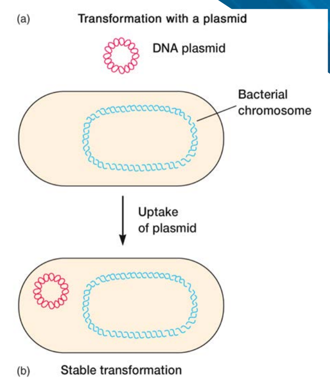

<p><span style="color: rgb(101, 220, 213);"><strong><em><u>Transformation</u></em></strong>: bacteria take up free DNA from their surroundings</span></p><p>DNA may be integrated into the chromosome or (if it is a plasmid) replicate freely</p><p></p><ul><li><p>A bacteria that can take up DNA is said to be “<span style="color: rgb(248, 178, 178);">competent</span>”</p><ul><li><p>Generally need to manipulate bacterial growth conditions to make bacteria competent</p></li></ul></li><li><p>Transformation is an important laboratory tool</p></li><li><p><span style="color: rgb(248, 178, 178);">Artificial transformation </span>(in the lab) achieved by a variety of techniques</p><ul><li><p>Chemicals such as CaCl<sub>2</sub> make membranes permeable</p></li><li><p>Electroporation uses voltage to generate pores in membrane</p></li></ul></li><li><p>Transformation of plasmid DNA is used in many <span style="color: rgb(248, 178, 178);">recombinant techniques</span></p><ul><li><p>Plasmid DNA is not easily degraded</p></li><li><p>Plasmids replicate autonomously so genes are expressed without recombination into chromosome</p></li><li><p>Can introduce foreign DNA/genes into cells</p></li></ul></li></ul><p></p>
87
New cards
<p>Transformation: The use of antibiotic resistance plasmids</p>

Transformation: The use of antibiotic resistance plasmids

  • Plasmids with antibiotic resistance markers are extensively used in molecular biology research

  • The antibiotic resistance marker allows selection of the recombinant bacteria

    • Rmbr electrically shocking the uptake of a plasmid that confers penicillin resistance, and then putting that culture on penicillin, only the culture that survives has successfully taken up the plasmid and can be extracted

  • Plasmids are used to express genes for purification or functional analysis


<ul><li><p>Plasmids with antibiotic resistance markers are extensively used in molecular biology research </p></li><li><p>The antibiotic resistance marker allows selection of the recombinant bacteria </p><ul><li><p><span style="color: rgb(156, 150, 150);"><em>Rmbr electrically shocking the uptake of a plasmid that confers penicillin resistance, and then putting that culture on penicillin, only the culture that survives has successfully taken up the plasmid and can be extracted</em></span> </p></li></ul></li><li><p>Plasmids are used to express genes for purification or functional analysis</p></li></ul><p></p>
88
New cards

Horizontal transfer: Conjugation

Conjugation: bacterial DNA is transferred by direct cell to cell contact


  • To be able to initiate conjugation a bacteria must have

    • DNA which encodes a sex pilus

    • Express proteins required for DNA transfer

    • DNA with a region that can begin transfer (origin of transfer oriT)


  • These factors may be expressed

    • From plasmids such as the F plasmid

    • Can also be expressed from genes on the chromosome


  • Conjugation positive cells often designated F+

  • Conjugation requires direct contact


<p><span style="color: rgb(101, 220, 213);"><strong><em><u>Conjugation</u></em></strong>: bacterial DNA is transferred by <u>direct</u> cell to cell contact</span></p><p></p><ul><li><p>To be able to initiate conjugation a bacteria must have</p><ul><li><p>DNA which encodes a sex pilus</p></li><li><p>Express proteins required for DNA transfer</p></li><li><p>DNA with a region that can begin transfer (origin of transfer oriT)</p></li></ul></li></ul><p></p><ul><li><p>These factors may be expressed</p><ul><li><p>From plasmids such as the F plasmid</p></li><li><p>Can also be expressed from genes on the chromosome</p></li></ul></li></ul><p></p><ul><li><p>Conjugation positive cells often designated F+</p></li><li><p>Conjugation requires direct contact</p></li></ul><p></p>
89
New cards
<p>Transfer of DNA requires direct contact between cells</p>

Transfer of DNA requires direct contact between cells

Experiment 1 (above image)

Experiment 2 (below image)

  • Same experiment as on the previous slide, but here the A and B strains are separated by a filter

    • No growth on minimal medium

    • Conjugation requires direct contact


<p>Experiment 1 (above image)</p><p>Experiment 2 (below image) </p><ul><li><p>Same experiment as on the previous slide, but here the A and B strains are separated by a filter</p><ul><li><p>No growth on minimal medium</p></li><li><p><span style="color: rgb(255, 190, 145);"><strong>Conjugation requires direct contact</strong></span></p></li></ul></li></ul><p></p>
90
New cards
<p>Mechanism of conjugation (F plasmid)</p>

Mechanism of conjugation (F plasmid)

Initiation of conjugation: forming the bridge

  1. Pilus tip of F+ cell binds to receptor on surface of F- cell

  2. Retraction of pilus brings F+ and F- cells close together

  3. Formation of the conjugative bridge, an opening between the cytoplasm of the two cells


DNA transfer steps

  1. One strand of DNA is nicked at the oriT site (only one of the two DNA strands is cleaved)

  2. One DNA strand is pushed through the conjugation bridge

  3. F+ cell repairs plasmid

  4. F- recipient uses incoming DNA as a template for DNA synthesis

  5. DNA transfer/synthesis completed and cells separate

  6. Both cells are now F+


*F⁺ cell contains the F (fertility) plasmid, which carries genes for forming the sex pilus and initiating conjugation. F⁻ cell does not have the F plasmid

*oriT stands for origin of transfer — it’s a specific sequence on the F plasmid where DNA transfer begins

<p>Initiation of conjugation: forming the bridge</p><ol><li><p>Pilus tip of F+ cell binds to receptor on surface of F- cell </p></li><li><p>Retraction of pilus brings F+ and F- cells close together </p></li><li><p>Formation of the conjugative bridge, an opening between the cytoplasm of the two cells</p></li></ol><p></p><p>DNA transfer steps</p><ol start="4"><li><p>One strand of DNA is nicked at the oriT site (only one of the two DNA strands is cleaved)</p></li><li><p>One DNA strand is pushed through the conjugation bridge</p></li><li><p>F+ cell repairs plasmid</p></li><li><p>F- recipient uses incoming DNA as a template for DNA synthesis</p></li><li><p> DNA transfer/synthesis completed and cells separate</p></li><li><p>Both cells are now F+</p></li></ol><p></p><p><span style="color: rgb(147, 143, 143);"><em>*F⁺ cell contains the F (fertility) plasmid, which carries genes for forming the sex pilus and initiating conjugation. F⁻ cell does not have the F plasmid</em></span></p><p><span style="color: rgb(147, 143, 143);"><em>*oriT stands for origin of transfer — it’s a specific sequence on the F plasmid where DNA transfer begins</em></span></p>
91
New cards
<p>Horizontal transfer: Transduction</p>

Horizontal transfer: Transduction

Transduction: A means of genetic exchange whereby DNA is transferred from the donor cell to the recipient cell via a bacteriophage


  • A repeat of the experiment of Lederberg and Tatum in Salmonella showed that genetic exchange could occur when the bacteria were separated by a filter → due to transduction not conjugation Phage can pass through the filter

    • You need to be very careful about how big the filter is and whether or not the bacteria is competent

    • E.g. 5nm filter that only allows the exchange of macromolecules with bacterial species competent → transformation

    • 2 um filter, bacterial species not competent → Transduction

  • Bacteriophage (or phage) are viruses that infect bacteria

  • Bacteriophages can exist outside cells but are inactive/inert while extracellular

  • When they contact a susceptible cell they – Attach → Inject their DNA → Replicate or integrate into genome


<p><span style="color: rgb(101, 220, 213);"><strong><em><u>Transduction</u></em></strong>: A means of genetic exchange whereby DNA is transferred from the donor cell to the recipient cell via a bacteriophage</span></p><p></p><ul><li><p>A repeat of the experiment of Lederberg and Tatum in Salmonella showed that genetic exchange could occur when the bacteria were separated by a filter → due to <em>transduction </em>not conjugation Phage can pass through the filter</p><ul><li><p>You need to be very careful about how big the filter is and whether or not the bacteria is competent </p></li><li><p>E.g. 5nm filter that only allows the exchange of macromolecules with bacterial species competent → transformation </p></li><li><p>2 um filter, bacterial species not competent → Transduction </p></li></ul></li><li><p>Bacteriophage (or phage) are viruses that infect bacteria</p></li><li><p>Bacteriophages can exist outside cells but are inactive/inert while extracellular</p></li><li><p>When they contact a susceptible cell they – Attach → Inject their DNA → Replicate or integrate into genome</p></li></ul><p></p>
92
New cards

What are the two ways that bacteriophages can replicate?

  • Lytic cycle (lytic and temperate phage)

    • Phage injects DNA, replicates, lyses the host cell and releases progeny phage


  • Lysogenic cycle (temperate phage only)

    • Phage injects its DNA and this integrates into the chromosome (no replication or release of progeny)

    • Phage DNA is then replicated along with the host chromosome


93
New cards

What is Generalised transduction?

  • Occurs when a bacteriophage incorrectly packages bacterial DNA into the phage particle

  • During assembly, it’s supposed to package its own viral DNA into new phage heads

  • Occasionally, it makes a mistake and packages fragments of the host bacterium’s DNA instead

  • That defective phage can still infect another bacterium, but since the DNA is bacterial, not viral, it will not result in the production of viable phage

  • Transferred genes will only be stably maintained in the new cell if they recombine into the chromosome, thus transferring genes between bacteria


94
New cards

How does the DNA survive after transfer?

  • If DNA is transferred to a new cell it will only survive if it

    • Can replicate by itself (e.g. a replicating plasmid)

    • Is integrated into the chromosome (or into an already present plasmid)


  • How can DNA be integrated into the host chromosome?

    • The DNA molecule may encode its own proteins for integration (integrases, recombinases)

      • Usually the case with mobile DNA (transposons)

    • The DNA may recombine into the chromosome by homologous recombination

      • Requires some level of DNA sequence similarity (homology)


95
New cards

What is Transfection?

Transfection is the introduction of foreign DNA into animal cells (similar to transformation of prokaryotic cells and other eukaryotes)

Stable (integrated into chromosome) + Transient (expressed from a replicating vector/plasmid)


  • Modification of the eukaryotic membrane by

    • Incubation of cells in certain chemicals

    • Application of an electric pulse (electroporation)


  • Addition of the DNA in liposomes

    • Relies on liposome/membrane fusion


  • Physical introduction using gene gun

    • DNA coupled to an inert solid is fired directly into nucleus


<p><span style="color: rgb(101, 220, 213);">Transfection is the introduction of foreign DNA into <em>animal cells</em></span> (similar to transformation of prokaryotic cells and other eukaryotes)</p><p>Stable (integrated into chromosome) + Transient (expressed from a replicating vector/plasmid)</p><p></p><ul><li><p><span style="color: rgb(255, 182, 166);">Modification of the eukaryotic membrane by</span></p><ul><li><p>Incubation of cells in certain chemicals</p></li><li><p>Application of an electric pulse (electroporation)</p></li></ul></li></ul><p></p><ul><li><p><span style="color: rgb(255, 182, 166);">Addition of the DNA in liposomes</span></p><ul><li><p>Relies on liposome/membrane fusion</p></li></ul></li></ul><p></p><ul><li><p><span style="color: rgb(255, 182, 166);">Physical introduction using gene gun</span></p><ul><li><p>DNA coupled to an inert solid is fired directly into nucleus</p></li></ul></li></ul><p></p>
96
New cards

What is Particle bombardment?

  • Also known as the gene gun

  • Coat plasmid with gene of interest on tungsten or gold particles (microprojectiles)

  • Shoot → Penetration of cell wall

  • Microprojectiles enter the cells, transgenes may incorporate into chromosomal DNA

  • Selectable markers used to identify cells that take up transgene

  • Regenerate plant cells


97
New cards

What is Electroporation?

  • Increase in permeability of cell plasma membrane caused by applied electrical field

  • Used for transformation of bacteria, yeast and protoplasts


<ul><li><p>Increase in permeability of cell plasma membrane caused by applied electrical field</p></li><li><p>Used for transformation of bacteria, yeast and protoplasts</p></li></ul><p></p>
98
New cards

Transfection by electroporation

  • Used for both bacteria and eukaryotes

  • Important to be chilled to maintain stability (?)


<ul><li><p>Used for both bacteria and eukaryotes </p></li><li><p>Important to be chilled to maintain stability (?) </p></li></ul><p></p>
99
New cards

What is Polymerase Chain Reaction?

  • Single-stranded DNA can be replicated by a DNA polymerase

  • PCR uses the fact that DNA strands separate at high temperatures above Tm

  • IF a primer is provided

    • In the cell: generated by a DNA primase

    • In PCR: provided in the reaction mix

  • Invented in 1985 by Kary B. Mullis


  • Intensity of labelling (dark areas) using a probe indicates amount of mRNA and therefore expression level


<ul><li><p><span style="color: rgb(36, 177, 177);">Single-stranded DNA </span>can be replicated by a DNA polymerase</p></li><li><p>PCR uses the fact that DNA strands separate at high temperatures above Tm </p></li><li><p>IF a primer is provided</p><ul><li><p>In the cell: generated by a DNA primase</p></li><li><p>In PCR: provided in the reaction mix</p></li></ul></li><li><p>Invented in 1985 by Kary B. Mullis</p></li></ul><p></p><ul><li><p><span style="color: rgb(165, 159, 159);"><em>Intensity of labelling (dark areas) using a probe indicates amount of mRNA and therefore expression level</em></span></p></li></ul><p></p>
100
New cards

What do you need for PCR?

  • Template DNA

    • The template is the original sample DNA that is to be amplified

    • The template contains the target sequence that primers will anneal to

    • PCR can amplify very small amounts of DNA however if there is too little starting material, the reaction will not amplify enough PCR product

    • In some cases, this can be prevented by increasing the number of cycles

    • Too much DNA can hinder the reaction by sequestering Mg2+ or exhausting primer concentrations

    • For a successful PCR reaction, it is critical that the DNA template is of good quality, free from contaminants


  • DNA polymerase (in a compatible buffer) - we are using Taq

    • Important characteristics to know about the DNA polymerase used is their working temperature and optimal buffer


  • Primers (this means the ends of the DNA templates must be known)

    • Specifically ssDNA


  • dNTPs (nucleotides + provides energy)

    • Deoxynucleotide triphosphates (dATP → adenine, dTTP → thymine etc.)

    • They have 3 phosphate groups → extra phosphates provide the energy needed for DNA polymerase to attach the nucleotide to the growing DNA strand

    • These are the nucleotide bases that are used to generate new strands: A, T, C and G. Without sufficient concentration of dNTPs, there will be insufficient material to synthesize the new DNA

    • If dNTP concentrations are too high, Mg2+ may be depleted as dNTPs can sequester Mg2+, reducing the concentration of free Mg2+ for enzymatic activity


  • PCR Buffer

    • The components of the PCR buffer are included to optimise the conditions for Taq polymerase

    • Tris-HCl and KCl are included to buffer the reaction, maintaining the pH and stabilizing DNA

    • Gelatin and triton-X 100 are included to help stabilize Taq polymerase

    • Finally, DMSO is included to assist with denaturing and inhibition of secondary-structure formation of the template

    • The concentrations of these components can therefore impact the overall efficiency of the reaction


  • Nuclease-free H2O

    • Water is added to dilute reagents to their final, working concentrations

    • Most importantly, water must be free from contamination and anything that can degrade DNA such as nucleases