Chapter 14: Brain Anatomy, CSF, and Cranial Nerves
 Call Kai
 Learn
 Practice Test
 Spaced Repetition
 Match
 Flashcards
 Knowt Play
1/254

There's no tags or description
Looks like no tags are added yet.
255 Terms
What is the average volume of an adult human brain?
1200 mL
Which gender typically has larger brains?
Males, roughly 10% larger than females
Does brain size correlate with intelligence?
No, brain size does not correlate with intelligence.
What are the four major regions of the brain?
Cerebrum, Diencephalon, Cerebellum, Brainstem

What is the largest part of the adult brain?
The cerebrum
Purpose of cerebrum
Conscious though, voluntary movement, sensation, memory, language
What are gyri?
Rounded elevations on the cerebral cortex that increase surface area.
What are sulci and fissures?
Sulci are shallow grooves, and fissures are deep grooves that separate gyri.
the diencephalon consists of
thalamus
hypothalamus
epithalamus
infundibulum
What is the function of the thalamus?
It acts as a relay and processing center for sensory information.

What does the hypothalamus regulate?
Emotions, autonomic functions, and hormone production.
What connects the hypothalamus to the pituitary gland?
The infundibulum.

Role of hypophyseal portal system and hypothalamo-hypophyseal tract
Deliver blood to infundibulum and hormones to pituitary. Also carry hormones away from pituitary.
What are the three major parts of the brainstem? Roles?
Midbrain,
Visual/auditory processing, reflex motor response, consciousness
Pons
relay sensory to cerebellum and thalamus, subconscious motor
Medulla oblongata
autonomic centers, sensory relay

What vital centers are housed in the medulla oblongata?
Cardiac, respiratory, and vasomotor centers.
regulate heart rate, breathing, and blood vessel
diameter (blood pressure control)
What is the role of the cerebellum?
It compares incoming sensory input with past experiences for smooth and coordinated movements.
What are the primary brain vesicles?
Prosencephalon (forebrain), Mesencephalon (midbrain), Rhombencephalon (hindbrain).

What does the telencephalon develop into?
The two cerebral hemispheres and lateral ventricles.
What does the diencephalon specialize to form?
Hypothalamus, Thalamus, Epithalamus, and the retina of the eye.
How does CSF move through the brain?
LV → Foramen of Monro → Third → aquaduct of sylvius → Fourth ventricle → arachnoid granulations
What is the function of the cerebral aqueduct? What embryonic structure is associated
It connects the third ventricle to the fourth ventricle.
mesencephalon
What is the role of cerebrospinal fluid (CSF)? How is it formed?
It protects delicate structure from trauma
transports
supports the brain - effectively drops weight to prevent crusing
Choroid plexus makes in each ventricle; ependymal cells surround choroid plexus capillaries finish and cleanse CSF
How is the brain protected?
Bones of cranium
Cranial meninges
CSF
Biochemical isolation via BBB
What layers make up the cranial meninges?
Dura mater, Arachnoid mater, Pia mater.
These are cont. w/ spinal meninges

What is unique about the dura mater in the cranium?
It has outer and inner fibrous layers and is fused to the periosteum of cranial bones.
periosteal and meningeal

Importance of dural folds? What are the 3
Part of meningeal layer of dura, give stabilizationa nd support and have venous sinuses for carrying blood away
falx cerebri
tentorium cerebelli
falx cerebelli
Arachnoid granulations consist of
Arachnoid villi that penetrate the meningeal layer of dura to absorb CSF into venous circulation
What is clinically associated with CSF?
Hydrocephalus - head enlarged when skull bones are unfused. Treated with a shunt in ventricles to drain
What is the blood-brain barrier (BBB)?
A biochemical isolation formed by astrocytes that protects the brain from general circulation.
Permeable - lipid-soluble, sulfisoxazole & salfadiazine, NH3
Permeable with transporter - water, ions, water soluble molecules, glucose
Note permeable - tetracycline needs intrathecal delivery, urea (water soluble)

What are the two types of tracts found in the pons?
Somatic and visceral motor control nuclei.
What does the medulla oblongata connect?
It connects the brain to the spinal cord.
What is the significance of the central canal in the medulla?
It resembles the spinal cord and is part of the ventricular system.
What is the function of the midbrain?
It contains nuclei for visual and auditory processing and helps maintain consciousness.
What are the anterior, posterior, and inferior horns of the lateral ventricles?
They are extensions of the lateral ventricles into different lobes of the brain.
What is the interventricular foramen?
The passage that connects the two lateral ventricles to the third ventricle.
What is the epidural space like in the cranium compared to the spinal cord?
There is no epidural space in the cranium.
What are dural folds?
Inward extensions of the meningeal layer of the dura that provide stabilization and support for the brain.
Name the three major dural folds.
Falx cerebri, tentorium cerebelli, falx cerebelli.
What do dural venous sinuses do?
They lie within dural folds and carry venous blood from the brain to the neck veins.
What is the function of the falx cerebri?
It projects between the cerebral hemispheres in the longitudinal fissure.
Where do the superior and inferior sagittal sinuses run?
Within the falx cerebri.
What is the role of the tentorium cerebelli?
It separates and protects the cerebellum from the cerebral hemispheres.
What does the falx cerebelli do?
It divides the two cerebellar hemispheres along the midsagittal line.
What is the subdural space?
A narrow gap that may separate the dura mater from the arachnoid mater.
Describe the arachnoid mater.
A thin, delicate membrane with a spider-web appearance that adheres tightly to the dura.
What is contained in the subarachnoid space?
Arachnoid trabeculae, cerebrospinal fluid (CSF), and various cells and fibers.

What is the function of the pia mater?
It closely adheres to the surface of the brain and accompanies cerebral blood vessels.
What is cerebrospinal fluid (CSF) primarily composed of?
Water, proteins, electrolytes, glucose, and other substances.

How does CSF support the brain?
It suspends the brain, reducing its effective weight to about 25 to 50 grams.
What is one function of CSF?
Cushioning delicate neural structures against physical trauma.
How is CSF produced?
By the choroid plexus in each ventricle from filtrate leaking from capillaries.
What is the role of ependymal cells in CSF?
They modify, secrete, and cleanse CSF, removing wastes and adjusting its composition.
What is hydrocephalus?
A condition caused by obstruction in CSF circulation, leading to accumulation and pressure on the brain.
What substances can diffuse freely across the blood-brain barrier?
Lipid-soluble substances like CO₂, O₂, ammonia, steroids, and small alcohols.
What role do astrocytes play in the blood-brain barrier?
They release signals that strengthen tight junctions and help control permeability.
What separates the left and right hemispheres of the cerebrum?
The longitudinal fissure.

What is the basic pattern of the CNS?
A central cavity surrounded by gray matter, with white matter external to it.
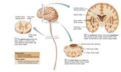
Where is gray matter located in the cerebrum?
In the cerebral cortex and deeper basal nuclei.
What primarily occupies the white matter of the cerebrum?
Myelinated and nonmyelinated axons.
What functions originate in the cerebral hemispheres?
Conscious thoughts and all intellectual functions.
What is the role of the cerebrum in sensory and motor information?
Much of the cerebrum is involved in processing somatic sensory and motor information contralaterally.
What part of the brain do the cerebral hemispheres form?
The superior part of the brain.
What percentage of total brain mass do the cerebral hemispheres account for?
About 83%.
What separates the cerebral hemispheres?
The median longitudinal fissure.
What are the elevated ridges of tissue on the cerebral surface called?
Gyri.

What are the shallow grooves that separate gyri called?
Sulci.
What is the function of gyri in the cerebral cortex?
They increase surface area available for cortical neurons.
What are deeper grooves in the brain called?
Fissures.
What are the five lobes of the cerebral hemisphere?
Frontal, parietal, temporal, occipital, and insula.
Which sulcus separates the frontal lobe from the parietal lobe?
The central sulcus.
What lobe is buried deep within the lateral sulcus?
The insula.
What lies in the anterior cranial fossa of the skull?
The frontal lobes.
What does the cerebral cortex consist of?
Gray matter, including neuron cell bodies, dendrites, glia, and blood vessels.
NO fiber tracts
What is the primary function of the cerebral cortex?
It is the executive center of the nervous system and the seat of conscious mind. Motor, sensory, association areas.
What are the three kinds of functional areas in the cerebral cortex?
Motor areas, sensory areas, and association areas.
What is lateralization in the context of the cerebral hemispheres?
The specialization of cortical functions between the two hemispheres.
What is the primary motor cortex responsible for?
Conscious control of precise or skilled voluntary movements of skeletal muscles.
What are pyramidal tracts?
Massive voluntary motor tracts formed by long axons from pyramidal cells. In motor areas of cerebral cortex.
What does somatotopy refer to?
The point-for-point correspondence between specific areas of the body and specific regions of the CNS.
What happens if the primary motor cortex is damaged?
It can paralyze the specific muscles it controls on contralateral side. Voluntary movement lost but reflex can remain as muscles are able to contract still.
What is the role of the premotor cortex?
It plans movements and organizes basic motor actions into complex sequences - typing, play guitar - skilled motor activities.
For learned/repetitive/patterned movement
What is the anterior association area responsible for?
Intellect, cognition, recall, and personality.
Working memory, judgment, reasoning, persistence, planning
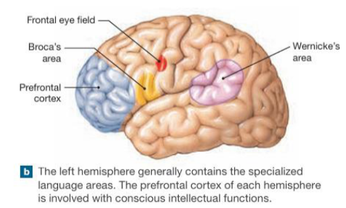
Where is Broca's area located?
Anterior to the inferior premotor cortex, usually in the left hemisphere.
What does the central sulcus separate?
The motor and sensory areas of the cortex.
What is the primary somatic motor cortex's location?
In the precentral gyrus of the frontal lobe of each hemisphere.
What is the function of the primary somatosensory cortex?
It processes sensory information from the body.
What is the significance of the motor homunculus?
It represents the relative amount of cortical tissue devoted to each body part based on motor control precision.
What is the role of association areas in the cerebral cortex?
They coordinate incoming and outgoing data from sensory and motor areas.
What is the primary function of Broca's area?
Regulates breathing and vocalization patterns required for normal speech.
respiratory, larynx, pharynx, tongue, cheek, lips, jaw muscle control
What muscles does Broca's area coordinate for speech?
Respiratory muscles, laryngeal and pharyngeal muscles, tongue, cheeks, lips, and jaw.
What is Broca aphasia?
A speech disorder caused by damage to the frontal lobe, characterized by hesitant and fragmented speech with little grammatical structure.
What is the frontal eye field responsible for?
Controls voluntary movement of the eyes and contributes to smooth following of moving objects. Visual attention.
Where is the primary somatosensory cortex located?
In the postcentral gyrus of the parietal lobe, just posterior to the primary motor cortex.
What does the somatosensory homunculus represent?
A distorted map of the human body laid across the primary somatosensory cortex, reflecting the density of sensory receptors.
What is the function of the somatosensory association cortex?
Integrates sensory inputs to produce an understanding of an object being felt, including its size, texture, and relationship of its parts.
What is the primary visual cortex responsible for?
Receives visual information that originates on the retina of the eye. It’s deep in the calcarine sulcus
What is the role of the visual association area?
Interprets raw visual data into meaningful objects, patterns, and spatial understanding.
What happens when the primary visual cortex is damaged versys the visual association area?
It results in functional blindness versus being unable to comprehend visual input
What is Wernicke's area essential for?
Understanding spoken and written language.
What type of aphasia is associated with damage to Wernicke's area?
Receptive, fluent aphasia, where speech lacks meaningful content.