Face and Neck Landmarks (LP3)
 Call Kai
 Learn
 Practice Test
 Spaced Repetition
 Match
 Flashcards
 Knowt Play
1/60

There's no tags or description
Looks like no tags are added yet.
61 Terms
Epicranial aponeurosis

Occipitofrontalis (Frontal Belly)
O: epicranial aponeurosis
I: Skin and subcutaneous tissue of the eyebrows and forehead
N: Facial
A: Wrinkles forehead and elevates eyebrows; protracts scalp, creating surprise and curiosity

Occipitofrontalis (Occipital Belly)
O: Lateral 2/3 of superior nuchal line
I: Epicranial aponeurosis
N: Facial Nerve
A: Retracts scalp

Temporoparietalis

Temporalis
O: Floor of the temporal fossa and deep surface of the temporal fascia
I: Tip and medial surface of the coronoid process; anterior border of the ramus of the mandible
N: CN V3 via the Temporal Nerve
A: Elevates and retrudes mandible

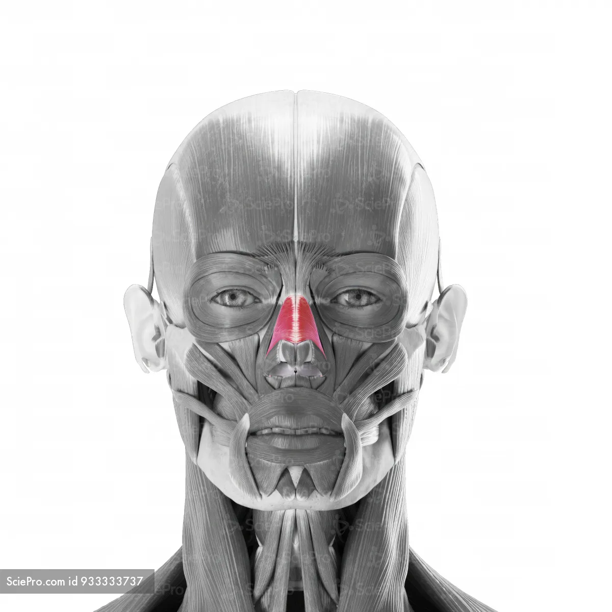
Procerus

Orbicularis Oculi
O: Medial orbital margin; medial palpebral ligament; lacrimal bone
I: Skin around margin of orbit; superior and inferior tarsal plates
N: Facial Nerve
A: Closes the eyelids and helps with winking

Nasalis
Levator Labii Superioris
O: Infraorbital margin (maxilla)
I: Skin of the upper lip
N: Facial Nerve
A: Elevates the upper lip; deepens the nasolabial sulcus (showing sadness)

Levator Anguli Oris

Zygomaticus Major
O: Angle of the mouth
I: Zygomatic bone
N: Facial Nerve
A: Draws the angle of the mouth superiorly and posteriorly; helps to sneer and smile

Zygomaticus Minor

Mentalis

Orbicularis Oris
O: Medial maxilla and mandible; angle of the mouth
I: Mucous membrane of the lips
N: Facial Nerve
A: Closes and protrudes the lips; resists tension (when blowing)

Depressor Labii Inferioris
O: Skin of the lower lip
I: Mandible
N: Facial Nerve
A: Depresses the lower lip; helps with pouting

Depressor Anguli Oris
O: Angle of the mouth
I: Mandible
N: Facial Nerve
A: Depresses the angle of the mouth; helps with frowning

Masseter
O: Inferior border and medial surface of the zygomatic arch
I: Lateral surface of the ramus and angle of the mandible
N: CN V3 via the Masseteric Nerve
A: Elevates and protrudes the mandible

Buccinator
O: Angle of the mouth
I: Pterygomandibular raphe and lateral surfaces of the alveolar processes of the maxilla and the mandible
N: Facial Nerve
A: Compresses the cheek against the molar teeth; keeps food on the occlusal surfaces of the teeth during chewing

Sternocleidomastoid (SCM)
O: Mastoid process and lateral half at the superior nuchal line
I: Anterior manubrium of the sternum and the superior medial 1/3 of the clavicle
N: Spinal Accessory Nerve
A: Laterally flexes the head; unilaterally rotates the face to the opposite and bilaterally extends the head

Trapezius (from face)
O: Superior nuchal line; external occipital protuberance; ligamentum nuchae; spinous processes of C7-T12
I: Lateral 1/3 of clavicle; spine of scapula; acromion
N: Spinal Accessory Nerve and Proprioception from C3-C4
A: Elevates, retracts, and depresses the scapula as it rotates to tilt the glenoid cavity superiorly

Platysma
O: Superficial fascia of the deltoid and pectoral region
I: Mandible forming the skin of the cheek to the angle of the mouth and the orbicularis oris muscle at the lips
N: Cervical branch of the Facial Nerve
A: Tenses the skin of the neck and depresses the mandible

Anterior Scalene
O: Anterior tubercles of the transverse processes of vertebrae C3-C6
I: Scalene tubercle and upper surface of 1st rib
N: Anterior rami of C4-C7
A: Elevates 1st rib
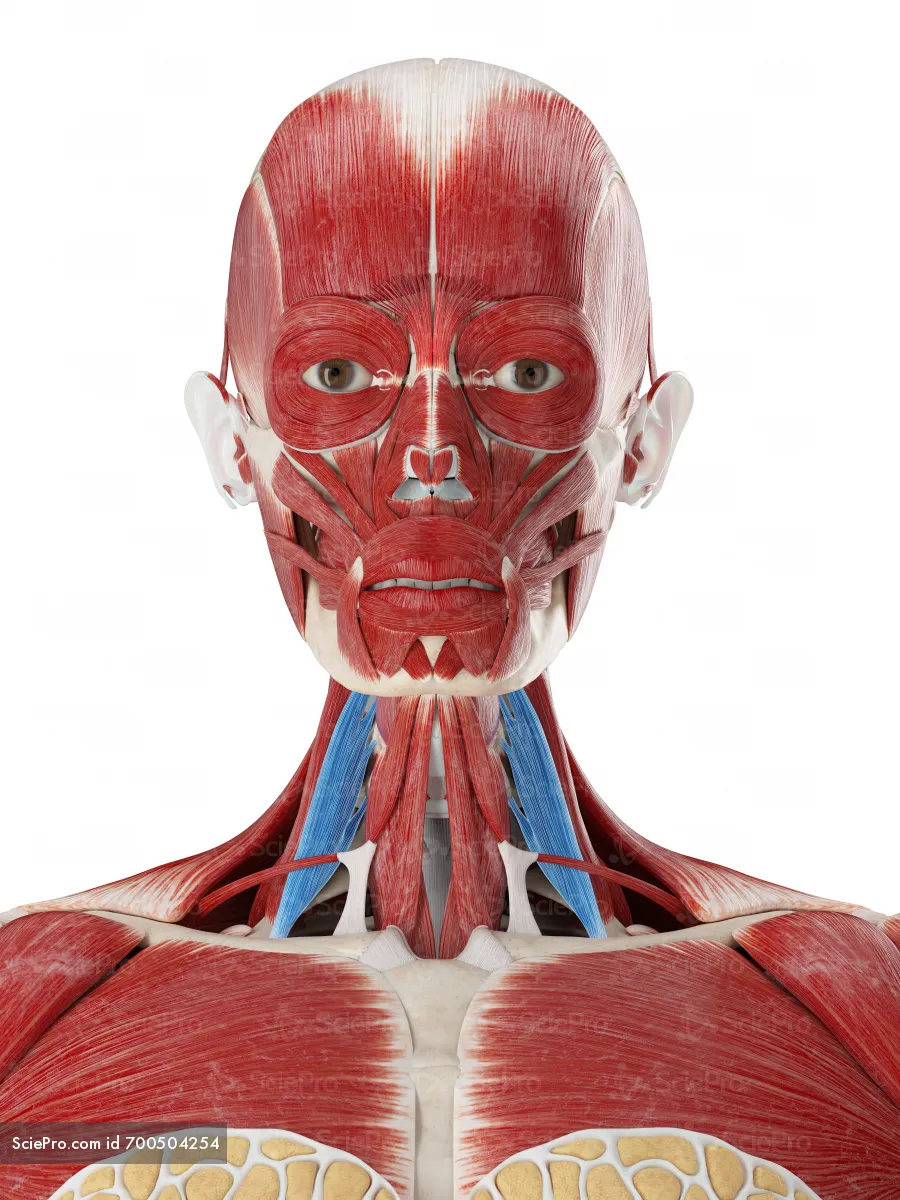
Middle Scalene
O: Transverse processes of vertebrae C2-C7
I: Upper surface of 1st rib posterior to the groove for the subclavian artery
N: Anterior rami of C3-C7
A: Elevates 1st rib

Posterior Scalene
O: Posterior tubercles of the transverse processes of vertebrae C4-C6
I: Upper surface of 2nd rib
N: Anterior rami of C5-C7
A: Elevates 2nd rib

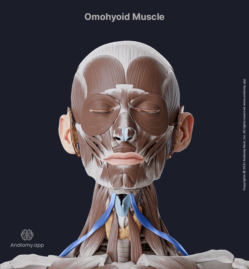
Omohyoid
O: Superior border of scapula medial to suprascapular notch
I: Lower border of body of hyoid bone
N: Anterior rami of C1-C3 through the ansa cervicalis
A: Depresses and fixes hyoid bone
Sternohyoid
O: Posterior aspect of sternoclavicular joint and adjacent manubrium of sternum
I: Body of hyoid bone medial to attachment of omohyoid muscle
N: Anterior rami of C1-C3 through the ansa cervicalis
A: Depresses hyoid bone after swallowing

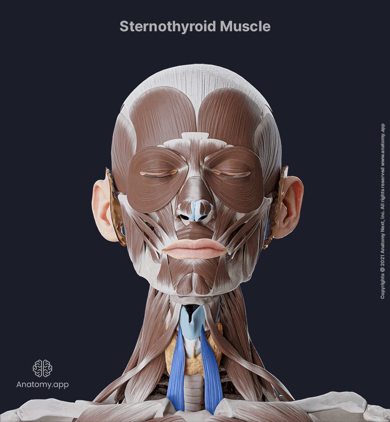
Sternothyroid
O: Posterior surface of manubrium of sternum
I: Oblique line on lamina of thyroid cartilage
N: Anterior rami of C1-C3 through the ansa cervicalis
A: Draws larynx downward
Thyrohyoid
O: Oblique line on lamina of thyroid cartilage
I: Greater horn and adjacent aspect of body of hyoid
N: Fibers from anterior ramus of C1 carried along Hypoglossal Nerve
A: Depresses hyoid bone, but when hyoid bone is fixed, raises larynx

Digastric
O: Anterior belly - Digastric fossa on lower inside of mandible; Posterior belly - Mastoid notch on medial side of mastoid process of temporal bone
I: Attachment of tendon between the two bellies to body of hyoid
N: Anterior belly - Mylohyoid nerve from inferior alveolar branch of Mandibular Nerve; Posterior belly - Facial Nerve
A: Anterior belly - opens mouth by lowering mandible; raises hyoid; Posterior belly - pulls hyoid bone upward and back

Mylohyoid
O: Mylohyoid line on mandible
I: Body of hyoid bone and fibers from muscle on opposite
N: Mylohyoid nerve from inferior alveolar branch of Mandibular Nerve
A: Supports and elevates the floor of the mouth; elevates the hyoid

Stylohyoid
O: Base of styloid process
I: Lateral area of body of hyoid bone
N: Facial Nerve
A: Pulls hyoid bone upward in a posterosuperior direction

Superficial Temporal Artery
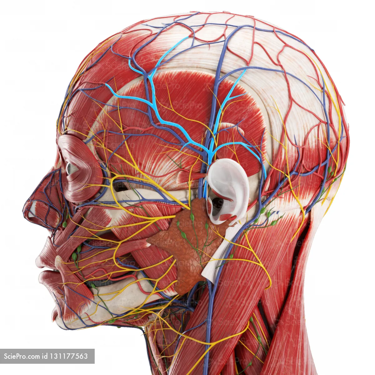
Occipital Artery

Internal Carotid Artery

Vertebral Artery

Inferior Thyroid Artery

Transverse Cervical Artery

Suprascapular Artery
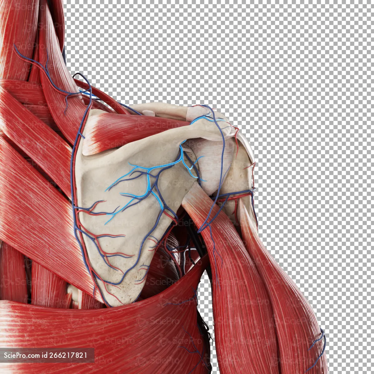
Subclavian Artery

Axillary Artery
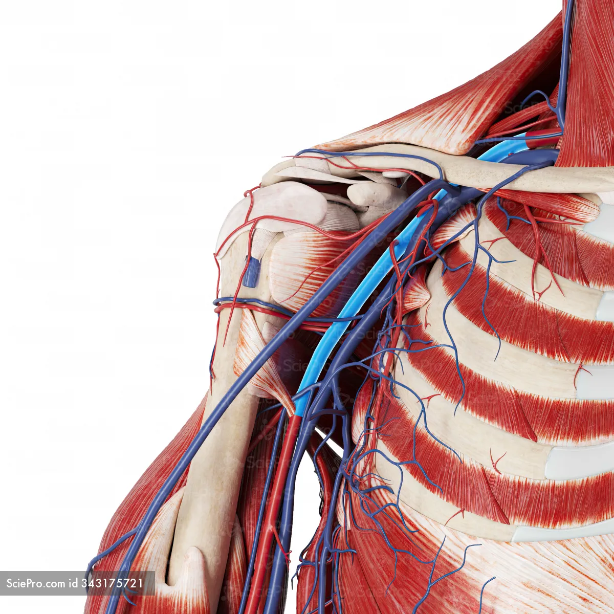
Ophthalmic Artery

Maxillary Artery

Facial Artery

Lingual Artery

External Carotid Artery

Carotid Sinus Artery

Common Carotid Artery

Thyrocervical Trunk Artery
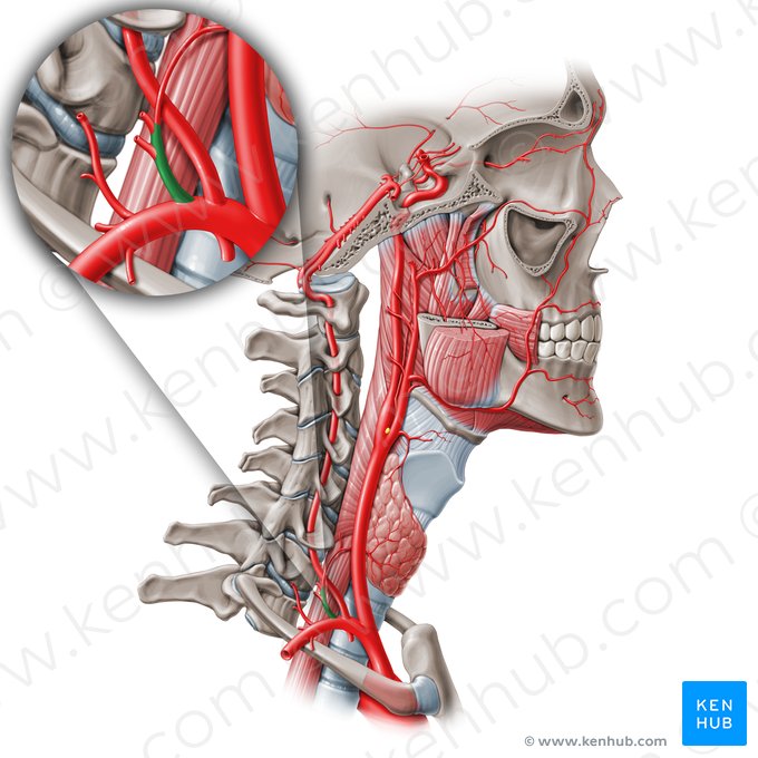
Internal Thoracic Artery

Brachiocephalic Trunk Artery

Occipital Vein

Vertebral Vein

External Jugular Vein

Axillary Vein
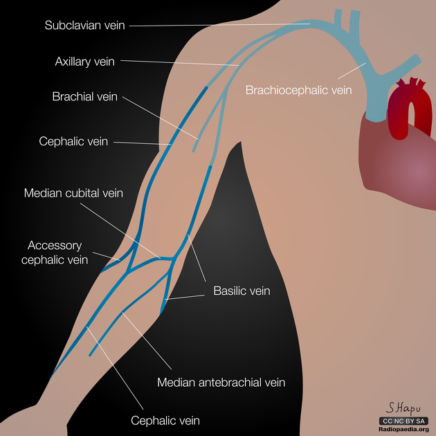
Subclavian Vein

Temporal Vein

Maxillary Vein

Facial Vein
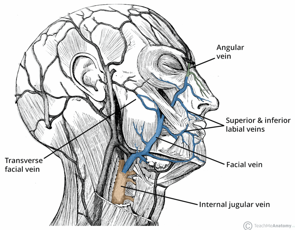
Internal Jugular Vein

Brachiocephalic Vein

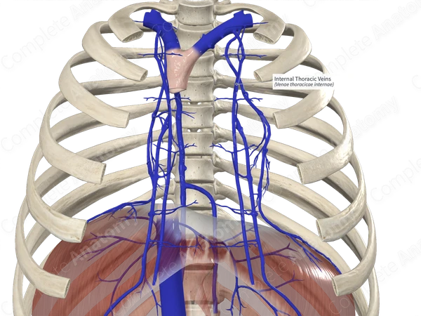
Internal Thoracic Vein