Micro Lab Quiz 1
 Call Kai
 Learn
 Practice Test
 Spaced Repetition
 Match
 Flashcards
 Knowt Play
1/14

There's no tags or description
Looks like no tags are added yet.
15 Terms
Gram positive vs. Gram negative
Gram positive
cell wall = 90% peptidoglycan + 10% periplasm
no outer membrane or LPS
contains teichoic acids + lipoteichoic acids
makes exotoxins
less resistant to antibiotics
gram stain color of purple/blue
Gram negative
cell wall = 45% outer membrane (LPS + phospholipids) + 5% peptidoglycan + 45% periplasm
outer membrane + LPS present
no teichoic acids + lipoteichoic acids
makes endotoxins
more resistant to antibiotics
outer membrane acts like a shield
gram stain color of pink/red

What is a gram stain
a laboratory technique that differentiates bacteria by their cell wall characteristics
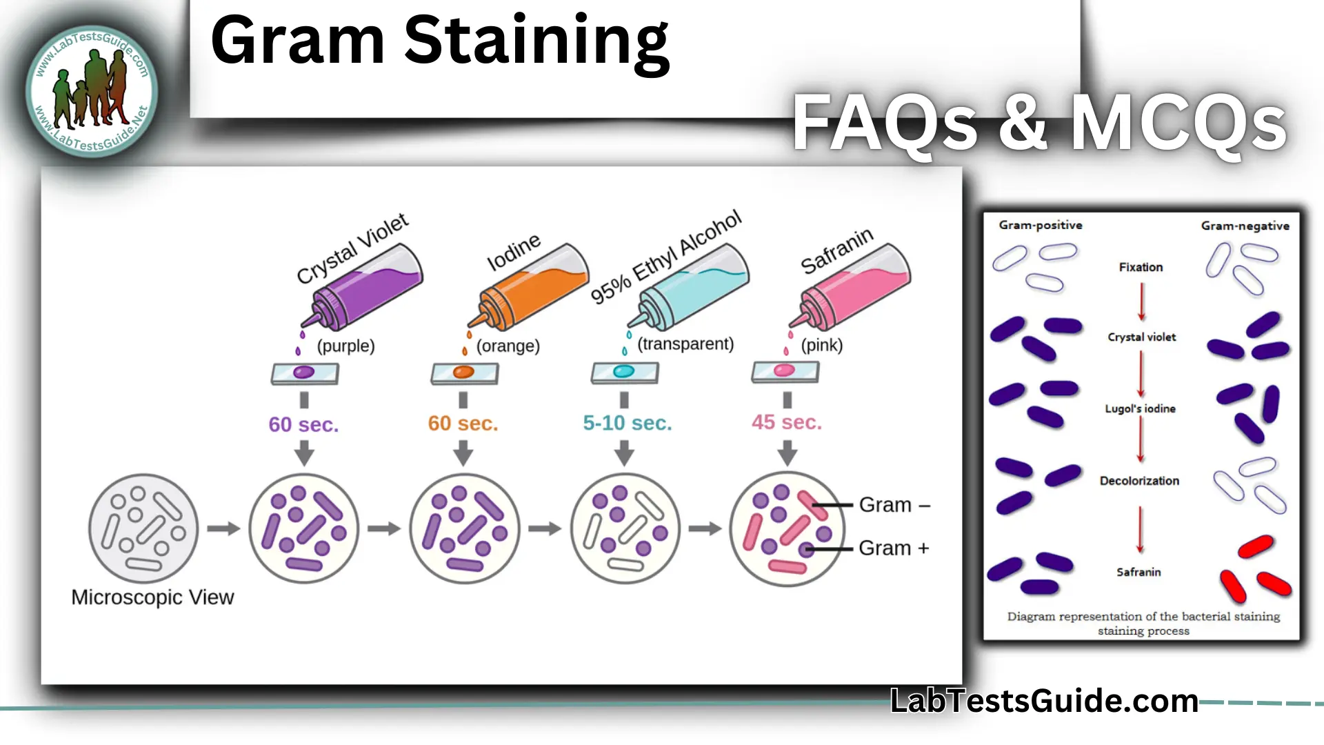
List the steps of the gram stain and their significance
Label the slide and heat-fix it
Add 2 loopfuls of culture in the center
Let it air dry and heat-fix 3 times
Add a small drop of water and mix it with the culture
Let it air dry
Flood with crystal violet for 30 seconds
stains all cells purple
Wash excess with water
Flood with Gram’s iodine for 1 minute
helps trap stain inside the cells
Wash with ethyl alcohol and then water
+ stays purple bc of thick wall, - turns colorless
Flood with safranin for 1 minute
+ stays purple, - turns pink
Wash with water
Blot with bibulous paper
Mordant
a chemical that binds the dye to the microbes
Primary Stain
the initial stain (crystal violet)
Decolorized
the ethyl alcohol dissolves the lipids found in the cell wall of the gram negative cell, allowing the crystal violet to escape & turn colorless
Counterstain
safranin increases the contrast by coloring the gram negative cells pink
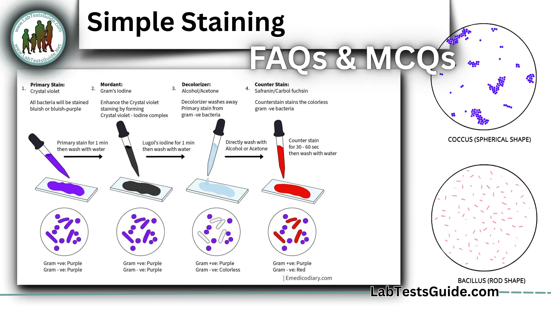
What is a simple stain
one stain is used to dye bacterial cells
Prepare a smear: Spread bacteria onto a microscope slide
Heat-fix: Gently heat the slide to attach the bacteria to it
Apply one dye: Add a stain, such as methylene blue, crystal violet, or safranin
Rinse and dry: Wash off excess dye and let the slide dry
Observe: View to see the shape, arrangement, size
What is a capsule stain
identifies capsules around a bacterial cell
Prepare a smear: Place bacteria on a microscope slide
Apply a negative stain: Use a dye, such as India ink or nigrosin, to darken the background
Apply a counterstain: Use a dye to color the bacterial cells
Observe under a microscope: The capsule appears as a clear halo surrounding the stained bacterial cell against a dark background
What is an acid-fast stain
identify bacteria with waxy cell walls containing mycolic acid (whether they can hold red stain)

What is an endospore stain
identifies endospores (tough & dormant structures that increase survival in harsh conditions)
add malachite green
rinse with water
add safranin
List how the acid-fast stain is done and why
add red stain w/ heat (heat helps penetrate the waxy cell wall)
add acid-alcohol (removes dye from non-acid)
add blue dye (acid-fast bacteria = red/pink + non-acid-fast bacteria = blue)
List how the acid-fast stain is done and why
List how the acid-fast stain is done and why
List the steps of the endospore stain and their significance