Approach to Weight Loss Cases
 Call Kai
 Learn
 Practice Test
 Spaced Repetition
 Match
 Flashcards
 Knowt Play
1/85

There's no tags or description
Looks like no tags are added yet.
86 Terms
why is weight loss important
Common diagnostic challenge
Can affect ability to perform and/or welfare
May involve multiple organs/systems
Multiple potential causes
Specific diagnosis often difficult and may not be reached
normal weight depends on
-size
-shape/breed/species
-repro status e.g. lactating, pregnancy
-season
-work
how to assess weight
BCS based on amount of fat deposited in specific areas

BCS scoring systems
-9 point henneke (professional) → use in records and collegues
-5 (6) point carroll and huntington (clients)
how to BCS
-hands on as big differences in subcut tissue
-assess horse from all angles
Regional adiposity associated with metabolic syndromes
-KEEP RECORD, photos
-re evaluate every month

what is regional adiposity assoc with
metabolic syndromes like PPID or EMS
henneke BCS
must have definitive answer, no 0.5
-best for notes

carroll and huntington BCS
-best for clients

cresty neck score
allows to be able to comment on marked adiposity in areas while also commenting on low BCS overall which makes good for diagnosis of things like PPID and EMS
regional adiposity in the neck

other ways to track weight
weigh bridges
weight tapes→ approx, not realyl used now, can track this number with trends tho
weight loss
general reduction in body fat from stores
muscle wasting
localised, often affecting specific muscle groups while fat stores remain relatively stable
muscles often affected by muscle wasting
-epaxial
-gluteal
-neck
causes of muscle wasting
NMSK, metabolic, less work
what should be assessed alongside bcs
Muscle condition should be assessed alongside BCS to evaluate muscle mass, independent of fat coverage
Significance: PPID, neuromuscular disorders
basics of history
-how long had horse
-previous meds history
-herd/social dynamics (stress inducing)
focused history questions
-duration of issue
-diet
-deworming
-dentistry work
mechanisms of weight loss
-insufficient consumption
-impaired digestion/absorption
-excessive loss
-increased requirements
-other
insufficient consumption
-detailed diet history (pasture? fibre? hay?)
-visit property to observe living conditions to see how much pasture
-watch eat and whether offered enough
if horse not offered enough, what to do
adjust diet to meet needs of 1.5-2% BW DM a day
if offered enough food, what to do next
-consuming enough?
causes of insufficient consumption
-dental disease esp shear mouth
-dysphagia
-pain
-stress/competition
shear mouth
when the occlusal surfaces are severely sloped on each individual tooth, so that, for example, the lingual side of one maxillary cheek tooth is severely worn down while the buccal side is not.
questions for impaired digestion/absorption
has faecal output been normal?
-amount?
-consistency?
causes of impaired digestion/absorption
Dental problems
Malabsorption (intestinal parasitosis, Lawsonia, sand enteropathy, IBD, etc.)
Liver disease (incl. toxicities)
Delayed gastric emptying?
can you determine increased losses from history
no
causes of increased losses
-protein losing enteropathy
-protein losing nephropathy
-third spacing e.g. peritonitis, pleuritis
third spacing
fluid moving from blood vessels to interstitium
what is needed to do to evaluate increased losses
diagnostics
increased requirements questions
-has work increased like exercise or breeding
-has food availability changed like reduced grazing
causes of increased requirements
-pregnancy/lactation
-adverse weather
-neoplasia
other (metabolic) causes of weight loss
inadequate distribution or muscle atrophy
-PPID
-EMS
-Equine Protozoal Myeloencephalitis
-selenium deficiency
physical exam in weight loss case
-thoracic and abod auscultation
can hear it just behind xiphoid→ sounds like a seashell (doesnt rule out tho)
-rectal palp
-sand investigation of faeces
-oral exam with speculum
sand investigation
-faeces out, suspended in water and see if sand drops
oral exam
-well sedated
-speculum, head torch, mirrors
what to check on oral exam
-denal pathology
-FB (often wires under tongue)
-masses (SCC)
what can be used to relax the smoot m in a rectal exam, when use it
-first without buscopan then with
is an anticholinergic, smooth m relaxant
why rectal first without buscopan
assess motility first
why rectal with buscopan
anticholinergic so causes smooth muscle relaxation
palpation findings on rectal
-thickened intestinal walls and colon
-enlarged LNs in retroperitoneal space
-increased motility
haem tests about weight loss
-PCV/TP
-CBC
whta will TP show
protein losing enteropathy
biochem renal profile
-creatinine
-urinalysis
-USG
hepatic biochem profile
-AST
-GGT
-GLDH
-SDH
-bile acids
-bilirubin
biochem of acute phase proteins
-albumin (-)→ acute
-fibrinogen (+)→ chronic
-SAA (+)→ acute
faecal testing
often parasitic burden in young that can cause weight loss
fewc, pcr
fwec dont show red worms → elisa
PCR on faeces
-infectious causes
-more acute than chronic
US show
-FLASH (fast localised abdo sonography of horses)
-more of colic exam

pathologies seen on US
-thickening intestine wall
-gastric dilation
-oedema in intestine
-distension of renal pelvis
-distension of biliary tree or changes in echogenicity of liver
what to assess on US
-motility
-wall thickness
-lumen characteristic
how to assess motility on US
-no sedation
-no buscopan
size of SI
<2-3mm
size of LI
<3mm
size of stomach
<5mm
lumen characteristics on us
large colon should be gas filled → not see it
colitis→ fluid filled colon
why thoracic US if weight loss
always assess as could be pneumonia as cause of weight loss
what to assess on thoracic US
-pleural surface for bronchopneumonia
-pleural effusion
-adhesions/fibrin tags
signs of bronchopneumonia
-comet tailing
-areas of consolidtion
clinical decision making factors to consider
-age (older=PPID. lymphoma)
-signalment
-chronicity (liver disease, IBD)
-presence of diarrhoea, colic, fever
-regional prevalence (e.g. strongyles, sand)
what test for malabsorption
oral glucose absorption test (OGAT)
oral glucose absorption test prep
-avoid stressors on day
-12 hour fast or only grass hay prior to test but allow water
nothing high in concentrates
-no sedation
-quiet
why don't sedate with oral glucose absorption test
-alpha 2 agonist cause glucose spike
how to oral glucose absorption test
-admin 1g/kg glucose as 20% dextrose via NGT
-blood samples for glucose at baseline then hourly for 6hr post glucose admin in oxalate flouride tubes
normal response to oral glucose absorption test
-approx doubling of baseline blood glucose at 2 hours post dosing

severely hypoalbuminaemic patients with oral glucose absorption test
-depressed peaks (colon oedema) so peak between 90-150 mins and may only be 60-70% above baseline
partial malabsoprtion on OGAT
15-65% rise
-insignificant and needs retest
-may be due to stress
-can progress in later tests as may show early signs
total malabsorption on OGAT
no greater than 15% rise in blood glucose, poor prognosis
positive OGAT can mean
-IBD
-lymphosarcoma
-chronic enteritis
-parasitic infestation
if worry peritonitis what do
peritoneal fluid analysis
how to collect abdominocentesis
EDTA and plain
EDTA of abdocentesis for
-TNCC
-cytology
-TP
-glucose
-lactate
plain tube from abdocentesus for
culture
interpreting peritoneal fluid
Test | Normal | Abnormal |
TNCC | < 5,000 cells/μL | > 5,000 cells/μL – indicative inflammation +/- infection |
Cytology | Predominantly non-degenerate neutrophils and macrophages. No bacteria or neoplastic cells | Degenerate neutrophils, reactive macrophages, bacteria (esp. intracellular), neoplastic indicators |
Total Protein | < 2.5 g/dL (transudate range) | > 2.5 g/dL – suggestive of exudative process e.g. peritonitis |
Glucose | Similar to blood glucose levels | Lower than blood glucose levels – indicative of bacterial process bacteria use up glucose |
Lactate | < 2 mmol/L | > 2 mmol/L – indicative of compromised perfusion or ischaemia of abdominal viscera |
what can cause ulcers
stress
equine gastric ulcer syndrome
-not often cause of weight loss but can coincide and proliferate
-may indicate stress
diagnosis of equine gastric ulcer syndrome
gastroscopy
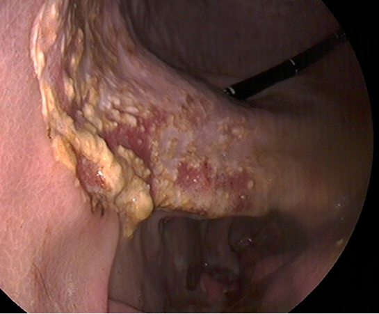

common gastric neoplasia
SCC
delayed gastric emtpying
-limit possible feed intake
-indicate intestinal disease
gastroscopy see
-gastric ulcers
-gastric neoplasia
-delayed gastric emptying
-evaluate oesophagus for strictures if history of choke
PPID testing
-basal ACTH
-TRH stim test
hyperthyroidism tests
-total T4/free T4/T3
-TRH basal/stim testing
renal testing
fractional excretion of electrolytes→ shows which part if affected
-look at USG
-chronic interstitial nephritis common from NSAID long term
transendoscopic biopsy
-gastric
-duodenal
partial thickness
manual biopsy
-liver
-rectal mucosa (IBD) → high risk
surgical biopsy
-full thickness and need time off
-midline exploratory celiotomy
-flank lap
-laparascopy
weight loss approach
