reconstructive 2- flaps, grafts, drains
 Call Kai
 Learn
 Practice Test
 Spaced Repetition
 Match
 Flashcards
 Knowt Play
1/26

There's no tags or description
Looks like no tags are added yet.
27 Terms
Cutaneous pedicle grafts/ skin flaps - purpsoe
Move skin + sc from area of low tension → high tension
Cutaneous pedicle grafts/ skin flaps - Ideal donor site
Low skin tension (head, neck, trunk)
Cutaneous pedicle grafts/ skin flaps - Flap size
Larger than defect
Avoid narrow base (BV)
Not > 3:1 length:width
Cutaneous pedicle grafts/ skin flaps - Flap removal
Undermine deep to panniculus m/ dermis
Cutaneous pedicle grafts/ skin flaps - How to ensure conformatiy
Fine suture + tacking sutures
Cutaneous pedicle grafts/ skin flaps - Recipient bed features
Free of inf, contamination, necrosis
Fresh sx wound/ healthy granulation bed
Cutaneous pedicle grafts/ skin flaps - Subdermal plexus flap def
BV from subdermal plexus + distant direct cutaneous arteries
Cutaneous pedicle grafts/ skin flaps - Transposition flap
Rectangle, within 90 degrees long axis defect
1 edge of defect forms flap border
Width flap = width defect
Length flap = pivot point → distant defect
Cutaneous pedicle grafts/ skin flaps - Unipedicle advancement flap
Cut along defect to enlarge → undermine → Stretch skin over defect → suture
Cutaneous pedicle grafts/ skin flaps - H plasty
Unipedicle advancement flap on 2edges defect
Cutaneous pedicle grafts/ skin flaps - axial pattern flap
Like subdermal plexus flap but Include direct cutaneous artery + vein
Cutaneous pedicle grafts/ skin flaps - Cause of failure
BV occlusion - thrombi, torsion (>90 degrees), stretch
Tension
Haematoma/ seroma
Inf
Cutaneous pedicle grafts/ skin flaps - How to assess flap health
Colour + temp compared to surrounding skin
Cutaneous pedicle grafts/ skin flaps - How to manage partial necrosis
Ointment (moist+protect)
Debride + open wound management, secondary/delayed primary, another flap
Free skin grafts - options
Full thickness meshed
Split thickness (epidermis)
pinch
Punch
Strip
Free skin grafts - Requirements for success
Suitable defect bed
Good contact
No movement (not joints)
No inf
Surgical drains - purpose
Remove excess fluid
Close dead space
Surgical drains - Types
Passive - drain by gravity
Active - drain by suction
Surgical drains - Principles of use
Asepsis
Nonadherent + absorbent dressing
Not near/ parallel to main wound sutures

Surgical drains - Drain type (fluid drains along outside)
Penrose

Surgical drains - Placement of passive
Place at ven end of wound

Surgical drains - active drain purpsoe
Suction so location on wound unimportant
Fluid gathered so dont need absorbent dressing

Surgical drains - Suction drain equipment to make own
IV extension set
Syringe
Large needle

Surgical drains - Steps to make own with IV + syringe
Fenestrate tube end
Insert into wound with forceps
Secure into wound + cover drain exit
Drain with syringe (negative P, needle into plunger)

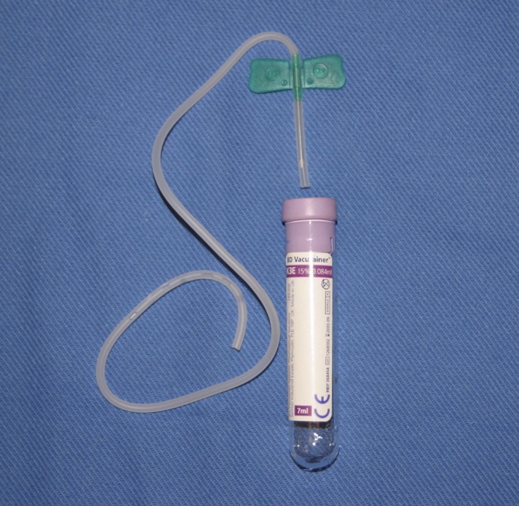
Surgical drains - Steps to make own with vacutainer (for small wounds + spaces)
Catheter into wound + vucutainer
Surgical drains - when to remove
Consistent small vol serosanguineous fluid produced
Surgical drains - When is fluid not collected
Drain blocked (FB → infl → always some fluid)