Intracorpuscular Defects: The Hemoglobinopathies
 Call Kai
 Learn
 Practice Test
 Spaced Repetition
 Match
 Flashcards
 Knowt Play
1/13

Earn XP
Description and Tags
Chapter 11
14 Terms
Heme Moiety
Combination of iron and protoporphyrin ring
Suspended in the center of polypeptide chains, so iron can exist in ferrous state
Globin Variants
Chromosome 16: alpha (α) and zeta (ζ)
Chromosome 11: beta, epsilon, gamma, delta

Sickle Cell Anemia
Substitution of valine for glutamic acid in the sixth position from the NH2 - terminal end of the β chain
HbS constitutes 80% of hemoglobin content
Expressed as α2β26Val
Prominent in african (Sickle Cell Disease is not immune Plasmodium malaria, only trait/carrier)
When oxygen tension decreases, cells become sickling. Repeated sickling permanently damage the membrane.
Hallmark features are hemolytic crisis, aplastic crisis particularly to parvoviruses, vaso-occlusion crisis(vessel blockage that are very painful), ischemic tissue injury (tissues & organs damage) including autosplenectomy (nonfunctional), dactylitis (swelling of hands & feet). Stroke occurs up to 22% of patients
Screening use high-performance liquid chromatography (HPLC) or isoelectric focusing (IEF), electrophoresis on cellulose acetate & citrate agar
Treat by stem cell transplantation, research into gene therapy with CRISPR
Susceptible to infections due to
MPS blockage by increased hemolysis
Blockage in sinusoids of the liver & spleen
Splenic dysfunction
Deficient opsonic activity
Three Principles in management of painful crisis
Adequate rehydration
Pain relief using analgesics
Antibiotic to treat underlying illness such as infection
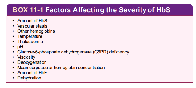
Vasculopathy
vaso-occlusion, and tissue ischemia can happen anywhere in the body.
Acute chest syndrome is the second most common cause of hospital admission and complication of surgery & anesthesia
Hyposthenuria
Inability of the kidney to concentrate urine (low specific gravity)
Earliest nephropathy in sickle cell disease
Hyperuricemia (high uric acid) and hematuria
Sickle Cell Trait
HbA to HbS ratio is 60:40, expressed as α2β1β16Val
Usually asymptomatic, but occasionally episodes of hematuria and hyposthenuria occur
Sickle Disease Morphology
Sickled cells, target cells, fragmented red cells, polychromasia, nucleated red cells, siderotic granules
Increased reticulocytes, except during an aplastic crisis
Leukocytosis with left-shift, thrombocytosis
Electrophoresis
Hemoglobin disease has no HbA, 80% HbS & ~20% HbF
Carriers have 60% HbA, 40% HbS
HbD and HbG both migrate to the same position as HbS at alkaline pH
HbE and HbOArab migrate to the same position as HbC at alkaline pH
Hemoglobin C Disease
Lysine replaces glutamic acid in the 6th position
Electrophoresis shows 95% HbC & HbA2, 7% HbF
Morphology includes bar crystal, microspherocytes, target cells, schistocytes
Hemoglobin D Disease & Trait
Glutamine replaces glutamic acid at position 121 of beta chain
Most common variant is HbDpunjab also called HbDLos Angeles
HbD is nonsickling soluble so it’s asymptomatic and peripheral smear is normal except for few target
HbD comigrates with HbS and HbG at alkaline pH, but comigrates with HbA at acid pH
Hemoglobin E Disease & Trait
Glutamic acid replaces lysine at position 26 of beta globin
Occurs commonly in Southeast Asia (Thailand, Cambodia, Laos)
Alkaline electrophoresis reveals approximately 95% to 97% HbE plus A2 , and the remainder of the hemoglobin is Hb
HbE comigrates with HbC and HbOArab at alkaline pH but migrates with HbA at acid pH
Hemoglobin OArab Disease
Glutamic acid replaces lysine at position 121 of beta globin
Common in North Africa, Balkan
Migrates electrophoretically with HbC, HbE, and HbA2 at alkaline, while comigrates HbA in acidic pH
Unstable Hemoglobins
Hemoglobin variants in which amino acid substitutions or deletions weaken the binding forces of the molecule structure. This causes denature and precipitate hemoglobin Heinz bodies
Cannot diagnose with electrophoresis or high-performance liquid chromatography. The best method is gene sequencing, or heat stability test
Methemoglobinemia
Clinical condition where methemoglobin is greater than 1%
3 common causes are hemoglobin M variant, NADH deficiency, toxic substances
Treat with methylene blue, hyperbaric oxygen therapy, RBC transfusion