Intestines 1 + 2
 Call Kai
 Learn
 Practice Test
 Spaced Repetition
 Match
 Flashcards
 Knowt Play
1/179

There's no tags or description
Looks like no tags are added yet.
180 Terms
State the regions of the small intestine giving their respective proportions of its entire length
Duodenum - ~15% of the entire length
Jejunum - ~75% of the entire length
Ileum - ~ 10% of the entire length

State the standard intestinal layers
Mucosa, submucosa, muscle (inner circular and outer longitudinal), serosa.
Explain how the small intestine is adapted for increasing its surface area for absorption
The surface area is increased by about 500x by:
-Mucosal folds
-Then villi
-Then microvilli (brush border in histology)
State the main cell types present in the small intestine
Goblet cells, enteroendocrine cells, paneth cells, enterocytes,
Describe the functions of goblet cells in the small intestine
Secrete mucus for lubrication and protection of mucosa, secrete HCO3- for neutralisation of stomach acid.

Describe the functions of enteroendocrine cells in the small intestine
Control digestive function via sensory mechanisms and the secretion of hormones.

Describe the functions of paneth cells in the small intestine
Possibly involved in the defence against microbial penetration.

Describe the functions of enterocytes in the small intestine
Responsible for absorption of molecules via transporter proteins. Contain brush border enzymes that remain attached to the epithelial membrane for digestion.

Which cells make up the majority of the small intestine?
Enterocytes.

Describe cell turn-over in the small intestine
Continuous migration from the crypts up to the villus where they are sloughed off at the villus tip. Takes ~ 2-5 days.
Describe the main mechanisms of absorption in the small intestine
Selective process via specific transporter proteins by:
-Diffusion down their concentration gradient (passive)
-Secondary active transport
Do body levels of monovalent (e.g. Na+, K+) ions affect their absorption in the small intestine?
No, they are absorbed irrespective of body requirements.
Do body levels of divalent (e.g. Cu2+) ions and trace elements affect their absorption in the small intestine?
Yes, they can be toxic if they are too high in concentration so are absorbed based on body requirements.
Can the intestine function if part of it is resected?
Yes, as long as at least 50% remains, digestion and absorption will remain unhindered.
Are all nutrients absorbed across the whole length of the small intestine?
Most nutrients are absorbed across the whole length of the small intestine however some (e.g. Vitamin B12, bile salts) are restricted to being absorbed in the ileum only.
Describe the main phases of digestion in the small intestine
Luminal - enzymes are secreted by salivary glands and the pancreas to digest food in the intestinal lumen.
Membranous - enzymes are attached to the epithelial surface of the small intestine cells so digestion occurs at the membrane surface.

What happens to remaining undigested food from the small intestine?
Passes into the large intestine where microbial fermentation occurs.
What are the main functions of small intestinal motility?
To mix luminal contents via segmental contracts and to propel luminal contents down the small intestine at an appropriate rate to allow maximum digestion and absorption via peristaltic contractions.
What type of contractions of the small intestine predominate in the digestive period?
Segmental contractions.
What type of contractions of the small intestine predominate in the inter-digestive period?
Peristaltic contractions.
Describe the circular muscle at the ileocolic junction
Well developed, functions as a physiological valve (sphincter) that is especially pronounced in the ileocaecal junction in horses.
What occurs in the small intestine as stomach motility increases due to feeding?
Ileal contractions increase and the ileocolic sphincter relaxes which facilitates the emptying of the small intestine contents into the colon. (gastroileal reflex)
What is the gastroileal reflex?
Contents in the stomach stimulate peristalsis in the ileum and relaxation of the ileocecal sphincter, which delivers the contents to the large intestine.
Describe what occurs in the small intestine during the digestive period
There are short periods of weak segmental contractions which allows weak peristaltic contractions to occur (however they eventually die out). This moves chyme slowly in an aboral direction.
What occurs when chyme reaches the distal small intestine?
A feedback mechanism occurs that inhibits proximal segmental contractions. This is mediated by neural and hormonal mechanisms. This is the major mechanism for coordinating small intestine transit to allow maximal digestion and absorption to occur.
How long is the transit of food through the small intestine in most species?
~3-4 hours regardless of size.
Describe the process of segmental contractions of the small intestine
Circular contractions occur along the distended intestine which divides the small intestine contents into smaller segments.
New contractions then occur in the centre of the distended segment resulting in the same division of contents.
This process is repeated many times causing mixing of contents with digestive juices and movement of contents towards the mucosal surface for digestion and absorption.

What is the inter-digestive period?
When absorption and digestion are complete and no further food has been ingested.
What is the digestive period?
When digestion is occurring during/shortly after food has been ingested.
Describe peristalsis that propagates food a short distance
Peristalsis is irregular and moderate that propagates a short distance before dying out.
Describe peristalsis that propagates food a long distance
Peristalsis is regular and strong that propagates over a long distance before dying out.
Describe the cycle of peristaltic contractions
When one peristaltic contraction reaches the ileum, a new one starts in the duodenum. However each contraction starts a little further down the small intestine.
What controls the cycle of peristaltic contractions?
The migrating myoelectric complex (MMC).
How long does it take for one peristaltic contraction to propagate down the entire small intestine?
~9 seconds.
What is the main function of peristaltic contractions?
To propagate food down the small intestine and empty small intestine contents. Also to prevent retrograde flow from the colon.
Describe how chyme is actually propagated down the small intestine through peristaltic contractions
Motor neurones of the ENS release Ach which stimulates contraction of smooth muscle.
Some inhibitory neurotransmitters are released too.
Distension of a small intestine segment causes inhibition and stimulation:
-Longitudinal muscle relaxes and circular muscle contracts behind chyme
-Longitudinal muscle then contracts and the circular muscle relaxes in front of the chyme

What cells control the motility of the small intestine?
Interstitial cells of Cajal (pacemaker cells).

Describe how interstitial cells of Cajal function for controlling the small intestine motility
They always have spontaneous oscillations (slow waves) that are always present which occur independently of neuronal and hormonal influences.
The presence of chyme in the duodenum causes distension.
This stimulates the interstitial cells of Cajal to become sufficiently depolarised causing multiple action potential spikes to occur causing the smooth muscle of the small intestine to contract.
What determines the amount of action potential spikes from the interstitial cells of Cajal
The degree of distension in the small intestine.
What part of the small intestine has the highest frequency of slow waves (from the interstitial cells of Cajal)?
Duodenum. Declines towards the ileum.
What is the strength of contraction (of the smooth muscle in the small intestine) determined by?
The number of action potential spikes occurring.
How does the small intestine protect itself from chyme causing too much distension?
The duodenum can inhibit stomach emptying.
What regulates the motility of the small intestine?
Enteric Nervous System.
State how the strength of contraction of smooth muscle in the small intestine is controlled by the autonomic nervous system
Parasympathetic nervous system - Increases strength
Sympathetic nervous system - Decreases strength
How is the motility of different parts of the GI tract coordinated?
By the ANS, via long reflex arcs.
Why is cellulose undigestible in the small intestine and stomach?
They do not contain the correct microbes to digest the beta glycosidic bonds in cellulose.
What monosaccharides are absorbed in the small intestine?
Glucose, fructose, galactose.
Describe what occurs during the luminal digestive phase
Starch/amylose converted to maltose by amylase.
Salivary amylase continues digestion in the stomach until acid inhibits/destroys it.
Amylase digestion continues in the small intestine with pancreatic amylase.
Describe what occurs during the membranous digestive phase
Digestion of disaccharides e.g:
maltose -> glucose + glucose
sucrose -> glucose + fructose
Lactose -> glucose + galactose
Are disaccharidases secreted into the lumen or attached to the membrane?
Attached to the enterocyte brush border so the membrane.
What are the typical levels of disaccharidases in a neonate?
High lactase and low maltase.
What are the typical levels of disaccharidases in an adult?
Low lactase and high maltase.
What are the typical levels of disaccharidases in ruminants?
No sucrase.
What is the transporter for glucose and galactose in the small intestine?
Sodium-glucose co-transporter (SGLT 1).
Describe the absorption of glucose and galactose
Secondary active transport.
Na+ and glucose (or galactose) bind to the transporter on the luminal side. A conformational change in the transporter moves Na+ and glucose (or galactose) into the cell and then releases them into the cytosol. The concentration of glucose (or galactose) in the cell is high so they diffuse down their concentration gradient into the bloodstream via facilitative transporter GLUT2 (passive facultative transport).
How are high levels of Na+ maintained in the gut lumen?
Digestive juice secretions (as they contain Na+).
Describe the absorption of fructose in the small intestine
Fructose is absorbed down its concentration gradient by facilitative transporter GLUT5 (passive transport). Fructose then diffuses out of the cytosol down its concentration gradient into the bloodstream via GLUT2.
What blood vessel do monosaccharides travel through to reach the liver from the small intestine?
Hepatic portal vein.
What happens to monosaccharides in the liver?
Stored as glycogen or continue in circulation to be metabolised by cells for energy.
Describe how the Na+ gradient is maintained into the enterocyte
By Na+/K+ ATPase on the basolateral membrane. This moves 3 Na+ ions out of the cell in exchange for 2 K+ ions using ATP.
Give an example of how some digestive and absorptive functions are regulated by diet
High carbohydrates in feed = high amount of monosaccharide transport proteins or more sucrase/lactase secretions.
Give an example of how some digestive and absorptive functions are pre-programmed to appear/disappear at certain phases of life, irrespective of the diet
Lactase levels are high at birth but over time, they gradually decrease to negligible levels.
Explain the SGLT 1 levels in omnivores
Diet contains high levels of hydrolysable carbohydrates so SGLT 1 levels remain high throughout the small intestine.
Explain the SGLT 1 levels in ruminants
SGLT1 expression in lambs is high.
As the rumen develops, less hydrolysable carbohydrates pass into the small intestine.
Therefore SGLT1 expression declines to negligible amounts in adult grazers.
SGLT1 expression is maintained in intermediates and browsers as some hydrolysable carbohydrates bypass fermentation and pass into the small intestine.
Explain the SGLT 1 levels in horses
SGLT1 is highest proximally in the small intestine, declining distally in wild horses on grass.
In order to compete, domestic horses are fed increased levels of hydrolysable carbohydrates so consequently, the small intestine adapts by increasing levels of SGLT1 overall but especially proximally
What is the main carbohydrate in milk?
Lactose.
What enzyme breaks down lactose?
Lactase.
What is lactose broken down into?
Glucose and galactose.
What occurs in the stomach if lactase is absent?
Lactose will accumulate in the gut. This increases osmotic force which decreases water absorption out of the gut lumen
What occurs in the intestines if lactase is absent?
Fermentation occurs. Lactic acid is produced which causes pH to decline, upsetting the microbial balance. Lactate is also produced which is poorly absorbed compared to VFAs thus creating an osmotic effect to further decrease water absorption. Gas products are also produced which causes distension of the large intestine causing pain and discomfort.
Where is protein digestion initiated in most species?
Stomach, then continues in the small intestine (luminal phase of digestion).
What are the end products of protein digestion?
Amino acids and di/tripeptides.
Describe the absorption of amino acids in the small intestine
Transported into enterocytes via Na+ co-transporter similar to monosaccharides (via secondary active transporter). They are transported out of the enterocyte down their concentration gradient via facilitative transporters into the bloodstream.
Describe the absorption of di/tripeptides in the small intestine
Transported into enterocytes via H+ co-transporter. They are transported out of the enterocyte down their concentration gradient via facilitative transporters into the bloodstream.
What is the general rate-limiting step in digestion
Absorption.
Why can't proteins be absorbed as intact proteins in neonates?
They would be regarded as foreign proteins by the immune system, and would hence evoke an immune response.
Which species does the placental transfer of antibodies occur in?
Primates, dogs, rodents, cats.
What adaptations does the neonate have to enable the absorption of intact antibodies within the first 24 hours of life?
Epithelial cells of the intestine are permeable to intact proteins.
The stomach produces negligible amounts of HCl.
Pancreatic enzyme secretion is low.
Colostrum contains trypsin inhibitors.
Consequently allows passive immunity of the neonate to be obtained.
What is the fat level in carnivore and omnivore diets?
High.
What is the fat level in herbivore diets?
Low.
What is the main source of fats in the diet?
Triglycerides, of 16C or 18C atoms.
What fats do plants contain?
Galacto-glycerides and phospholipids.
What fats does animal tissue contain?
Cholesterol esters and phospholipids.
What enzyme breaks down triglycerides into monoglycerides and free fatty acids?
Lipase.
Where is most lipase secreted?
Pancreatic juice from the pancreas into the small intestine.
Summarise and explain the main functions of bile salts
Fat enters the small intestine as large globules meaning digestion of fat in this form would be slow. Lipase is water-soluble whereas fat is water-insoluble. Bile salts reduce the globules to much smaller sizes (emulsification) and keep these droplets separate from each other to increase SA:V. They also have 2 portions to allow the correct interactions between fat and lipase.
Describe the structure of bile salts
2 sections:
A lipid-soluble part that dissolves into the fat droplet.
A water-soluble part that protrudes outside the droplet and interacts with surrounding water (and hence lipase).
Describe how large globules of fat are broken down into smaller, more digestible globules
Segmental (mixing) contractions of the small intestine break fat droplets into smaller droplets and in the presence of bile salts, these remain separated as smaller droplets due to the repelling action of the negative charge of the water-soluble end. Lipases can then attach to the water-soluble part of the emulsified droplet and digest the lipids within the droplet.

Describe the structure of a micelle
Micelles are collections of lipids with their hydrophobic ends oriented toward the centre and their charged ends oriented toward the aqueous environment. Micelles collect lipids within their hydrophobic centres.
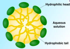
Describe the process of fat absorption in the small intestine
Segmental contraction breaks fat globules down into smaller globules. Bile salts surround the fat globules to form aggregates called micelles. Micelles absorb monoglycerides and volatile fatty acids from nearby emulsified fat droplets. Once the micelles come in contact with the membrane of an enterocyte, the products inside diffuse into the enterocyte (no specific transporter proteins required). The micelles then return to the luminal contents to absorb more monoglycerides and FFAs from emulsified fat droplets and the process repeats. See next question for how lipids are circulated into the bloodstream
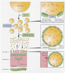
Describe the formation of chylomicrons within an enterocyte
Monoglycerides and FFAs are re-esterified to form triglycerides in the endoplasmic reticulum. Triglycerides can then coalesce along with cholesterol and phospholipids, and the exterior lipoprotein coat forms to create a chylomicron.
What component of the chylomicron allows their transport in the lymphatic and circulatory system?
The exterior protein coat made of lipoproteins.
How do chylomicrons enter circulation?
They are formed in the endoplasmic reticulum and packaged in the Golgi apparatus. They are then transported to the basolateral membrane and released into the ECF via exocytosis. They are too large to pass into the capillaries and are hence transported into lymphatic vessels. They are then transported via the thoracic duct into the junction of the left subclavian and left jugular vein. This way, they avoid the liver to enter the systemic circulation.
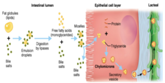
Which food substances enter the bloodstream without passing through the liver?
Fat-soluble vitamins and chylomicrons.
State the main routes that water is transported through
Para-cellular and trans-cellular.
Describe the para-cellular water transport route
Across tight junctions between epithelial cells. This route is restrictive and little water is moved this way.
Describe the trans-cellular water transport route
Across the cell membrane via transporter proteins specific to water on the luminal membrane (e.g. aquaporins). This route is more commonly used to transport water.
What is the largest route for water absorption?
Osmosis through Na+/glucose or amino acid transporters (passive transport).
What digestive component is most water reabsorbed from?
Digestive juice secretions (80-90%). The rest is food and drink and occurs principally in the small intestine.
Approximately, how much water in the SI do carnivores and omnivores absorb?
~90%.