Immune Patho II
 Call Kai
 Learn
 Practice Test
 Spaced Repetition
 Match
 Flashcards
 Knowt Play
1/52

There's no tags or description
Looks like no tags are added yet.
53 Terms
Part 1: Host Defenses, Infection, & Disease Transmission
3 lines of defense
1. Barriers (skin, mucous membranes, secretions)
2. Inflammatory response (phagocytic leukocytes, antimicrobial proteins, inflammatory response, fever)
3. Adaptive immunity (lymphocytes, antibodies, memory cells)
Skin - epidermis
Superficial outer layer
Continuously renews itself
Has a lot of keratin
0.05-0.01mm thick
No vascular structures

Skin - dermis
0.05mm-4mm thick
Elastin & collagen fibers
Gives skin strength & helps store water & electrolytes
Nerves & blood vessels
Location of sensory receptors

Skin - hypodermis/subcutaneous
Connective tissue, fat cells, immune cells, blood vessels & nerves
Connects skin to rest of body

Dermal appendages
Nails
Hair
Sebaceous glands → oil producing, protects skin
Sweat glands → temp regulation, toxin removal (minor)
**All within the dermis**
Resident microbiota
Average human has ~30 trillion human cells
Also has ~39 trillion bacterial cells
Live in nonsterile areas of body, including skin
Provide many benefits & usually don’t cause harm
Take up space
Compete for nutrients
Antimicrobial properties (can destroy microbes)
Infection
Invasion or overgrowth of a micoorgansim within the body, resulting in cellular, tissue, & organ destruction or even death
To cause infection, organism must penetrate the 3 lines of defense
Primary pathogens:
Bacteria, virus, fungi, parasites
Pathogens
Any microorganism that produces disease
2 types:
1. True pathogens: cause disease in healthy people
Bypass normal defenses & cause infection
Associated w/ a specific & recognizable disease (Covid, TB)
2. Opportunistic pathogens: part of our normal flora but can cause illness if the conditions are right
Often developed by immunocompromised people
Ex. Antibiotic-associated diarrhea or yeast infections
Pathogenicity - how easy is it to catch?
Communicability: ability to spread from person to person
Portal of entry is important
Infectivity: ability to invade & multiply in host
Pathogenicity - how well does it evade host defenses?
Immunogenicity: ability to produce an immune response
Antigenic variability: ability to alter antigens recognized by immune cells
Pathogen defenses: ability to avoid destruction
Pathogenicity - how bad can it harm us?
Mechanism of action: how a micoorganism damages tissue
Virulence: capacity to cause severe disease (potency)
Toxigenicity: production of toxins
Coinfection: 2+ pathogens transmitted together
Superinfection: new infection develops after an initial infection
Transmission
Infectious organisms reside in reservoirs
3 types:
1. Direct transmission → must have physical contact
2. Indirect transmission → fecal-oral, droplets/airborne, vectors (ex. mosquitos, fleas)
3. Vertical (parent to offspring in utero) vs. horizontal (spread from one subject to another)
Transmission - Zoonotic
Infectious disease that can spread from animals to humans
Chain of infection
Direct contact: touch
Droplet: large respiratory particles
Airborne: small suspended respiratory particles
**By breaking chain of infection, we can limit pathogen spread**
Universal precautions: all blood & body fluids treated as potentially infected

Phases of acute infection (important)
1. Exposure: contact w/ pathogen
2. Incubation: time b/w when a person get infected, & when they start experiencing symptoms
Pathogen is multiplying inside body
3. Prodrome: mild, vague, nonspecific symptoms
Ex. fatigue, low grade fever, nausea
4. Clinical illness: specific disease symptoms
Can have local manifestations, systemic manifestations, or both
5. Convalescence: recovery, fatigue
Gradual improvement of symptoms

Tests to identify correct pathogen
Direct culture: take 2-3 days, some organisms are difficult to grow
Antigenic tests: rapid, some tests have high false +/- rates
Ex. at-home Covid test
Antibody tests: doesn’t necessarily mean you have an active infection
Genetic tests (PCR): very accurate but must know genetic sequence to look for, need expensive equipment
Serum labs: basic blood cultures
Ex. WBC count
Part 2: Major Infectious Organisms
Bacteria
Bacterial characteristics
Bacteria are prokaryotes
Single cell w/ cell wall
Different DNA/RNA replication
Different ribosomes
Folic acid synthesis
Classification (shapes):
Cocci, Bacilli
Staphylo- (cluster), Strepto (chain), etc.
Can produce endospores
Resistant to heat, drying, chemicals (harsh conditions)
Ex. Clostridioides difficile
Various transmission routes & ways to avoid immune detection
Biofilms
Protective coating around bacteria that protects against antibiotic or host immunity → so they’re resistant to antibiotics
Highly difficult to remove
Ex. plaque, implanted devices

Bacterial toxins - Exotoxins vs. Endotoxins
Exotoxins: enzymes released during growth
Damages cell membranes, activates second messengers, & inhibits protein synthesis
Endotoxin aka Lipopolysaccharide (LPS):
Part of Gram-Negative cell wall
LPS activates a very strong immune response, which is what damages tissues (inflammation)
This is one of the patterns that innate immune cells recognize

Tissue damage from toxins - direct & indirect
Direct: exotoxin production
Pathogen → immune response → inflammation → leads to much of the tissue damage & symptoms
Indirect: induction of the immune response (endotoxin)
Indirect damage = immune-mediated damage
Staphylococcus aureus
Gram positive cocci
Often a normal colonizer (lives in body w/out causing issues)
Around 30% of people are colonized
Sits on skin → so must disinfect skin before procedures
Is an opportunistic pathogen (usually harmless, but can turn bad)
Numerous virulence factors (toxins, immune-evasion, biofilms)
Common cause of healthcare-associated infections
Antibiotic resistance
MRSA → specific type of Staphylococcus aureus that’s mutated & is resistant to certain antibiotics, so very difficult to treat
S. aureus skin conditions - Folliculitis
Superficial inflammation of hair follicles
Can be infectious or noninfectious
S. aureus skin conditions - Furuncles
Boils
Deeper inflammation of hair follicles
Develop from preceding folliculitis; spread through follicular wall into surrounding dermis
S. aureus skin conditions - Carbuncles
Collection of infected hair follicles that may develop into an abscess
Erthematous, painful, swollen mass that drains through many openings
Commonly causes systemic symptoms (fever, malaise) & often required medical intervention
S. aureus - Cellulitis
Infection/inflammation of the dermis &/or subcutaneous tissue
Occurs when bacteria gets into deeper layers of skin
Frequently occurs when early infections go untreated
Clinical manifestations:
Erythema, edema, warmth, pain
Risk factors:
Bacterial entry: bug bites, cuts, pressure injuries
Impaired healing: diabetes, poor circulation, immunosuppression
Increased exposure: crowded living conditions, poor sanitation
Clostridioides Difficile (C. diff)
Gram-positive, anaerobic, spore-forming bacillus
Exotoxin
2-5% of population are colonized, but not “infected”
Transmission by fecal-oral route
Common cause of healthcare-associated diarrhea
Spores can survive on fomites (contaminated surface) for months
Hospitalized patients require enteric isolation

C. diff - clinical manifestations, complications, & risk
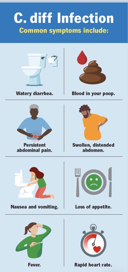
Clinical manifestations:
Frequent, watery diarrhea (up to 10-15x/day)
Fever, loss of appetite, nausea/vomiting
Abdominal pain, tenderness. or cramping
Complications:
Toxic megacolon (severely dilated colon, stops working), colon perforation, sepsis, death
Greater risk for:
Antibiotic use
65+ years old
>14 days in hospital
Immunocompromised
Fungi
Fungal infection (mycosis)
Large eukaryotes:
Thick cell walls
Single-celled yeasts
Mulicellular molds
Dimorphic (changes forms) → very common
Transmission:
Inhalation, contamination of wounds, opportunistic (present in body)
Candiasis
Most common fungal infection, typically due to commensal overgrowth
Oral thrush (yellowy-white coating on tongue)
Vaginal candidiasis
Risk factors:
Moist environment
Recent antibiotic use
Pregnancy
Diabetes
Infants/Immunocompromised

Dermatophytes
Fungi that infects keratinized tissues
Dermatophyte infection are called tinea, followed by body area infected
Tinea capitis: scalp
Tinea pedis: foot (athletes foot)
Tinea corporis: body (ringworm)
Tinea cruris: groin (jock itch)
Tina unguium: nail (onychomyosis)
Often presents as patches that are dry, scaly, erythematous, & pruritic

Parasites
Parasites patho
Organism that lives on/in host & lives at the expense of the host
3 types:
1. Protozoa
2. Helminths
3. Ectoparasites
Protozoa
Eukaryotic, unicellular
Amoebas, Flagellates, Ciliates, Sporozoan
Helminths
Parasitic worms
Large, multicellular
Intestinal & tissue nematodes (ex. hookworm, roundworm)
Flatworms (ex. liver fluke, lung fluke, tapeworm)
Ectoparasites
Arthropods that depend on a blood meal from humans
Fleas, ticks, lice, mites
Parasite transmission
Rarely transmitted from human to human; transmitted mainly through vectors
Malaria by mosquito bites
Trypanosomes by tsetse fly
Leishmania spp. by sand fleas
Others found in contaminated water or food (ex. Giardia duodenalis)
Typically have complex life cycles that require multiple hosts to complete their life cycle
Toxoplasma Gondii
Causative agent of toxoplasmosis
Parasitic protozoa
Reservoirs: undercooked contaminated meat, cat feces, contaminated water
**Pregnant woman should not change cat litter**
Transmission: fecal-oral route or eating contaminated meat
Many infections are asymptomatic, but can also cause flu-like symptoms
Fever, headache, myalgias, pharyngitis (sore throat), rash, cervical lymphadenopathy (swollen neck lymph nodes)

Viruses
Virus patho
Basic structure: nucleic acid surrounded by protective protein (capsid)
Virion only contains bare necessities:
Genetic material, viral-specific proteins/enzymes, protective coating
Cannot replicate on their own
Disease occurs when virus takes over host cell, interfering w/ normal function & sometimes causing cellular death
Viral infection stages
1. Attachment
Virus into body
Finds right cell w/ right receptor (lock & key)
2. Uncoating
Virus takes off its coat surrounding genetic material
Genetic material is released into host cell
3. Replication
Virus takes over
Uses host cell as a factory
4. Release
Multiplied virus is released
Through budding or by rupturing host cell

Viral latency
Survival strategy where a virus remains dormant in a cell w/out producing more viruses → can last for years
Viral genome is left behind in the cell
IF the virus can integrate its DNA into host cell, it can be transmitted to daughter cells
The daughter cells can then make new viruses at a later time
Examples of latent viruses
Herpes Simplex Virus 1 & 2 (cold sores, genital warts)
**Varicella Zoster Virus (chicken pox = shingles when reactivated)**
Human Immunodeficiency Virus (HIV, AIDS)
Epstein Barr Virus (infectious mononucleosis, various lymphomas)
Influenza A
Highly contagious respiratory virus spread through respiratory droplets
Single-stranded RNA virus w/ a range of hosts
Humans, birds, pigs, bats, horses, dogs
Mutates quickly & changes
Antigenic Drift → small genetic changes that slowly change over time
Antigenic Shift → major genetic changes that occur very quickly
Flu pandemic, Covid-19 pandemic

Influenza A - Clinical Manifestations
Localized:
Dry cough, sore throat, nasal congestion, nasal drainage, shortness of breath
Symptoms caused by inflammation of immune response, not virus
Systemic:
Chills, fever, body aches, anorexia, weakness, malaise
Can progress to pneumonia, sepsis, & death in severe cases
Viral Hepatitis
Group of viral infections that cause liver inflammation & damage
A & E = fecal-oral route, most commonly through food
B, C & D = body fluids (sex, childbirth, vaccinations)
Must have B to get D

Acute Viral Hepatitis stages
Incubation
Phase lasts 2-6 weeks for HepA, & up to 6 months for HepB
Asymptomatic
Prodrome
Nonspecific GI symptoms (nausea, vomiting, right upper quadrant abdominal pain, fatigue, malaise, fever)
Icteric
Jaundice, dark urine, clay-colored stools, significantly elevated liver enzymes
Recovery
Symptoms resolve as pathogen is destroyed, typically occurs 4-8 weeks after symptom onset
Chronic Hepatitis
Liver inflammation lasting longer than 6 months
HepB & HepC
Persistent infection can lead to progressive liver damage
Fibrosis (due to inflammation)
Cirrhosis (long term fibrosis)
Portal hypertension
Ascitis
Hepatocellular carcinoma
Can be asymptomatic
Herpes Simplex Virus (HSV)
HSV-1
Contact w/ infected saliva
Cold sores or fever blisters
HSV-2
Genital infections
Skin-to-skin mucous membrane contact during viral shedding
Lesions are painful, erythematous vesicles
Both can go latent! (inactive, no more symptoms)
Comes down same nerve route (so after going away, it’ll come back in the same spot)
Varicella Zoster Virus
Varicella (chickenpox)
Initial infection transmitted by respiratory droplets
Full body pruritic fluid-filled blisters that crust over time
Herpes Zoster (shingles)
Reactivated zoster virus (usually 50+ years old)
Pain & paresthesia localized to a dermatome, followed by vesicular eruptions
Can’t “catch” shingles, but if exposed to vesicles the person can get chickenpox if not vaccinated or previously infected