L24 - Laboratory Assessment of Platelets
 Call Kai
 Learn
 Practice Test
 Spaced Repetition
 Match
 Flashcards
 Knowt Play
1/45

There's no tags or description
Looks like no tags are added yet.
46 Terms
What are the Primary Components of Hemostasis?
TO STOP BLEEDING
Vascular endothelium
Platelets
Coagulation factors
How do Primary and Secondary Hemostatic Defects Differ?
Sepsis and DIC can affect both systems
Primary: Defects in →
Platelet number/function
von Willebrand factor (vWF)
Endothelium
Secondary: Defects in →
Coagulation factors
What is the Initial Approach to a Bleeding Patient?
First exclude physical causes (trauma, tumors), then evaluate:
History: Bleeding location, duration, previous episodes, medications/toxins, and travel
Signalment: Species, breed, and age
Physical examination: Bleeding pattern and distribution
Screening tests:
CBC/platelet count and BMBT for primary hemostasis
PT/PTT for secondary hemostasis
How do Bleeding Patterns Differ between Primary and Secondary Hemostatic Defects?
Both → Excessive bleeding after trauma or surgery.
Primary: Superficial/mucosal bleeding →
Petechiae
Ecchymoses
Epistaxis
Hematuria
Melena
Secondary: Deep/high-volume bleeding →
Cavitary hemorrhage
Large hematomas
Hemarthrosis

What are the Four Major Differential Diagnoses for Primary Hemostatic Defects?
Thrombocytopenia → decreased platelet number
Thrombopathia → abnormal platelet function
von Willebrand disease (vWD) → abnormal vWF quantity/function
Vasculopathy → vascular/endothelial abnormalities
Vasculopathy is often a diagnosis of exclusion
What Bleeding Pattern Suggests a Coagulation Factor Deficiency?
Deep, high-volume bleeding, including hemoabdomen, hemothorax, large hematomas, and hemarthrosis
Example: Hemophilia A (factor VIII deficiency) can cause joint hemorrhage
What Tests are Used to Evaluate Primary Hemostasis?
Platelet metrics:
Platelet count, mean platelet volume (MPV), plateletcrit (PCT)
Buccal mucosal bleeding time (BMBT):
Overall primary hemostatic function
vWF testing:
Detects von Willebrand disease
Specialized platelet function assays:
Evaluate thrombopathia
What is the Most Important Initial Test for Suspected Primary Hemostatic Disorders?
CBC with platelet count
Because platelet number is the easiest component of primary hemostasis to assess
What is the Platelet Count, and How is It Measured?
Number of platelets per µL of blood, measured using
EDTA-anticoagulated blood
Automated hematology analyzer
Blood smear platelet estimate

How does Platelet Clumping Affect Automated Platelet Metrics?
Platelet clumping causes:
Falsely decreased platelet count → pseudothrombocytopenia
Falsely increased MPV
Common in cats and horses
Citrate-anticoagulated blood may provide a more accurate count
Why Must a Blood Smear Always Be Reviewed When Evaluating Platelets?
To identify platelet clumps, assess platelet density and size, and verify automated counts
Clumps commonly occur along the feathered edge and prevent an accurate numerical estimate, although numerous clumps may suggest adequate platelet numbers

How is Platelet Count Estimated from a Blood Smear?
At 1000× oil immersion, average the number of platelets across multiple monolayer fields
Average PLTs/field × 20,000 = Estimated PLT count
Example: 7 platelets/field × 20,000 = 140,000 platelets/µL

Calculate Estimated Platelet Count
Average PLTs/field × 20,000 = Estimated PLT count
7 platelets/field × 20,000 = 140,000 platelets/µL

What is Mean Platelet Volume (MPV)?
The average size of circulating platelets measured by an automated analyzer
MPV is analogous to MCV for erythrocytes
What are Macroplatelets, and What Do They Indicate?
Unusually large platelets visible on a blood smear
Younger platelets are LARGER → if have thrombocytopenia, tells us the bone marrow megakaryocytes are creating newer platelets (regenerative thrombocytopenia)
Useful for determining mechanism/cause of thrombocytopenia

How is MPV Interpreted with Platelet Count?
Low PLT + high MPV: Suggests…
Regenerative thrombocytopenia (increased marrow response)
Commonly due to destruction or consumption
Low PLT + normal/low MPV: Suggests…
Decreased production or sequestration
Important: Normal MPV does not exclude regeneration → interpret with smear and clinical findings

What is Congenital Macrothrombocytopenia?
An inherited beta-tubulin 1 mutation causing low platelet numbers with unusually large platelets
Common in Cavalier King Charles Spaniels; also occurs in other breeds
Typically does NOT CAUSE ABNORMAL BLEEDING ISSUES because total platelet mass is maintained
Can be mistaken for severe acquired thrombocytopenia
What is Plateletcrit (PCT), and How Does It Differ from Platelet Count?
PCT: Proportion of whole blood occupied by platelets
Reflects total platelet mass
PLT count: Number of platelets per µL
Used more routinely
In congenital macrothrombocytopenia, PLT count may be low while PCT remains normal
What Tests Can Evaluate Platelet Function?
If suspect a patient has platelet function disorder (thrombopathia) → These are specialized tests and are less routinely available than platelet counts
PFA-100: Detects marked platelet functional abnormalities
Thromboelastography: Evaluates whole-blood clot formation kinetics and can identify platelet-related abnormalities
What is Buccal Mucosal Bleeding Time (BMBT), and How is It Interpreted?
A functional assessment of primary hemostasis
Normal: Less than 4 minutes in most species
Pathologic increase of BMBT if:
Thrombocytopenia
Thrombopathia
vWD
Vasculopathy
BMBT confirms impaired primary hemostasis but does not identify the specific cause
What Tests are Used to Diagnose von Willebrand Disease?
vWF antigen assay (vWF:Ag): ELISA measuring vWF quantity relative to healthy canine plasma; reported as a percentage of normal
vWF collagen-binding assay
Multimer analysis (Western blot)
Breed-specific genetic testing
How are Canine vWF Antigen Results Interpreted?
vWF:Ag
70–180%: Normal
50–69%: Borderline
0–49%: Consistent with vWD
Less than 35%: Increased hemorrhage risk
What are the Three Types of von Willebrand Disease?
Type I: Decreased but present vWF
Most common inherited hemostatic disorder, classically in Dobermans
Type II: Abnormal/inadequate vWF multimers
Type III: Complete absence of vWF
Often severe bleeding
What are the Four Major Mechanisms of Thrombocytopenia?
Increased platelet consumption (utilization)
Increased platelet destruction
Increased platelet sequestration
Decreased platelet production
What Causes Increased Platelet Consumption, and What is the Expected MPV?
Platelets are consumed faster than the marrow can replace them
Causes: Severe trauma, vasculitis, DIC, sepsis, and viper envenomation
MPV: May increase if the marrow responds (regenerative thrombocytopenia)
What Causes Increased Platelet Destruction, and What is the Expected MPV?
Platelets are abnormally destroyed
Causes: Immune thrombocytopenia (ITP), tick-borne infections, and neoplasia (e.g., hemophagocytic histiocytic sarcoma)
MPV: May increase with a regenerative marrow response
What is Immune Thrombocytopenia (ITP), and When Should It Be Suspected?
Immune-mediated platelet destruction, analogous to IMHA affecting RBCs
ITP is an especially important differential in dogs with severe thrombocytopenia below 50,000 platelets/µL, after excluding other causes
What Causes Increased Platelet Sequestration, and What are Its Key Features?
Platelets redistribute outside circulating blood rather than being destroyed
Causes: Splenic dilation (rest, anesthesia, neoplasia), hypothermia, and pulmonary congestion
Usually mild thrombocytopenia
Generally no spontaneous hemorrhage
Normal MPV because platelet mass is maintained
What Causes Decreased Platelet Production, and What is the Expected MPV?
Bone marrow cannot adequately produce platelets
Causes: Medications, infections, myelophthisis, leukemia, and immune destruction of megakaryocytes
MPV: Typically normal or low (nonregenerative)
May occur with pancytopenia (anemia + leukopenia + thrombocytopenia)
What Platelet Count Thresholds are Clinically Important for Bleeding Risk?
These are clinical risk thresholds, not absolute predictions for every patient.
Below 20,000/µL:High risk of spontaneous hemorrhage
Below 50,000/µL: Increased risk of excessive surgical or trauma-associated bleeding
What is Thrombopathia, and How Can It Cause Bleeding?
A platelet function disorder that impairsprimary hemostasis despite potentially normal platelet numbers
It may be acquired (more common) or inherited (rare)
What are the Major Causes of Acquired Thrombopathia?
These interfere with platelet activation, signaling, or aggregation
Medications: Aspirin and clopidogrel
Uremic acids (kidney disease)
Ketoacids (diabetic ketoacidosis)
Significant hypergammaglobulinemia (e.g., multiple myeloma)
What are Examples of Inherited Thrombopathia?
The professor emphasized recognizing that these rare disorders exist rather than memorizing every example.
Glanzmann thrombasthenia: GPIIb/IIIa defect → impaired fibrinogen-mediated aggregation
Chediak–Higashi syndrome: Abnormal granule movement
Delta storage pool disease: Abnormal dense granule production
Scott syndrome: Defective phosphatidylserine exposure → impaired coagulation-factor assembly
What are the Major Mechanisms and Differentials of Thrombocytosis?
Excitation: Epinephrine-mediated splenic contraction
Inflammation: Sterile or infectious inflammation
Drug-induced: Chronic corticosteroid therapy
Iron deficiency anemia: Common associated finding
Neoplasia: Paraneoplastic thrombocytosis or rare essential thrombocytosis
What are the Most Important Initial Differentials for Thrombocytosis?
Prioritize inflammation and corticosteroid exposure → then evaluate for iron deficiency anemia and neoplasia
Inflammation and drug-associated thrombocytosis are particularly common
What is the Difference between Paraneoplastic and Essential Thrombocytosis?
Paraneoplastic: Another tumor stimulates increased platelet production through cytokines
Essential thrombocytosis: Neoplastic megakaryocytes directly overproduce platelets
Why Must Platelet Counts Be Interpreted Using Species-Specific Reference Intervals?
Normal platelet counts vary substantially between species
Ruminants and rodents may naturally have much higher platelet counts than dogs, so species-specific intervals prevent misdiagnosing thrombocytosis
TABLE 1. Primary vs. Secondary Hemostatic Disorders
Feature | Primary Hemostasis | Secondary Hemostasis |
Defect |
| Coagulation factors |
Bleeding pattern | Superficial/mucosal | Deep, high-volume |
Petechiae | Common | Uncommon |
Ecchymoses | Common | Large hematomas more typical |
Epistaxis | Common | Possible |
Hemarthrosis | Uncommon | Characteristic |
Cavitary hemorrhage | Uncommon | Characteristic |
Initial tests | CBC/PLT count, BMBT | PT, PTT |
Examples | ITP, vWD, thrombopathia | Hemophilia A, anticoagulant rodenticide toxicity |
Key distinction:
Primary = Problem forming the initial platelet plug
Petechiae suggest a primary hemostatic defect
Secondary = Problem stabilizing the plug with fibrin
Hemarthrosis and cavitary hemorrhage suggest a secondary defect
TABLE 2. Platelet Metrics & Interpretation
Metric | What It Measures | Clinical Interpretation |
PLT count | Platelets per µL | Low = thrombocytopenia High = thrombocytosis |
MPV | Average platelet size | Increased MPV may suggest increased platelet production |
PCT | Total platelet volume as a proportion of blood | Reflects platelet mass |
Blood smear | Platelet density, size, clumping | Confirms automated results and identifies artifacts |
Important PLT + MPV Combinations
PLT Count | MPV | Interpretation |
↓ | ↑ | Increased marrow response, often destruction or consumption |
↓ | Normal/↓ | Decreased production or sequestration |
↓ | Falsely ↑ | Platelet clumping (pseudothrombocytopenia) |
↓ | ↑ with normal PCT | Congenital macrothrombocytopenia |
Remember:
MPV is supportive, not definitive
Always interpret it alongside the blood smear and clinical findings
TABLE 3. Diagnostic Tests for Primary Hemostatic Disorders
Test | Evaluates | Important Findings |
CBC / platelet count | Platelet number | First test to perform |
Blood smear | Platelet morphology and density | Detects clumping and macroplatelets |
BMBT | Overall primary hemostatic function | Prolonged with primary hemostatic defects |
vWF:Ag | vWF quantity | Decreased with quantitative vWF deficiency |
vWF multimer analysis | vWF multimer distribution | Helps characterize vWD |
Platelet function analyzer | Platelet function | Detects functional abnormalities |
Thromboelastography | Whole-blood clot formation | Can reveal platelet-related abnormalities |
Important values from L24:
BMBT: Normal is generally less than 4 minutes.
PLT below 20,000/µL: High risk of spontaneous hemorrhage.
PLT below 50,000/µL: Increased bleeding risk with surgery or trauma.
The professor emphasizes beginning with the CBC and platelet count before pursuing more specialized testing.
TABLE 4. Four Mechanisms of Thrombocytopenia
Mechanism | What Happens | Important Differentials | Expected MPV |
Consumption | Platelets USED faster than replaced | DIC, sepsis, trauma, vasculitis, envenomation | May ↑ |
Destruction | Platelets DESTROYED prematurely | ITP, tick-borne infections, neoplasia | May ↑ |
Sequestration | Platelets REDISTRIBUTED OUTSIDE circulation | Splenic dilation, anesthesia, hypothermia | Usually normal |
Decreased production | Bone marrow is NOT MAKING enough | Drugs, infections, leukemia, myelophthisis | Normal/↓ |
How to Interpret These Mechanisms
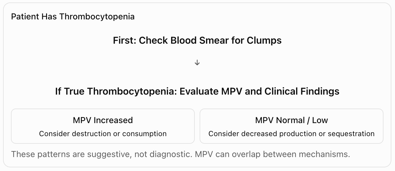
TABLE 5. Thrombocytopenia vs. Thrombopathia vs. vWD
Disorder | Primary Problem | Platelet Count | Key Clue |
Thrombocytopenia | Insufficient platelets | ↓ | Petechiae, mucosal bleeding |
Thrombopathia | Dysfunctional platelets | Often normal | Bleeding despite adequate platelet number |
von Willebrand disease | Abnormal vWF quantity/function | Usually normal | Impaired platelet adhesion |
Vasculopathy | Endothelial abnormalities | Often normal | Diagnosis of exclusion |
This table is particularly useful for clinical questions where an animal has primary hemostatic bleeding but a normal platelet count.
TABLE 6. Major Mechanisms of Thrombocytosis
Mechanism | Important Differentials | Explanation |
Excitation | Epinephrine release | Splenic contraction |
Inflammation | Infectious or sterile inflammation | Cytokine stimulation |
Drug-induced | Chronic corticosteroid therapy | Increased platelet count |
Iron deficiency | Iron deficiency anemia | Reactive thrombocytosis |
Paraneoplastic | Neoplasia | Tumor-associated cytokine stimulation |
Essential thrombocytosis | Megakaryocytic neoplasia | Autonomous platelet overproduction |
Key distinction: Paraneoplastic thrombocytosis occurs secondary to another tumor, whereas essential thrombocytosis results from neoplastic platelet-producing cells.
How These Tables Connect
