Exam 3 Health Assessment
 Call Kai
 Learn
 Practice Test
 Spaced Repetition
 Match
 Flashcards
 Knowt Play
 Podcast
1/143

There's no tags or description
Looks like no tags are added yet.
144 Terms
Diastole
AV valves are open during the ventricular filling period, when the heart is relaxing. During diastole, the aortic and pulmonic valves are closed to prevent regurgitation of blood from the aorta and pulmonary artery into the ventricles.
Systole
AV closes during ventricular contraction; contraction of the heart. During systole, the closed mitral and tricuspid valves prevent regurgitation (back flow) of blood into the atria
Precordium
The region or the thorax immediately in front of the heart
Point of Maximum Impulse
It is a term used to describe the area where the apical pulsation can be seen or palpated. Commonly found at the intersection of the 5th ICS mitral area & left MCL.
Apex
Is the lower portion of the heart
Atria
Are 2 upper chambers of the heart (right or left) that receive blood coming from the heart
Ventricles
Are 2 lower chambers of the heart that pump blood out of the heart. The right ventricle sends blood to the lungs, and the left ventricle sends blood to the rest of the body.
SA Node (Sinoatrial)
A small electrical impulse that fires in the right atrium generates the normal heartbeat and serve as the pacemaker of the heart
The heart is a large muscle
That acts as two pumps in one — the right side and the left side.
The right side of the heart
Sends deoxygenated blood (low in oxygen) to the lungs through the pulmonary circulation, where the blood picks up oxygen and releases carbon dioxide.
The left side of the heart
Then receives that oxygen-rich blood from the lungs and pumps it out to the rest of the body through the systemic circulation to deliver oxygen and nutrients to tissues.
The pulmonary artery
Is unique because it carries deoxygenated blood to the lungs, while the pulmonary veins carry oxygenated blood back to the heart — the opposite of how arteries and veins usually work in the rest of the body.
The systemic circulation sends oxygenated blood to all parts of the body:
- Carotid arteries and jugular veins serve the brain
- Subclavian arteries and superior vena cava serve the upper limbs
- Thoracic aorta and the inferior vena cava serve the lower limbs
S1: or "lub
Results from the closure of the mitral and tricuspid valves
S2: or "dub"
Results from the closure of the aortic and pulmonic valves.
Preload
Volume in the right atrium at the end of diastole; an indicator of how much blood will be forwarded to and ejected from the ventricles
Afterload
It is the pressure in the great vessel.
Cardiac output
The ability of the heart to shorten its muscle fibers, producing a contraction during systole. It is the amount of blood ejected from the left ventricle each minute.
CO = HR x SV
Stroke Volume
How much blood is ejected with each beat or stroke.
Contractility
The ability of the heart to shorten its muscle fibers, producing a contraction during systole.
Arrhythmia
Abnormal heart rhythms with early (premature), delayed or irregular beats that can arise from atria, AV junction, or ventricles.
Bruit
Are swooshing sounds similar to the sound BP makes. They result from turbulent blood flow related to atherosclerosis; audible when the artery is partially obstructed.
Thrill
A vibrating, palpable sensation felt on the skin that accompanies a loud cardiac murmur or turbulence in a blood vessel.
Patent Ductus Arteriosus
The continuous murmur peaks just before & after S2. It has a rough, harsh, mechanical quality with a 2nd left ICS palpable thrill.
Atrial Septal Defect
This continuous murmur is altered by the Valsalva maneuver. It is a systolic ejection murmur with a medium pitch
Pulmonary embolus
A condition where a blood clot travels to the lungs and blocks one or more pulmonary arteries.
Arterial ulcer
A sore that develops when there is inadequate blood flow to the affected area, typically the lower extremities.
Edema
Any swelling in the arm or legs.
D-dimer
Is a blood test procedure to assess a patient with a possible clot, such as DVT.
Intermittent claudication
Pain brought on by exertion and relieved by rest.
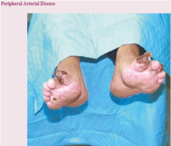
Peripheral Arterial disease
picture
Raynaud phenomenon
picture

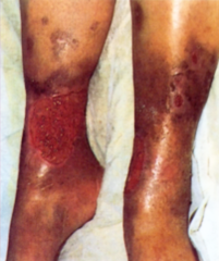
Chronic Venous Insufficiency
A condition where the veins in the legs do not function properly, leading to poor blood flow back to the heart.
Deep Vein Thrombosis
A condition where blood clots form in the lower extremities.

Thrombophlebitis
A condition where a blood clot (thrombus) forms in a vein and causes inflammation. This can lead to pain, swelling, redness, and warmth in the affected area.

Lymphedema
Occurs when lymph channels or nodes are obstructed; results from injury, scarring excision of lymph nodes.

Hepatomegaly
Enlargement of the liver.
Splenomegaly
Enlargement of the spleen
Ascites
Buildup of fluid in the abdomen, making it look swollen or bloated.
Striae
Known as stretch marks, they are linear, narrow bands of skin that appear as reddish or silvery lines. They are caused by the rapid stretching of the skin, often during pregnancy, puberty, or weight gain.
Borborygmi
The rumbling or gurgling sounds your stomach makes when gas or fluid moves through the intestines.
Tympany
A drum-like sound heard when tapping (percussing) the abdomen — it usually means there is air or gas underneath.
Referred pain
Pain that is felt in a different area of the body than where it is actually coming from (for example, shoulder pain caused by a gallbladder problem).
Epigastric
The upper middle part of the abdomen, just below the breastbone (sternum).
Cardiovascular System
consists of the heart, blood vessels, and blood
Blood vessels are arranged in two continuous loops
1. pulmonary circulation
2. systemic circulation
4 chambers
1. Right atrium
2. Left atrium
3. Right ventricle
4. Left ventricle
Great Vessels
- Superior and inferior vena cava
- pulmonary artery
- pulmonary veins
- aorta
Pericardium Structure
The tough, outermost layer is composed of dense connective tissue. It is a conical-shaped sac anchored to the diaphragm and connected to the sternum via ligaments.
Pericardium Function
Provides structural support, anchors the heart in the mediastinum, prevents the heart from over-expanding with blood volume, and protects it from infection.
Myocardium Structure
Is the middle, muscular layer of the heart, composed of specialized cardiac muscle cells called cardiomyocytes
Myocardium Function
Is to contract and relax to pump blood throughout the body.
Endocardium Structure
Is the thin, innermost layer of the heart wall, composed of endothelial cells, that lines the heart chambers and valves.
Endocardium Function
To provide a smooth surface for blood flow, minimize turbulence, and act as a barrier between the blood and the myocardium (heart muscle)
Epicardium Structure
The outer, protective layer of the heart that functions as a barrier, secretes factors crucial for myocardial development and repair, and serves as a source of cells that differentiate into other heart tissues.
Epicardium Function
It is a single layer of mesothelial cells on the outer surface of the heart wall, connected to the underlying muscle by connective tissue.
Heart wall, chambers, and valves structure and function
Four chambers separated by valves, whose main purpose is to prevent backflow of blood
Four valves
- Two atrioventricular (AV) valves
- Two semilunar (SL) valves
Cardiac Cycle
The rhythmic flow of blood through the heart is cardiac cycle
Diastole phase one
The relaxation phase of the heartbeat occurs when the heart's ventricles fill with blood.
Systole phase two
The period when the heart's ventricles contract and pump blood out to the lungs and the rest of the body.
Bundle of His
A specialized collection of nerve fibers located in the heart that plays a crucial role in the electrical conduction system of the heart.
Purkinje fibers
A specialized collection of nerve fibers located in the heart that plays a crucial role in the electrical conduction system of the heart.
Preload
The amount of blood that fills the heart's ventricles (chambers) before they contract.
Contractility
The force and velocity with which the heart muscle (myocardium) contracts and pumps blood.
Afterload
The resistance that the heart must overcome to eject blood during systole (contraction)
Cardiac output =
heart rate x stroke volume
To increase circulating blood (cardiac output)
- Increase heart rate
- Increase stroke volume
- Increase heart rate + stroke volume
S1 definition
The first of the two main heart sounds, typically described as a "lub" sound. It occurs when the mitral and tricuspid valves close at the beginning of systole (the contraction phase of the heart) happens at the apex of the heart
S2 definition
A sound heard during a heartbeat, occurring when the aortic and pulmonary valves close. It happens at the base of the heart .
S3 definition
A low-frequency "gallop" sound that occurs in early diastole and is caused by the rapid filling of the ventricles.
S4 definition
An extra heart sound that occurs just before the first heart sound (S1). It is a low-pitched, dull sound that can be heard with a stethoscope. Heard at the end of diastole.
Arrhythmias
A condition in which the heart beats with an irregular or abnormal rhythm. (Afib)
Extra heart sounds?
Murmurs (whooshing, blowing, or clicking noise)
Conditions resulting in a murmur
- Velocity of blood increases (flow murmur), e.g., in exercise, thyrotoxicosis
- Viscosity of blood decreases, e.g., in anemia
- Structural defects in valves, narrowed valve, incompetent valve
- Unusual openings occur in chambers, dilated chambers, wall defects
Fetal heartbeat
3 weeks' gestation (Fetal circulation compensates for nonfunctional lungs and Oxygenation)
Inflation and aeration of the lungs at birth produce what
circulatory changes
Blood volume for pregnant women
- Increases significantly to meet the needs of the growing fetus
- 30-40%
Stroke volume/cardiac output for pregnant women
- Both stroke volume and cardiac output increase to support the fetus, with cardiac output rising by
- 30-50%
Pulse rate for pregnant women
- Likely to see an increase between 12 and 16 weeks and again when you reach 32-36 weeks
- Increase 10-15bpm
Blood pressure for pregnant women
It is crucial to monitor blood pressure as it can indicate potential health risks for both the mother and baby. (especially last week of pregnancy)
Aging Adult
- CAD (coronary artery disease)
- Hemodynamic changes
- Arrhythmias
- Left Ventricular Wall
- Decreased activity tolerance
- Syncope (fainting spells)
- Angina (used to decreased heat pain)
Aging Adult: Blood pressure common with aging
- Systolic (raises about 20 mmHg)
- Diastolic (will rise)
- Orthostatic hypotension (drop when sitting and standing)
- Carotid artery (decreases)
Cultural Prevalence
- In the U.S., more than 1 in 3 have one or more forms of cardiovascular heart disease (CVD)
- > prevalence in >65 y/o, men, American Indians/Alaska Natives
- Causes of CVD
- Evidence shows that potentially modifiable risk factors contribute to the overwhelming majority of cardiac risk
Hypertension
- Prevalence of hypertension is 55% for African Americans
- 25.3% for American Indians or Alaska Natives
- 23.3% for whites
- 21% for Hispanics and Asians
Subjective Data (cardiovascular)
- Chest pain
- Dyspnea (shortness of breathe)
- Orthopnea (shortness of breathe while sleeping)
- Cough
- Fatigue
- Cyanosis or pallor
- Edema (swelling/pitting/nonpitting)
- Nocturia (having to pee at night)
- Past cardiac history
- Family cardiac history
- Personal habits (cardiac risk factors)
Subjective: Pediatrics (cardiovascular)
- Mother's health: Any unexplained fever, rubella in: first trimester, other infection, hypertension, or drugs taken
- Cyanosis: nursing or crying? Is the baby able to eat, nurse, or finish a bottle without tiring?
- Growth: Growth charts
- Activity: tire during play, squatting
- Strep infections, chromosomal abnormalities
Subjective: Pregnant Women (cardiovascular)
- History of hypertension during pregnancy?
- Usual blood pressure?
- Any treatments?
- Associated symptoms?
- Faintness or dizziness?
Subjective: Aging Adult (cardiovascular)
- Hypertension, CAD, chronic emphysema, or bronchitis
- Medications: Side effects? Compliant?
- Environment: Stairs? How often?
Objective Data (cardiovascular): Preparation
- Provide privacy for females
- Warm, quiet room
- Stethoscope
- Warm hands
Objective Data (cardiovascular): Inspect
- Anterior Chest: PMI (Point of Maximal Impulse)
- Carotid arteries-sitting
- Jugular veins and precordium: supine with head and chest slightly elevated (45 degrees)
- Right side
Objective Date (cardiovascular): Palpate
- PMI
- carotid artery: Location
- Heart valve areas: Thrills/vibrations
Objective Data (cardiovascular): Auscultation
- Second right intercostal space
- Second left intercostal space
- Left lower sternal border
- Fifth intercostal space/mid clavicular line
- S1 and S2
- Rate/rhythm
- Extra sounds
Abnormal Findings: Congenital Heart Defects: Patent ductus arteriosus
A heart defect where a temporary fetal blood vessel between the aorta and pulmonary artery stays open after birth.
Abnormal Findings: Congenital Heart Defects: Atrial septal defect
A hole in the wall between the two upper chambers of the heart.
Abnormal Findings: Congenital Heart Defects: Ventricular septal defect
A hole in the wall between the lower chambers of the heart.
Tetralogy of Fallot
A birth defect involving four heart problems that disrupt normal blood flow.
Coarctation of the aorta
A birth defect where the aorta is narrowed, restricting blood flow to the body.
Patent foramen ovale
A small opening in the wall between the heart's two upper chambers.
Abnormal Findings: Murmurs Due to Valvular Defects
- Aortic stenosis
- Pulmonic stenosis
- Mitral regurgitation
- Tricuspid regurgitation
- Mitral stenosis
- Tricuspid stenosis
- Aortic regurgitation
- Pulmonic regurgitation