Neck + Abdomen + Chest
 Call Kai
 Learn
 Practice Test
 Spaced Repetition
 Match
 Flashcards
 Knowt Play
1/98

There's no tags or description
Looks like no tags are added yet.
99 Terms
how many compartments are in the neck
4

red compartment
vertebral

yellow compartment
visceral

purple compartments
R/L vascular
what surrounds and separates neck compartments
fascia
what between the neck compartments and importance
potential spaces, space for pathogens and cancerous cells
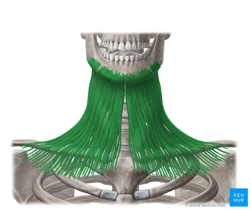
muscle (innervation) (origin, insertion)
platysma (facial nerve) (pectoralis major/deltoid, mandible)
2 types of fascia in the neck
superficial and deep

how many layers of the neck are there
9

layers of the skin (superficial to deep)
skin, subcutaneous fat, superficial fascia (platysma), investing cervical fascia, pretracheal fascia, muscles (sternocleidomastoid, infrahyoid muscles), carotid sheath (blood vessels and nerves), viscera layer (thyroid, trachea, esophagus), prevertebral fascia

subcutaneous fat
what is subcutaneous fat
cutaneous nerves, lymphatic vessels, lymph nodes

space (includes what?)
carotid space (carotid sheath, common carotid artery, internal jugular vein, nerves)

retropharyngeal space

space (importance)
danger space (cancer spreading)

muscle (origin, insertion)
sternocleidomastoid (sternum/clivical, mastoid process/occipital bone)

trapezius

levator scapulae
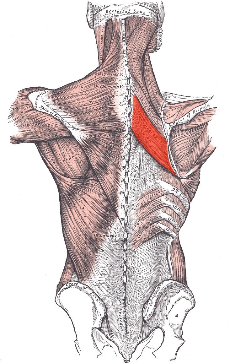
muscle (origin, insertion)
rhomboid major (vertebrae T2-5, medial border of scapula)

muscle (origin, insertion)
rhomboid minor (vertebrae C7-T1, medial border of scapula)

muscle (origin, insertion)
latissimus dorsi (thoracolumbar fascia, intertubercular sulcus)

splenius capitis

splenius cervicis
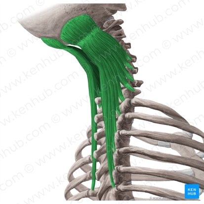
semispinalis capitis

posterior triangle

levator scapulae

middle scalene

anterior scalene
vein landmarks for SCM
internal jugular (underneath), external jugular (on top), anterior jugular (runs along anterior)

term, importance
lesser supraclavicular fossa, includes inferior portion of interior jugular vein
when does the mastoid develop
when infants start using that level of movement in SCM

term (innervation)
suboccipital triangle (posterior ramus of C1 spinal nerve)
importance of suboccipital triangle
carry the arteries and nerves that go directly from vertebrae to the brain

green
obliques capitis superior
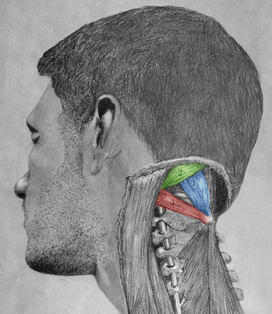
red
obliques capitis inferior

blue
rectus capitis posterior major
shoulder droop is damage to which nerve
CN 11 (accessory)
describe shoulder droop
damage to nerve, no tension in trapezius, clavicle/scapula cannot pull muscle up
what muscle separates the posterior triangle into what and what
omohyoid separates into occipital and subclavian
what runs through occipital triangle, why concerning
CN 11 (most superficial), external jugular vein
what runs through the subclavian triangle, what’s its use
subclavial artery, pressure point preventing bleeding in the arm

anterior triangle
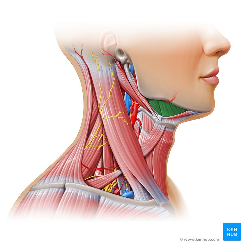
term (including?)
submandibular triangle (lymph nodes, hypoglossal/facial nerves)
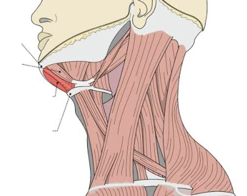
submental triangle

carotid triangle
importance of carotid triangle
carotid artery, too much pressure and pt ko

muscle triangle

external obliques

rectus abdominus
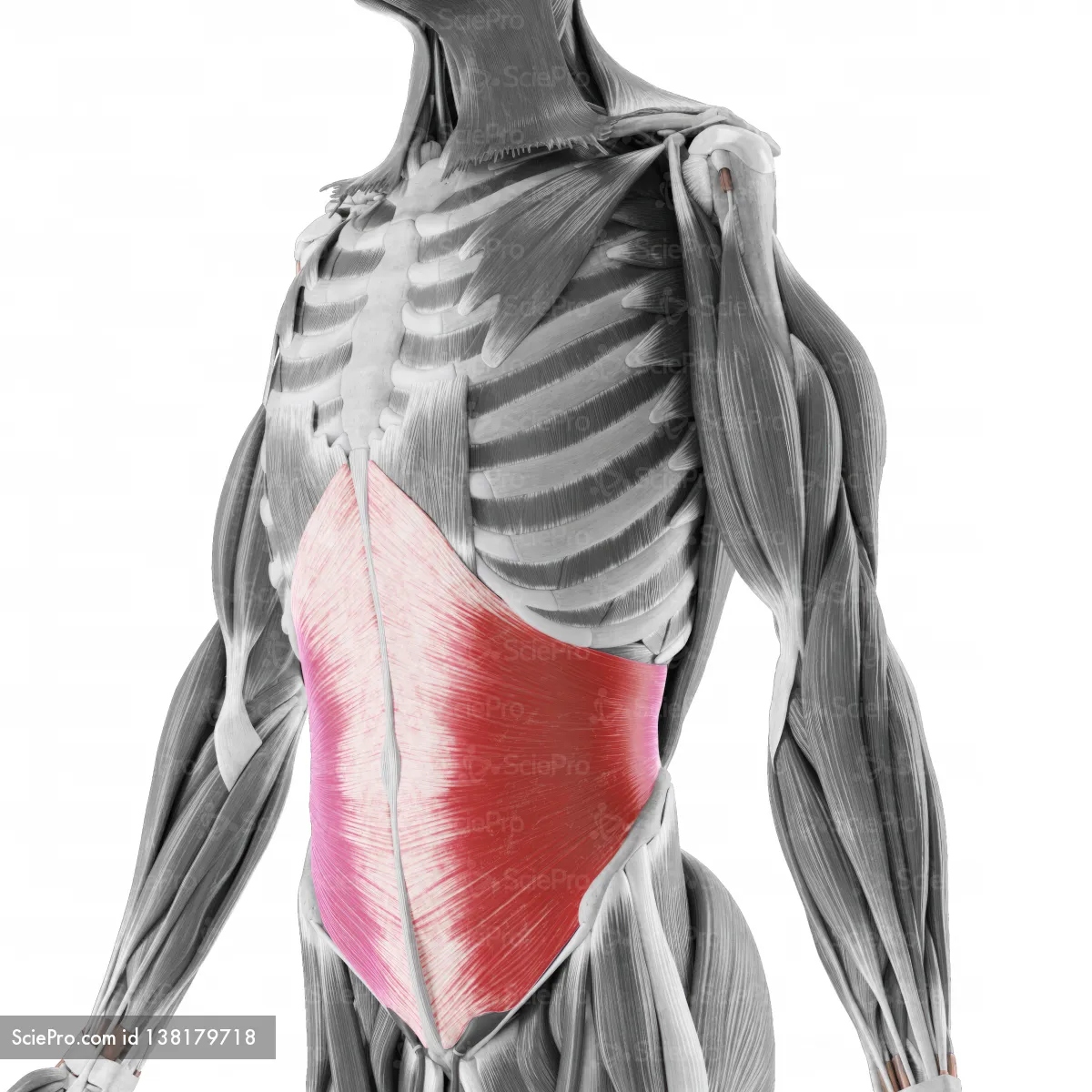
internal obliques

transverse abdominis

pectoralis major

deltoid

subclavius

linea alba
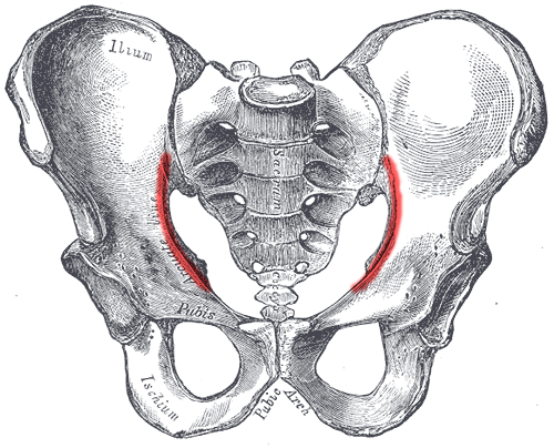
arcuate line
what differs in the aponeuroses of the abdomen above/below the arcuate line
above - internal obliques split and form around the rectus abdominis, transverse abdominis runs posterior
below - internal obliques and trasnverse abdominis runs anterior
significance of anterior lateral abdominal wall
lots of hernias
describe what a hernia is
separate of muscle fibers, space in between for organ/tissue petrusion
describe the singular physiological hernia (others are pathological)
week 10 of fetal life, fast growth of GI tract (intestines) spreads outside the abdominal wall
borders of anterolateral wall
coastal cartilage of ribs 7-10, pelvis/external obliques
layers of anterolateral abdominal wall (superficial to deep)
skin, superficial fatty (camper fascia), deep membranous (scarpa fascia), external/interal obliques, transverse abdominis, deep fascia (endoabdominal), fatty tissue (extraperitoneal fascia), parietal peritoneum
aponeurosis of what muscle for what important ligament (what’s the importance)
external obliques turn into the inguinal ligament (finds the femoral artery, hernias)
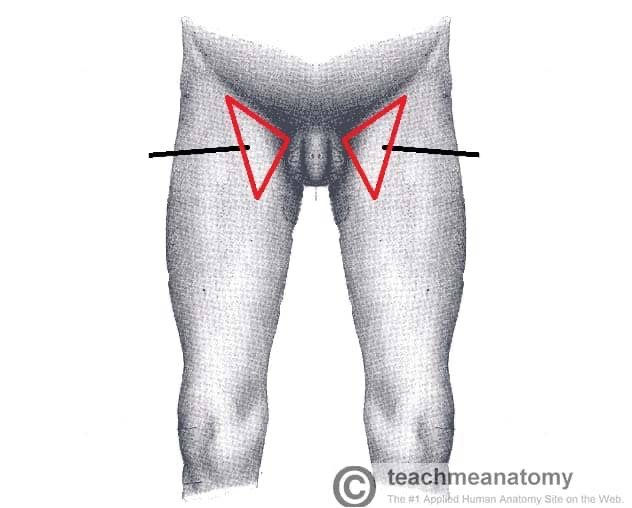
femoral triangle
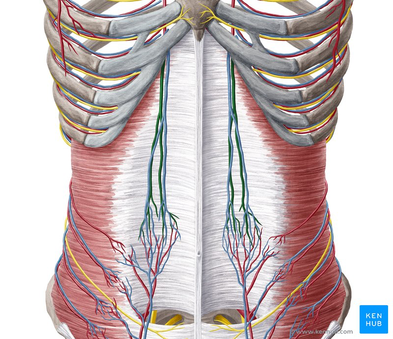
arteries (importance), location
superior/inferior epigastric arteries (supply abdominal muscles), deep to rectus abdominis
course of vessels/nerves in the abdomen
between internal obliques and transverse abdominis
folds of the posterior aspect of the anterolateral abdominal wall
median umbilical, 2 median umbilical, 2 lateral umbilical

fold (structures)
middle umbilical fold (urinal bladder, umbilicus)
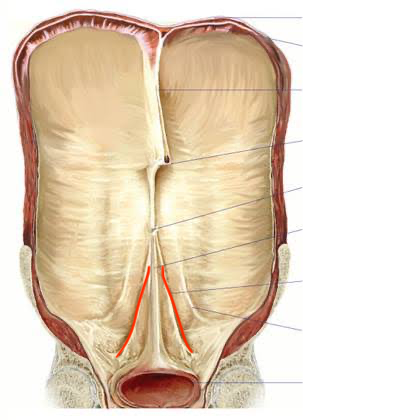
fold (structures)
anterior/posterior middle umbilical folds (embryo umbilical arteries

fold (structures)
anterior/posterior lateral umbilical folds (inferior epigastric vessels)
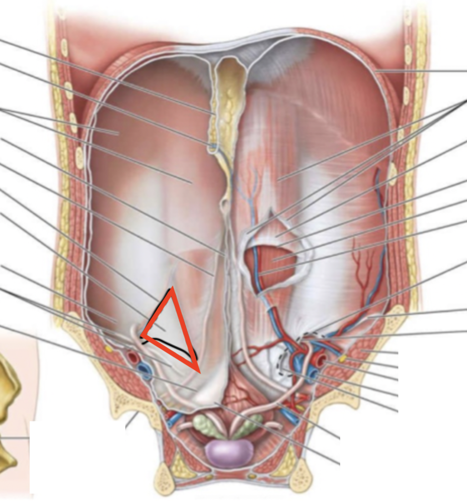
structure (importance)
Hesselbach Triangle (weak spot, inguinal canal/hernia)

lateral inguinal fossa
importance of inguinal canal for men
passageway of testes from posterior abdominal wall scrotum, through superficial inguinal ring (covering from wall)
importance of tested being outside of the abdominal cavity
temperature/spermatogenesis, high risk of testicular cancer

muscles (function)
cremaster muscles (pulls up testes)
importance of inguinal canal for women
framework to prevent overdescent of ovaries from the anterolateral wall to lesser pelvis. ligamentun teres goes through to attach itself to teres major and pubic
inguinal hernias are more common in ____ (why)?
men (bigger canal from testes than ligaments)
what goes through the inguinal canal along with the testes/ligamentum teres? (importance)
arteries, veins, nerves, lymphatic vessels (first sight of lymphatic drainage, no palpable in cases of cancer)

inguinal ligament

vas deferens

inguinal canal

epigastric vessels

deep inguinal ring
what separates the parietal cavity and the inguinal canal (importance)?
parietal peritoneum, transversalis fascia (thin layers, more likely to have a direct inguinal hernia
structures commonly involved in inguinal hernia
ilium, greater augmentum
what type of inguinal hernia is congenital?
indirect
describe indirect inguinal hernia
structure must pass through the deep inguinal ring into the inguinal canal and out the superficial inguinal ring into the scrotum
describe direct inguinal hernia
structures go through the Hesselbach triangle and put pressure on the inguinal canal, occasionally it pushes into the canal and out the superficial inguinal ring
why do inguinal hernias typically go unnoticed
symptoms include diarrhea, slight discomfort, and bulging, no pain
locate femoral canal
find the inguinal ligament from anterior superior iliac spine, posteriorly retroinguinal space that holds the femoral canal, including the femoral artery/vein and inguinal lymph node
why is the femoral canal not really a canal
its surrounded by loose connective tissue
what’s the space inside the femoral canal called? used for the diameter extension of the femoral artery
retrofemoral space
what covers the femoral ring (contents)?
femoral septum (connective and fatty tissue)
femoral septum differences men vs women (why)
fibers are more separate in women vs men (wider pelvis), hernia is more likely in women
which nerve innevates the anterior scalene
phrenic nerve
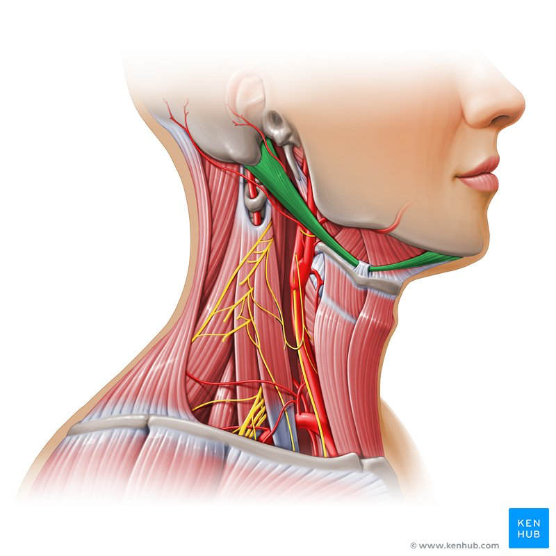
muscle (innervations)
anterior (trigeminal, mandibular)/posterior (facial) belly of the digastric muscle
describe corono mortis
vascular branch connecting the deep inferior epigastric vessels with the obturator vessels, commonly hit when repairing a femoral hernia
hesselbach triangle borders
medial (rectus abdominis), lateral (inferior epigastric vessels), inferior (inguinal ligament)
exit of a femoral hernia (importance)
saphenous opening in the fascia lata (same exit point for saphenous vein)