MRI Procedures for TMJ and Soft Tissue Neck: Techniques, Indications, and Imaging Protocols
 Call Kai
 Learn
Learn Practice Test
 Spaced Repetition
 Match
Match Flashcards
 Knowt Play
1/24

There's no tags or description
Looks like no tags are added yet.
Name | Mastery | Learn | Test | Matching | Spaced | Call with Kai | Chat |
|---|
No analytics yet
Send a link to your students to track their progress
25 Terms
What is TMJ?
Temporomandibular joint, the joint connecting the jaw to the skull.
What imaging coils are used for TMJ MRI?
Bilateral 3-inch coils or multi-channel head coil.

What is the patient position for TMJ MRI?
Supine, head first.
What landmark is used for TMJ imaging?
External auditory meatus (EAM).
What is the sequence of imaging for TMJ?
Closed mouth performed first, then open mouth.
What are the indications for TMJ MRI?
Painful TMJ clicking, masticatory muscle tenderness, limitation of opening, and frozen jaw.
What does a sagittal image setup for TMJ involve?
Angle perpendicular to the long axis of the mandibular condyles.
What is the purpose of dynamic MRI for TMJ?
To capture the movement and function of the TMJ.

What structures are included in soft tissue neck imaging?
Pharynx, larynx, thyroid and parathyroid glands, and salivary glands.
What coil is used for soft tissue neck MRI?
Multi-channel neurovascular coil, referred to as NV Array.
What is the patient position for soft tissue neck MRI?
Supine, head first.
What landmark is used for soft tissue neck imaging?
Sternal notch.
What are the indications for soft tissue neck MRI?
Undiagnosed tumors, congenital anomalies, salivary gland duct stones, obstructive thyroid disease, and abscesses.
What imaging technique can help reduce motion artifacts during neck scans?
Using gradient moment nulling and an inferior saturation band.
What is the recommended imaging protocol for TMJ?
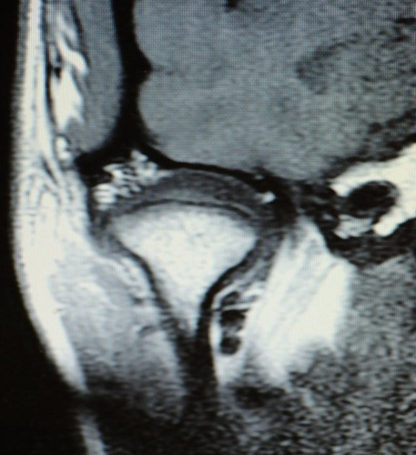
Sagittal T1, PD, T2 + Fat/Sat for open and closed mouth.
What type of images are taken for soft tissue neck MRI?
Coronal, axial, and sagittal images.
What is the significance of using saline bags during neck imaging?
To improve fat saturation.
What is the purpose of inversion recovery (STIR) in neck imaging?
To replace T2 fat/saturation images if fat is not uniformly saturated.
What is the anatomical coverage for coronal imaging of the neck?
From the anterior hyoid bone to the posterior aspect of the spinous processes.
What is the imaging setup for coronal neck scans?
Set up off sagittal images, scanning anterior to posterior or posterior to anterior.
What is a common protocol for soft tissue neck MRI post-contrast?
Axial T1 + Fat/Sat + Contrast, Sagittal T1 + Fat/Sat + Contrast.
What does the axial image setup for TMJ involve?
Setup off a coronal image with slices straight through the mandibular condyles.

What is the imaging technique for assessing TMJ disc function?
Sagittal imaging with open and closed mouth positions.

What is the importance of immobilization during soft tissue neck scans?
To minimize swallowing and avoid artifacts.
What are the two types of mouth positions used in TMJ MRI?
Open mouth and closed mouth.