Diseases of Skin and Eyes Chapter 16
 Call Kai
 Learn
Learn Practice Test
 Spaced Repetition
 Match
Match Flashcards
 Knowt Play
1/133

There's no tags or description
Looks like no tags are added yet.
Name | Mastery | Learn | Test | Matching | Spaced | Call with Kai | Chat |
|---|
No analytics yet
Send a link to your students to track their progress
134 Terms
Epidermis
top layer
replaced every 25-45 days

Stratum corneum
layer in epidermis
dead cells that have migrated from deeper layers

Dermis
contain sebaceous glands and hair follicles

blister formation
result of separation of epidermis and dermis
sebaceous glands
secrete sebum and are associated with hair follicles
antimicrobial peptides
positively charged chemicals in the skin that disrupt the negatively charged membranes of the bacteria
keep microbial count on skin relatively low
Sebum
low pH (acidic) makes skin inhospitable to microorganisms
high lipid concentration serve as nutrients for normal microbiota
Sweat
inhibit microorganisms due to low pH (acidic) and high salt concentration
Lysozyme
enzyme found in sweat, tears, and saliva
break down peptidoglycan
works best on Gram positive bacteria
Normal skin biota
streptococcus
staphyloccus
corynbacterium
propionibacterium
pseudomonas
lactobacillus
yeasts such as Candida
Staphylococcus sp. general info
gram positive cocci
S.aureus is main pathogenic species
white-bronze colored colonies on agar
withstands high salt, extreme pH, and high temps
Staphylococcus physiology and structure
capsules
peptidoglycan outer layer (endotoxin like effect)
Protein A (binds IgG)
Characteristics of MRSA
resistant to multiple antibiotics
Staphylococcus aureus
remains viable after air drying
signs and sx of MRSA
raised, red, tender localized lesions
pus
hot to touch
may localize around a hair follicle
fever
trasmission of MRSA
direct or indirect contact
any surface
culture/diagnosis of MRSA
PCR
isolation on SBA or MSA
positive catalase distinguishes S.aureus from other streps
MRSA prevention/treatment
good hygiene
incision of lesion and pus drainage
multiple antimicrobial treaments
MRSA toxins
Alpha, beta, gamma, delta, hemolysins
Exfoliative toxins: cause skin to blister + peel
MRSA enzymes
coagulase, catalase, hyaluronidase, fibrinolysin, nuclease, penicillinase
MRSA virulence factors
Leukocidin: kills neutrophils
Catalase: inhibits antibacterial activities
Hyaluronidase: dissolves cell-cell connection
Scalded skin syndrome (SSS): exfoliative toxin
Toxic shock syndrome (TSS)
Slime production: capsule like layer
Hemolysin: rupture RBC
Protein A: bind Fc region of IgG antibodies/ attachment
antibiotic resistance: penicillinase and beta lactimase
Scalded skin syndrome
pediatric, large bullae (blister) formed under the skin and rupture
Toxic shock syndrome sx
high fever, headache, confusion, macular rash, conjunctival injection, vomiting, diarrhea
caused by S aureus
Penicillinase
inactivates penicillin
usually associated with a plasmid
85% of S aureus are resistant to _______-
penicillin G
what is used to treat S aureus
vancomycin
starring to see VRSA
S aureus coagulase/catalase
coagulase: +
catalase: +
Characteristics of impetigo
superficial bacteria that causes the skin to flake
Bacteria that cause impetigo
Staphyloccoccus aureus or Streptococus pyogenes
which bacteria begins every case of impetigo
streptococcus pyogenes
which bacteria often takes over/ outcompetes in cases of impetigo
Staphylococcus aureus
Signs and sx of impetigo
peeling skin, honey-colored crusts
lesions around mouth, face, and extremities
Impetigo caused by Staphylococcus aureus
exfoliative toxins A and B cause blistering and spread of the bacterium
Streptococcus pyogenes differentials
Gram positive coccus
Beta hemolytic on SBA
negative catalase
Diseases caused by streptococcus pyogenes
streptococcus pharyngitis, scarlet fever, pneumonia, puerperal fever, necrotizing fasciitis, bacteremia
which organism is more likely to cause impetigo in newborns
Streptococcus pyogenes
which organism is more likely to cause impetigo in children
Staphylococcus aureus
Mupirocin
inhibits protein synthesis
Gram + and some Gram negative
Impetigo antibiotic treatments
mupirocin
Cephalosporins, Amocicillin-Clavulonic Acid
Penicillin (only if S. pyogenes)
Cephalosporins
inhibit cell wall synthesis
Amoxicillin-Clavulonic Acid
covers Beta-lactamase producing strains
Macropapular diseases
Measles, Rubella, Fifth Disease, Roseola
Measles (rubeola) signs and sx
sore throat
dry cough
headache
conjunctivitis
lymphadenitis
fever
red, macropapular exanthem that begins on head and spreads to trunk and extremities
Kopliks spots
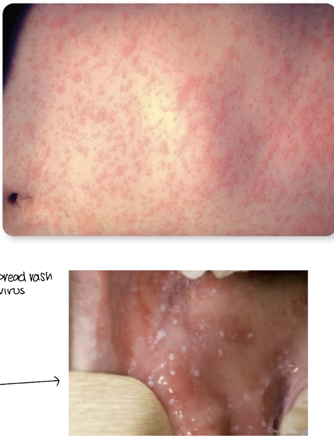
exanthem
widespread rash from a virus or bacteria
Subacute sclerosing panencephalitis
degeneration of the cerebral cortex, white matter, and brainstem
involves a defective measles virus
measles pathogenesis and virulence factors
implant on respiratory mucosa, travel to lymphatic system and enters bloodstream which then goes to the skin
causes host cells to fuse together and not preform properly
transmission of mealses
respiratory droplets
how to test for measles
ELISA to test for IgM antigens
attenuated
modified weaker viruses in vaccines
treatment for measles
treating sx
rubella causative agent
rubivirus
Rubella virulence
stop mitosis in fetuses
apoptosis of tissue cells, causing harm to organs
damages vascular endothelium
characteristics of rubella
minor rash with few complications
serious damage in utero (teratogen)
german measles
rubella
how is rubella diagnosed
IgM antibody detection through ELISA
when is rubella virus shed
through prodromal phase and up to a week after rash appears
Fifth Disease
Parvovirus B19
Erythema infectiosum
fifth disease
how is fifth disease diagnosed
clinical presentation and ruling out rubella through testing for IgM antibodies
Fifth disease prevention and treatment
no vaccine and no treatment
Roseola causative agent
human herpesvirus 6 (HHV-6)
roseola transmission and epidemiology
it is though 100% of US population is infected by adulthood
roseola prevention and treatment
no vaccine and no treatment
Cellulitis
fast spreading infection in the dermis and subcutaneous tissue
lymphangitis
red lines leading away from the area of cellulitis
result of microbes being carries by the lymphatic system
cellulitis signs and sx
pain, tenderness, swelling, warmth
fever and swelling of the lymph nodes
cellulitis causative agents
in healthy individuals: staphylococcus aureus or streptococcus pyogenes
immunocompromised individuals: any bacteria, some fungi
infants: group B streptococci
group B strep
can naturally occur in the body
typically only cause disease in immunocompromised/infants/elderly
cellulitis transmission
introduction of bacteria into dermis, trauma ect.
cellulitis diagnosis
clinical sx
cellulitis treatment
oral or IV antibiotics
Styaphylococcal scalded skin syndrome (SSSS)
dermolytic skin condition caused by S. aureus
affects mostly newborns and babies
SSSS signs and sx
bullous lesions (fluid filled blister), widespread dequamation of skin
SSSS causative agent
staphylococcus aureus
exfoliative toxins A and B
SSSS transmission
from baby to baby or caregivers may be asymptomatic carriers
SSSS diagnosis
clinical sx
SSSS prevention and treatment
prevent contact w carriers
systemic antibiotics
Vesicular or Pustular rash diseases
chickenpox and smallpox
pox
individual lesions that contain fluid
Chickenpox causative agent
Alphaherpesvirus 3 aka Varicella-Zoster Virus
Chickenpox signs and sx
fever and rash that starts on scalp/face and trunk and radiates to extremeties
macules
flat spot
papules
raised bump
centripetal
more lesions in the center of the body than in the extremities
Shingles causative agent
Herpes zoster that enter sensory endings of spinal nerves after chickenpox
can be reactivated by stress, immunosuppressive therapy
shingles signs and sx
persistent vesicles
pain and tenderness
eye inflammation and facial paralysis
chickenpox/shingles pathogenesis
enters respiratory tract and invades bloodstream then moves to skin
virus then enters sensory nerves and remains latent
chickenpox/shingles prevention and treatment
vaccines
treat sx, no aspirin as may lead to Reye’s syndrome
smallpox signs and sx
fever, rash that starts in pharynx and then spreads to face and extremities
variola major
highly virulent
causes toxemia, shock, and intravascular coagulation
rash w bumps filled w thick, opaque fluid
variola minor
minor, less dense rash
smallpox causative agent
variola virus
smallpox transmission
droplets, contaminated bedding and clothing
smallpox prevention/treatment
vaccine, no treament
Cutaneous leishmaniasis causative agent
leishmania tropica
mucocutaneous leishmaniasis causative agent
leishmania brasiliensis
leishmaniasis transmission
female sand flies
leishmaniasis diagnosis
microscope visualisation, giemsa stain
leishmaniasis prevention/treatment
no vaccine, avoiding sand fly is the only way
leishmaniasis
parasitic disease
Cutaneous anthrax causative agent
bacillus anthracis